RESUMO.-
[
Investigação sorológica de anticorpos contra
vírus respiratórios em bezerros de propriedades familiares
brasileiras e sua relação com os sinais clínicos da doença
respiratória dos bovinos.
] A doença respiratória dos bovinos
(DRB) é responsável por importantes perdas econômicas
para a produção bovina, com maior impacto na agricultura
familiar. Os vírus são comumente caracterizados como
agentes etiológicos primários devido a mudanças na mucosa
respiratória, na produção de citocinas e no funcionamento do
sistema imune. Os objetivos deste estudo transversal foram
avaliar a presença de anticorpos contra o vírus da diarreia
ABSTRACT
.
-
Gaeta N.C., Ribeiro B.L.M., Alemán M.A.R., Yoshihara E., Marques E.C., Hellmeister
A.N., Pituco E.M. & Gregory L. 2018.
Serological investigation of antibodies against
respiratory viruses in calves from Brazilian family farming and their relation to
clinical signs of bovine respiratory disease.
Pesquisa Veterinária Brasileira 38(4):642-648.
Departamento de Clínica Médica, Faculdade de Medicina Veterinária e Zootecnia, Universidade
de São Paulo, Avenida Prof. Dr. Orlando Marques de Paiva 87, Cidade Universitária, São Paulo,
SP 05508 270, Brazil. E-mail: lgregory@usp.br
Bovine respiratory disease (BRD) is responsible for economic losses in cattle production.
Viruses are categorized as primary etiological agents. The aims of this study were to evaluate
the presence of antibodies against bovine viral diarrhea virus (BVDV), bovine herpes virus
type 1 (BoHV-1), and bovine respiratory syncytial virus (BRSV) in healthy and BRD calves
from family farming in relation to clinical signs of BRD. Hundred and forty-five calves
were randomly selected and physical examination was performed. Only 123 animals were
classified as healthy and BRD calves. Antibodies were evaluated by virus neutralization
test. Person’s Chi-square test and Fisher’s exact test were performed as univariate analysis.
Binary Logistic Regression was applied as multivariate analysis. Variables with
P
<0.10 were
considered statistically significant. Variables with 0.15<
P
<0.10 were considered as statistical
tendencies. Antibodies against BoHV-1, BVDV, and BRSV were detected in 32%, 23% and
37% animals. Antibodies against both three viruses were detected in 26.8% of calves.
The presence of antibodies against BRSV were associated to normal heart rates (
P=
0.018)
and unilateral airflow (
P=
0.035). Tendency was observed to unilateral airflow (
P=
0.06)
Statistical tendencies were observed to Ab-BoHV-1 and body temperature (
P=
0.119) and
breathing pattern (
P=
0.123). The profile of antibodies against respiratory viruses in calves
from Brazilian family farming was firstly described. The results confirmed the importance
of some clinical signs to viral infection.
INDEX TERMS: Serology, antibodies, respiratory viruses, calves, cattle, bovine respiratory disease, BVDV, BRSV, BoHV-1, viroses.
Vet 2561 pvb-5234 LD
Serological investigation of antibodies against
respiratory viruses in calves from Brazilian family
farming and their relation to clinical signs of bovine
respiratory disease
1Natália C. Gaeta
2*, Bruno L.M. Ribeiro
2, Mario A.R. Alemán
2,
Eidi Yoshihara
3, Eduardo C. Marques
2, Adriana N. Hellmeister
4,
Edviges M. Pituco
4and Lilian Gregory
21 Received on January 20, 2017.
Accepted for publication on April 29, 2017.
2 Departamento de Clínica Médica, Faculdade de Medicina Veterinária e Zootecnia, Universidade de São Paulo, Avenida Prof. Dr. Orlando Marques de Paiva 87, Cidade Universitária, São Paulo, SP 05508 270, Brazil. *Corresponding author: natalia.gaeta@hotmail.com
3 Agência Paulista de Tecnologia dos Agronegócios, Departamento de Descentralização do Desenvolvimento, Pólo Regional de Desenvolvimento Tecnológico dos Agronegócios da Alta Sorocabana, Cx. Postal 298, Rod. Raposo Tavares Km 563, Presidente Prudente, SP 19015-970, Brazil.
viral bovina (VDVB), herpes vírus bovino tipo 1 (HVbo-1)
e vírus respiratório sincicial bovino (VRSB) em bezerros
sadios e com DRB de assentamentos em associação com a
presença sinais clínicos de DRB. Cento e quarenta e cinco
animais foram randomicamente selecionados e exame físico
foi realizado. Somente 123 animais foram classificados em
sadios e com DRB. Anticorpos foram identificados pelo teste
de virusneutralização. Teste de qui-quadrado e teste exato de
Fisher foram utilizados como análise univariada. A análise
multivariada foi realizada pela regressão binária logística
com o método Backward conditional. Variáveis com
P<
0,10
foram considerados significantes. Variáveis com 0,15<
P
<0,10
foram consideradas tendências. Anticorpos contra HVbo-1,
VDVB e VRSB foram detectados em 32%, 23% e 37% dos
animais. A presença concomitante de anticorpos contra os
três vírus foi detectada em 26,8% dos bezerros. A presença
de anticorpos contra VRSB foi associada à frequência cardíaca
normal (
P=
0,018) e fluxo de ar nasal unilateral (
P=
0,035).
Tendência estatística observada para anticorpos contra
VDVB temperatura corporal (
P=
0,119) e padrão respiratório
(
P=
0,123). Ao nosso conhecimento, este é o primeiro estudo
que objetivou a avaliação da presença de anticorpos contra
VDVB, VRSB e HVbo-1 em bezerros de assentamentos do
estado de São Paulo, Brasil e sua relação com as manifestações
clínicas da DRB.
TERMOS DE INDEXAÇÃO: Sorologia, anticorpos, vírus respiratório, bezerros, bovinos, doença respiratória bovina, complexo respiratório bovina, VDVB, VRSB, HVbo-1, viroses.
INTRODUCTION
Bovine respiratory disease (BRD) is responsible for great
economic losses in cattle production (USDA 2013), with major
impact on family farming due to poor sanitary control. Viruses
are commonly categorized as primary etiological agents of
BRD, by changing respiratory mucosa, producing of cytokines
and functioning of immune system cells (Bosch et al. 2013).
Bovine viral diarrhea virus (BVDV), bovine herpes virus
type 1 (BoHV-1), bovine respiratory syncytial virus (BRSV)
are important species to the pathogenesis of BRD.
Bovine viral diarrhea virus (BVDV) belongs to family
Flaviviridae
and to Genus
Pestivirus
(ICTV 2014). BVDV is
responsible for diarrhea, reproductive disorders, mucosal
disease and pneumonia. Seroprevalence studies are frequently
conducted on adults due to its importance as causative agent
of reproductive disorders (Samara et al. 2004, Quincozes et al.
2007, Brito et al. 2010, Piovesan et al. 2013, Pasqualotto et al.
2015), while researches with calves showing respiratory
diseases are uncommon.
Bovine Herpes Virus type 1 (BoHV-1), as well as BVDV,
it is an important etiological agent of bovine reproductive
disorders (infectious pustular vulvovaginitis and infectious
balanoposthitis), in addition to respiratory diseases (infectious
bovine rhinotracheitis - IBR). BoHV-1 belongs to Genus
Varicellovirus,
Family
Herpesviridae,
subfamily
Alphaherpesvirinae
,
and Order
Herpesvirales
(ICTV 2014). BoHV-1 has the ability to
lead latent infections. During stress periods (e.g. transportation,
lactation and calving), the reactivation of the infection is
observed, and the transmission of the virus (Ackermann &
Wyler 1984) occurs. Young calves are more susceptible to
respiratory disease infection by BoHV-1, showing unspecific
clinical symptoms such as fever, inappetence and depression,
besides mucopurulent nasal discharge (Oliveira 2011).
As well as BVDV, seroprevalence studies about BRD calves is
uncommon, while same studies about adults are usual due to
its importance for reproductive disorders. The prevalence of
BoHV-1 in Brazilian herds ranges between 41,9% and 92,95%
(Médici et al. 2000, Poletto et al. 2004).
Bovine Respiratory Syncytial Virus (BRSV) belongs to
Order
Mononegavirales
, Family
Paramyxoviridae
and Genus
Pneumovirus
(ICTV 2014). Clinical signs of BRSV infection
are milder in adults compared to calves. The infection is
characterized by clinical signs frequently associated to other
respiratory agents such as depression, fever, cough, anorexia,
and tachypnea (Gershwin 2007). Although few studies reported
BRSV infection in cattle (Driemeier et al. 1997), only one
describes the occurrence of antibodies against BRSV in Brazil.
In São Paulo State, Affonso et al. (2011) observed prevalence
between 46% and 84%, which suggests its importance to the
respiratory diseases in this state.
According to São Paulo Land Institute Foundation (ITESP),
milk production on family farming farms in São Paulo State,
from 2010 to 2011 was 71.635.36 liters. Family farming
influences the local economy (Mazzini et al. 2011) since
producers supply their milk to local dairies and indirectly
produces new jobs every day.
Bovine respiratory disease affects all ages, especially young
calves. Besides having few Brazilian studies that evaluated the
prevalence of antibodies against the most important viruses
related to the respiratory diseases, researches that estimated
their prevalence in calves of family farming are not available.
Thus, the aim of this cross-sectional study was to determine
the antibodies against bovine viral diarrhea virus, bovine
herpes virus type 1, and bovine respiratory syncytial virus
in healthy and BRD calves from family farming farms located
in Caiuá, Presidente Epitácio and Mirante de Paranapanema,
São Paulo State, Brazil in relation to clinical signs of BRD.
MATERIALS AND METHODS
The present study was conducted at the Laboratory of Bovide Viruses, Biologic Institute and at the Department of Internal Medicine, School of Veterinary Medicine and Animal Science, University of São Paulo, Brazil, from August 2014 until March 2016. All procedures were carried out in agreement with the guidelines Committee of Ethics on Animal Use (Protocol number: 7973040214).
Area definition. The State of São Paulo is located in the southeast region of Brazil (21°49’S 49°12’W). It has 645 cities and a total area of 248,222,362 Km2. The cattle population is 10.126.223 animals, from which 1,287,509 are dairy cattle (IBGE 2014). Pontal do Paranapanema is located in the extreme west region of the São Paulo State. It has 32 municipalities with family farming distributed along the region. The present study was carried out in Caiuá (21°49’55”S 51°59’52”O), Presidente Epitácio (21°45’53”S 52°06’19”O) and Mirante de Paranapanema(22°17’31”S 51°54’21”O), important cities located in Pontal do Paranapanema.
Animals and case definition. Hundred and forty-five
base diet. Mostly herds received vaccination against clostidiosis, foot-and-mouth disease and brucelosis. No vaccination against respiratory viruses was performed. Calves were randomly selected
in each farm and they were classified as healthy and BRD calves after physical examination. BRD was defined when calves showed two or
more of the following clinical signs (Benesi et al. 2013, Lima et al. 2016, Gaeta et al. 2017): mucopurulent or purulent nasal discharge, cough, crackle/snoring/whistle, respiratory rate >40 breaths/min and rectal temperature >39.5oC. An experienced veterinarian member of our research team performed the physical examination in all calves.
Total blood collection. Eight mililiters of total blood was obtained by jugular vein punction using a dry BD vacutainer tube. All tubes
were centrifuged at 600xg for 15 minutes and the serum obtained was stored in tubes at -20°C until processing.
Virus neutralization for BVDV, BoHV-1 and BRSV. Serum diagnosis to BVDV and BoHV-1 was performed by virus neutralization (VN) according to OIE (2004). Antibodies against BRSV were also detected by virus neutralization, but according to Affonso (2012). Serological response was evaluated in relation to BVDV-Ia NADL strain, BoHV-1 Los Angeles strain and ATCC VR-1485 incubated in Madin-Darby Bovine Kidney cells (MDBK) in 96 well plates. All plates were analyzed after incubation at 37oC/5% CO
2. Antibody titers were expressed as the inverse of the dilution that the absence of cytopathic effect was observed. Samples with titers equal or higher than three were considered reactive. VN tests were validated through the control of negative and positive sera anti-BVDV antibodies, control of cells, and retrotitration (log10), according to Reed-Muench method (1938). All tests were performed at the Laboratory of Bovine Viruses, Biologic Institute, São Paulo, Brazil. Laboratory personnel
were blinded to calves classification.
Statistical analysis. Descriptive results were determined by relative and absolute frequencies. Pearson’s chi-square test and Fisher’s exact test were applied to evaluate the association between the presence of antibodies against BVDV, BoHV-1 and BRSV and health status (healthy and BRD calves), besides the clinical signs of BRD. Binary logistic regression with Backward conditional method was also performed. Variables related to clinical signs were grouped to obtain
more significant analysis (altered nasal discharge: mucopurulent
or purulent; altered percussion: coarse and thin crackle, whistle, and snoring; altered percussion: submassive or massive sounds). Odds Ratio was also determined. Analyzes were performed using SAS (SAS Institute Inc., NC). Variables with P<0.10 were considered statistically different. Variables with 0.15<P<0.1 were considered as statistical tendencies.
RESULTS
Serum diagnosis to BVDV, BRSV and BoHV-1 was performed
in 145 samples, revealing 23% (34/145), 37% (54/145) and
32% (46/145) of reactive calves, respectively. The lack of some
clinical information allowed the classification of 123 calves
as healthy and BRD calves. No differences between healthy
and BRD calves were detected to BoHV-1 (
P=
0.281), BVDV
(
P=
0.982), and BRSV (
P=
0.485). Calves showing antibodies
against BRSV were numerically increased in BRD calves
(Table 1).
The concomitant presence of antibodies against two or
three viruses was observed in 22.8% calves, out of which
BoHV-1+BRSV was numerically higher compared to the others
(Table 2). The concomitant detection of antibodies against
BVDV+BRSV was increased in BRD calves compared to healthy
calves (
P
=0.011). Antibodies against all three viruses were
detected in healthy calves only.
The frequency of calves showing antibodies titers against
BRSV and BoHV-1 were numerically increased between 2 and 16.
The highest titers (2048-8192) were noted in calves showing
antibodies against BVDV, only (Table 3).
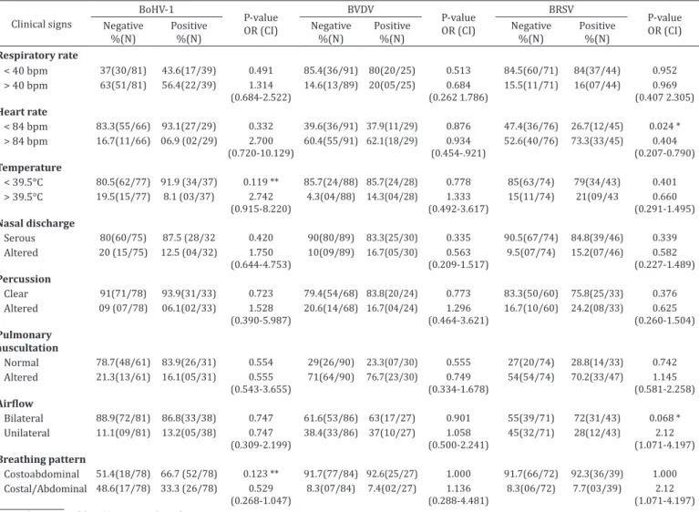
Table 4 shows the results from odds ratio and Chi-square
test /Fisher’s exact test. The variables of heart rates (
P=
0.024),
and unilateral airflow (
P=
0.068) were selected for multivariate
analysis. Statistical tendencies were observed to Ab-BoHV-1
and body temperature (
P=
0.119) and breathing pattern
(
P=
0.123). In multivariate analysis, statistical significance was
still observed for heart rate (
P=
0.18) and airflow (
P=
0.035).
Table 1. Antibodies against BVDV, BRSV and BoHV-1 according to health status of calves of family farming farms from Caiuá, Presidente Epitácio and Mirante de Paranapanema, São Paulo state, Brazil, 2016
Antibodies Healthy%(N) %(N)BRD %(N)Total OR (CI 90%) P-value
BoHV-1 34.4 (31/90)a 24.2 (08/33)a 31.7 (39/123) 1.642 (0.767-3.515) 0.281
BVDV 24.4 (22/90)a 24.2 (08/33)a 24.4 (30/123) 1.011 (0.463-2.207) 0.982
BRSV 35.6 (32/90)a 42.4 (14/33)a 37.4 (46/123) 0.485 (0.378-1.483) 0.485
a,b Different letters indicate statistical difference (P<0.10).
Table 2. Simultaneous frequency of antibodies against BVDV, BRSV and BoHV-1 according to healthy status of calves reared in settlements of Caiuá, Presidente Epitácio and Mirante de Paranapanema, São Paulo state, Brazil, 2016
Antibodies Healthy% (N) % (N)BRD % (N)Total CI (90%) P-value
BoHV-1+BRSV 14.40 (13/90)a 6.10 (02/33)a 12.20 (15/123) 0.729 (0.188-2.833) 0.350
BVDV+BRSV 3.30 (03/90)a 18.20(06/33)b 7.30 (09/123) 5.833 (1.7111-9.886) 0.011
BVDV+BoHV-1 4.40 (04/90)a 3.00 (01/33)a 4.10 (05/123) 0.729 (0.111-4.777) 1.000
BVDV+BoHV-1+BRSV 5.60 (05/90)a 0.00 (00/33)a 4.10 (04/123) - 0.573
Total 22.20(20/90) 27.30 (09/33) 22.80(28/123)
Table 3. Antibody titers frequency according healthy status of calves from settlements in Caiuá, Presidente Epitácio and Mirante de Paranapanema, São Paulo state, Brazil, 2016
Antibody titer Healthy
%(N)
BRD %(N)
Total %(N)
BoHV-1
Negative 65(59/90) 76 (25/33) 68 (84/123)
2-16 20 (18/90) 18 (06/33) 19 (24/123)
32-128 10 (09/90) 06 (02/33) 09 (11/123)
256-1024 04 (04/90) - 03 (04/123)
2048- 8192 - -
-BVDV
Negative 75 (68/90) 76 (25/33) 76 (93/123)
2-16 03 (03/90) 03 (01/33) 03 (04/123)
32-128 13 (12/90) 09 (03/33) 12 (15/123)
256-1024 05 (05/90) 09 (03/33) 06 (08/123)
2048- 8192 02 (02/90) 03 (01/33) 02 (03/123)
BRSV
Negative 64 (58/90) 54 (18/33) 62 (76/123)
2-16 32 (29/90) 42 (14/33) 35 (43/123)
32-128 03 (03/90) 03 (01/33) 03 (04/123)
256-1024 - -
-2048- 8192 - -
-Table 4. Antibodies against respiratory viruses in relation to clinical signs detected after physical examination of healthy and BRD calves
Clinical signs
BoHV-1
P-value OR (CI)
BVDV
P-value OR (CI)
BRSV
P-value OR (CI) Negative
%(N)
Positive %(N)
Negative %(N)
Positive %(N)
Negative %(N)
Positive %(N)
Respiratory rate
< 40 bpm 37(30/81) 43.6(17/39) 0.491 85.4(36/91) 80(20/25) 0.513 84.5(60/71) 84(37/44) 0.952
> 40 bpm 63(51/81) 56.4(22/39) 1.314 (0.684-2.522)
14.6(13/89) 20(05/25) 0.684
(0.262 1.786)
15.5(11/71) 16(07/44) 0.969
(0.407 2.305) Heart rate
< 84 bpm 83.3(55/66) 93.1(27/29) 0.332 39.6(36/91) 37.9(11/29) 0.876 47.4(36/76) 26.7(12/45) 0.024 *
> 84 bpm 16.7(11/66) 06.9 (02/29) 2.700 (0.720-10.129)
60.4(55/91) 62.1(18/29) 0.934 (0.454-.921)
52.6(40/76) 73.3(33/45) 0.404 (0.207-0.790) Temperature
< 39.5°C 80.5(62/77) 91.9 (34/37) 0.119 ** 85.7(24/88) 85.7(24/28) 0.778 85(63/74) 79(34/43) 0.401
> 39.5°C 19.5(15/77) 8.1 (03/37) 2.742 (0.915-8.220)
4.3(04/88) 14.3(04/28) 1.333 (0.492-3.617)
15(11/74) 21(09/43 0.660
(0.291-1.495) Nasal discharge
Serous 80(60/75) 87.5 (28/32 0.420 90(80/89) 83.3(25/30) 0.335 90.5(67/74) 84.8(39/46) 0.339
Altered 20 (15/75) 12.5 (04/32) 1.750 (0.644-4.753)
10(09/89) 16.7(05/30) 0.563 (0.209-1.517)
9.5(07/74) 15.2(07/46) 0.582
(0.227-1.489) Percussion
Clear 91(71/78) 93.9(31/33) 0.723 79.4(54/68) 83.8(20/24) 0.773 83.3(50/60) 75.8(25/33) 0.376
Altered 09 (07/78) 06.1(02/33) 1.528
(0.390-5.987)
20.6(14/68) 16.7(04/24) 1.296 (0.464-3.621)
16.7(10/60) 24.2(08/33) 0.625 (0.260-1.504) Pulmonary
auscultation
Normal 78.7(48/61) 83.9(26/31) 0.554 29(26/90) 23.3(07/30) 0.555 27(20/74) 28.8(14/33) 0.742
Altered 21.3(13/61) 16.1(05/31) 0.555 (0.543-3.655)
71(64/90) 76.7(23/30) 0.749 (0.334-1.678)
54(54/74) 70.2(33/47) 1.145
(0.581-2.258) Airflow
Bilateral 88.9(72/81) 86.8(33/38) 0.747 61.6(53/86) 63(17/27) 0.901 55(39/71) 72(31/43) 0.068 *
Unilateral 11.1(09/81) 13.2(05/38) 0.747 (0.309-2.199)
38.4(33/86) 37(10/27) 1.058
(0.500-2.241)
45(32/71) 28(12/43) 2.12
(1.071-4.197) Breathing pattern
Costoabdominal 51.4(18/78) 66.7 (52/78) 0.123 ** 91.7(77/84) 92.6(25/27) 1.000 91.7(66/72) 92.3(36/39) 1.000
Costal/Abdominal 48.6(17/78) 33.3 (26/78) 0.529 (0.268-1.047)
8.3(07/84) 7.4(02/27) 1.136 (0.288-4.481)
8.3(06/72) 7.7(03/39) 2.12
(1.071-4.197)
DISCUSSION
Viral infection are important determinant factor to respiratory
infection due to significant changes in the respiratory
environment (Bosch et al. 2013) and it is frequently reported
as a predisposing factor to bacterial infections (Hartel et al.
2004, Autio et al. 2007).
In the present study, the prevalence of antibodies against
BoHV-1 (Ab-BoHV-1) was 31.7%. Results reported in the state
of Rio Grande do Sul, Brazil, by Poletto et al. (2004), were
similar to the present research (32.4%). Lower frequencies
were detected in the same state by Becker et al. (2015), in
which study frequencies ranged from 26.4% in 2013 to 21.7%
in 2014. Many studies have detected high frequencies of
Ab-BoHV-1 in Brazil. In the Northeast region, Silva et al. (2015)
and Sousa et al. (2013) detected prevalence of 79.5% and 67.5%
in the states of Pernambuco and Maranhão, respectively. In the
Center-south region, Oliveira et al. (2016) and Santos et al.
(2014) reported prevalence of 61.4% and 66.75% in the
states of São Paulo and Espírito Santo. The high frequency
of animals with Ab-BoHV-1 in several Brazilian states could
be related to the virus’ ability to remain latent in the nerve
ganglia, allowing its recurrent shedding and the maintenance
of the infection inside the herd (Médici et al. 2000). As well
as observed in Brazil, the occurrence of Ab-BoHV-1 outside
the country is variable (Boelaert et al. 2000, Raaperi et al.
2010, 2014, Szeredi et al. 2010).
The prevalence of antibodies against BVDV (Ab-BVDV)
observed in the present research was 24.4%. Recent national
studies showed higher occurrence compared to our results.
In the States of São Paulo and Minas Gerais, Alexandrino et al.
(2011) detected 69.70% of reagents. Chaves et al. (2010)
reported 61.5% of animals showing Ab-BVDV in Maranhão.
Piovesan et al. (2013) and Quincozes et al. (2007) detected
85.4% and 66.32% of reagents, respectively, in the State of
Rio Grande do Sul. In Brazilian’s central region, Brito et al.
(2010) observed prevalence of 64% in the State of Goiás.
In the Northeast, Fernandes et al. (2016) reported lower
prevalence. Finally, Oliveira et al. (2016) reported 55.35%
of positive samples in São Paulo State, especially in healthy
calves. On the other hand, higher frequencies in BRD calves
were detected in the present study (33%).
In our research, the occurrence of antibodies against
BRSV (Ab-BRSV) was 37.4%. Affonso et al., (2011) reported
prevalence ranged from 45.61% to 84.42% in São Paulo State.
In different locations in the same state, Oliveira et al. (2016)
detected 93.10%. In the South, Campalans & Arns (1997) ,
reported prevalence of 75%. In order to evaluate the role
of BRSV in cases of pneumonia in calves from the state of
Minas Gerais, Flores et al. (2000) observed Ab-BRSV in 8/41
pulmonary tissue by indirect immunofluorescence.
Discrepancies between serological results observed in
the present research and those reported elsewhere occurs
due to differences between studied locations, production
type, management, ages and diagnostic methods performed
(Silva et al. 2015).
Many studies report the clinical signs of bovine respiratory
disease by viral infection. Elvander (1996) described an
outbreak of pneumonia in cattle by BRSV in Sweden. Animals
showed fever, respiratory distress, coughing, a serous
nasopharyngeal discharge, tearing and salivation. Similarly, a
spontaneous BRSV infection in Brazilian cattle was described
by Peixoto et al. (2000) who referred fever, dry cough and
serous ocular discharge, besides loud and severe breathing
sounds detected by pulmonary auscultation. In the present
study, the presence of Ab-BRSV was associated to serous nasal
discharge, confirming the importance of this clinical sign on BRSV
infections. BVDV is considered a relevant predisposing factor
to respiratory disease particularly by bacterial infection due
to its ability to develop immunossuppression (Ridpath 2010).
Many clinical signs of BRD showed by calves infected by BVDV
are mostly due to secondary bacterial infection (Fulton et al.
2002). In the present research, calves showing abnormal
sound on percussion were numerically increased in Ab-BVDV
calves. On the other hand, a tendency to Ab-BVDV and normal
pulmonary sounds on auscultation were detected. Finally, an
experimental infection by BoHV-1 leads to red appearance of
nasal mucosa, coughing and serous to mucopurulent nasal
discharge (Muylkens et al. 2007). Results from the present
study revealed a tendency to Ab-BoHV-1 and normal heart
rates and unilateral nasal airflow. Abnormal nasal discharge
signs were numerically more detected in calves with Ab-BoHV1.
Besides, serous nasal discharge was also numerically more
detected in Ab-BoHV-1 calves, confirming the possibility of
observing both characteristics in infected calves.
CONCLUSIONS
To the best of our knowledge, this is the first study that
aimed to evaluate the profile of antibodies against BVDV,
BRSV and BoHV-1 in calves from Brazilian family farming.
The results contribute to better understand the epidemiology
of these three viruses in Pontal do Paranapanema.
The results also confirmed the importance of some clinical
signs to a specific infection, especially for BRSV.
The epidemiological and clinical studies should be expanded
to better understand the role of the important microorganisms
in the respiratory diseases of calves.
Acknowledgements.- We would like to thank São Paulo Research Foundation (protocol #2014/03188-3) and Coordination of Improvement of Higher Education Personnel (CAPES) for the financial support, and to São Paulo Land Institute (ITESP) for the opportunity to study herds from settlements.
REFERENCES
Ackermann M. & Wyler R. 1984. The DNA of an IPV strain of bovid herpesvirus 1 in sacral ganglia during latency after intravaginal infection. Vet. Microbiol. 9(1):53-63. http://dx.doi.org/10.1016/0378-1135(84)90078-6. PMid:6326378.
Affonso I.B., Gatti S.P., Alexandrino B., Oliveira M.C., Medeiros A.S.R., Buzinaro M.G. & Samara S.I. 2011. Detection of antibodies against bovine respiratory syncytial virus (BRSV) in dairy cattle with different prevalences of bovine herpesvirus type 1 (BoHV-1) in São Paulo State, Brazil. Semina, Ciênc. Agrárias 32(1):295-300. http://dx.doi.org/10.5433/1679-0359.2011v32n1p295. Affonso I.B., Gatti S.P., Medeiros A.S.R., Buzinaro M.G. & Samara S.I. 2012.
Atividade viral do vírus respiratório sincicial bovino (BRSV) em bezerros leiteiros. Ciênc. Anim. Bras. 13(2):253-258. http://dx.doi.org/10.5216/ cab.v13i2.10318.
Alexandrino B., Dias F.C., Oliveira M.C., Affonso I.B., Pereira G.T. & Samara S.I. 2011. Herpesvirus bovino associado à diarreia viral bovina e à leucose enzoótica bovina. Ars Vet. 27(3):168-174.
virus isolated in Brazil. Braz. J. Med. Biol. Res. 36(2):213-218. http://dx.doi. org/10.1590/S0100-879X2003000200008. PMid:12563523.
Autio T., Pohjanvirta T., Holopainen R., Rikula U., Pentikäinen J., Huovilainen A., Rusanen H., Soveri T., Sihvonen L. & Pelkonen S. 2007. Etiology of respiratory disease in non-vaccinated, non-medicated calves in rearing herds. Vet. Microbiol. 119(2/4):256-265. http://dx.doi.org/10.1016/j. vetmic.2006.10.001. PMid:17084565.
Becker A.S., Rodrigues M.G., Olandin J.R., Menezes P.Q., Matos C.S., Wilsmann D.E., Viana A.E. & Rodrigues P.R.C. 2015. Anticorpos neutralizantes contra o herpesvirus bovino tipo 1 e o vírus da diarreia viral bovina em bovinos vacinados e não vacinados da região sul do estado do Rio Grande do Sul. Sci. Anim. Health 3(2):209-220.
Benesi F.J., Bertagnon H.G., Wachholz L., Leal M.L.R., Fernandes W.R., Benites N.R. & Melville P.A. 2013. Microbiota bacteriana e citologia da região traqueobrônquica de bezerros no período neonatal. Pesq. Vet. Bras. 33(6):700-704. http://dx.doi.org/10.1590/S0100-736X2013000600002. Boelaert F., Biront P., Soumare B., Dispas M., Vanopdenbosch E., Vermeersch J., Raskin A., Dufey J., Berkvens D. & Kerkhofs P. 2000. Prevalence of bovine herpesvirus-1 in the Belgian cattle population. Prev. Vet. Med. 45(3/4):285-295. http://dx.doi.org/10.1016/S0167-5877(00)00128-8. PMid:10821966. Bosch A.A.T.M., Biesbroek G., Trzcinski K., Sanders E.A.M. & Bogaert D.
2013. Viral and bacterial interactions in the upper respiratory tract. PLoS Pathog. 9(1):e1003057. http://dx.doi.org/10.1371/journal.ppat.1003057. PMid:23326226.
Brito W.M.E.D., Alfaia B.T., Caixeta S.P.M.B., Ribeiro A.C.C., Miranda T.D.M.T., Barbosa A.C.V.C., Barthasson D.L., Linhares D.C. & Faria B.O. 2010. Prevalência da infecção pelo Vírus da Diarréia Viral Bovina (BVDV) no Estado de Goiás, Brasil. Revta Patol. Trop. 39(1):7-20.
Chaves N.P., Bezerra D.C., Sousa V.E., Santos H.P. & Pereira H.M. 2010. Frequência de anticorpos e fatores de risco para a infecção pelo vírus da diarreia viral bovina em fêmeas bovinas leiteiras não vacinadas na região Amazônica Maranhense, Brasil. Ciência Rural 40(6):1448-1451. http:// dx.doi.org/10.1590/S0103-84782010005000089.
Driemeier D., Gomes M.J.P., Moojen V., Arns C.W., Vogg G., Kessler L. & Costa U.M. 1997. Manifestação clínico-patológica de infecção natural pelo Vírus Respiratório Sincicial Bovino (BRSV) em bovinos de criação extensiva no Rio Grande do Sul, Brasil. Pesq. Vet. Bras. 17(2):77-81. http://dx.doi. org/10.1590/S0100-736X1997000200006.
Elvander M. 1996. Severe respiratory disease in dairy cows caused by infection with bovine respiratory syncytial virus. Vet. Rec. 138(5):101-105. http:// dx.doi.org/10.1136/vr.138.5.101. PMid:8650902.
Fernandes L.G., Nogueira A.H., De Stefano E., Pituco E.M., Ribeiro C.P., Alves C.J., Oliveira T.S., Clementino I.J. & Azevedo S.S. 2016. Herd-level prevalence and risk factors for bovine viral diarrhea virus infection in cattle in the State of Paraíba, Northeastern Brazil. Trop. Anim. Health Prod. 48(1):157-165. http://dx.doi.org/10.1007/s11250-015-0937-x. PMid:26498460. Flores E.F., Weiblen R., Medeiros M., Botton S.A., Irigoyen L.F., Driemeier
D., Schuch L.F. & Moraes M. 2000. A retrospective search for bovine respiratory syncytial virus (BRSV) antigens in histological specimens by immunofluorescence and immunohistochemistry. Pesq. Vet. Bras. 20(4):139-143. http://dx.doi.org/10.1590/S0100-736X2000000400002. Fulton R.W., Cook B.J., Step D.L., Confer A.W., Saliki J.T., Payton M.E., Burge L.J., Welsh R.D. & Blood K.S. 2002. Evaluation of health status of calves and the impact on feedlot perfomance: assessment of a retained ownership program for postweaning calves. Can. J. Vet. Res. 66(3):173-180. PMid:12146889. Gaeta N.C., Lima S.F., Teixeira A.G., Ganda E.K., Oikonomou G., Gregory L.
& Bicalho R.C. 2017. Deciphering upper respiratory tract microbiota complexity in healthy calves and calves that develop respiratory disease using shotgun metagenomics. J. Dairy Sci. 100(2):1445-1458. http:// dx.doi.org/10.3168/jds.2016-11522. PMid:27988122.
Gershwin L.J. 2007. Bovine respiratory syncytial virus infection: immunopathogenic mechanisms. Anim. Health Res. Rev. 8(2):207-213. http://dx.doi.org/10.1017/S1466252307001405. PMid:18218161.
Härtel H., Nikunen S., Neuvonen E., Tanskanen R., Kivelä S.-L., Aho R., Soveri T. & Saloniemi H. 2004. Viral and bacterial pathogens in bovine respiratory disease in Finland. Acta Vet. Scand. 45(3/4):193-200. http://dx.doi. org/10.1186/1751-0147-45-193. PMid:15663079.
IBGE 2014. Censo Agropecuário. Available in <www.ibge.org.br> Accessed on Apr 13, 2018.
ICTV 2014. International Committee on Taxonomy of Viruses. Available in <ictvonline.org> Accessed on Jan. 20, 2017.
Lima S.F., Teixeira A.G.V., Higgins C.H., Lima F.S. & Bicalho R.C. 2016. The upper respiratory tract microbiome and its potential role in bovine respiratory disease and otitis media. Sci. Rep. 6(1):29050. http://dx.doi.org/10.1038/ srep29050. PMid:27363739.
Mazzini E.J.T., Martin E.S. & Fernandes B.M. 2011. Assentamentos rurais no pontal do Paranapanema/SP: uma política de desenvolvimento regional. Revta Formação 14(1):56-66.
Médici K.C., Alfieri A.A. & Alfieri A.F. 2000. Prevalência de anticorpos neutralizantes contra o herpesvírus bovino tipo 1, decorrente de infecção natural, em rebanhos com distúrbios reprodutivos. Ciência Rural 30(2):347-350. http://dx.doi.org/10.1590/S0103-84782000000200025. Muylkens B., Thiry J., Kirten P., Schynts F. & Thiry E. 2007. Bovine herpesvirus
1 infection and infectious bovine rhinotracheitis. Vet. Res. 38(2):181-209. http://dx.doi.org/10.1051/vetres:2006059. PMid:17257569.
OIE 2014. Manual of Diagnostic Tests and Vaccines for Terrestrial Animals 2014. Available in <http://www.oie.int/en/international-standard-setting/ terrestrial-manual/access-online/> Accessed on Apr. 13, 2018.
Oliveira B.A.D., Gaeta N.C., Ribeiro B.L.M., Alemán M.A.R., Marques L.M., Timenetsky J., Melville P.A., Marques J.A., Marvulle V. & Gregory L. 2016. Determination of bacterial aetiologic factor on tracheobronchial lavage in relation to clinical signs of bovine respiratory disease. J. Med. Microbiol. 65(10):1137-1142. http://dx.doi.org/10.1099/jmm.0.000345. PMid:27582268. Oliveira M.T. 2011. Detecçao de Herpesvirus bovino tipo 1 (BoHV-1) e tipo 5 (BoHV-5) em amostras de sêmen & construção de um BoHV-5 com uma deleção do gene UL49.5. Dissertação de Mestrado em Microbiologia Agrícola e do Ambiente, Instituto de Ciências Básicas da Saúde, Universidade Federal do Rio Grande do Sul, Porto Alegre. 135p.
Pasqualotto W., Sehnem S. & Winck C.A. 2015. Incidência de rinotraqueíte infecciosa bovina (IBR), diarreia viral bovina (BVD) e leptospirose em bovinos leiteiros da região oeste de Santa Catarina, Brasil. Revta Agroneg. Meio Amb. 8(2):249-270. http://dx.doi.org/10.17765/2176-9168.2015v8n2p249-270.
Peixoto P.V., Mota R.A., Brito M.F., Corbellini L.G., Driemeier D. & Souza M.I. 2000. Infecção natural pelo Vírus Sincicial Respiratório Bovino (BRSV) no Estado de Alagoas. Pesq. Vet. Bras. 20(4):171-175. http://dx.doi. org/10.1590/S0100-736X2000000400008.
Piovesan M., Fernandes M.H.V., Corrêa R., Prado M.H.J., Camargo A.D. & Rodrigues P.R.C. 2013. Anticorpos contra o herpesvírus bovino tipo 1, vírus da diarreia viral bovina e vírus da leucose enzoótica bovina na região da campanha do estado do Rio Grande do sul. Sci. Anim. Health 1:38-49. Poletto R., Kreutz L.C., Gonzales J.C. & Barcellos L.J.G. 2004. Prevalência
de tuberculose, brucelose e infecções víricas em bovinos leiteiros do município de Passo Fundo, RS. Ciência Rural 34(2):595-598. http://dx.doi. org/10.1590/S0103-84782004000200043.
Quincozes C.G., Fischer G., Hübner S.D.O., Vargas G.D.A., Vidor T. & Brod C.S. 2007. Prevalência e fatores associados à infecção pelo vírus da diarréia viral bovina na região Sul do Rio Grande do Sul. Semina, Ciênc. Agrárias 28(2):269-276. http://dx.doi.org/10.5433/1679-0359.2007v28n2p269. Raaperi K., Nurmoja I., Orro T. & Viltrop A. 2010. Seroepidemiology of bovine herpesvirus 1 (BHV1) infection among Estonian dairy herds and risk factors for the spread within herds. Prev. Vet. Med. 96(1/2):74-81. http://dx.doi. org/10.1016/j.prevetmed.2010.06.001. PMid:20598386.
Ridpath J. 2010. The contribution of infections with bovine viral diarrhea viruses to bovine respiratory disease. Vet. Clin. N. Am., Food Anim. Pract. 26(2):335-348. http://dx.doi.org/10.1016/j.cvfa.2010.04.003. PMid:20619188. Samara S., Dias F. & Moreira S. 2004. Ocorrência da diarréia viral bovina
nas regiões sul do Estado de Minas Gerais e nordeste do Estado de São Paulo Occurence of the bovine viral diarrhoea in the south region of the state of Minas Gerais and northeast region of the state of São Paulo. Braz. J. Vet. Res. Anim. Sci. 41(6):396-403. http://dx.doi.org/10.1590/S1413-95962004000600007.
Santos M.R., Ferreira H.C.C., Santos M.A., Saraiva G.L., Tafuri N.F., Santos G.M., Tobias F.L., Moreira M.A.S., Almeida M.R. & Silva Júnior A. 2014. Antibodies against Bovine herpesvirus 1 in dairy herds in the state of Espirito Santo, Brasil. Revta Ceres 61(2):280-283. http://dx.doi.org/10.1590/S0034-737X2014000200017.
Silva F.S., Oliveira J.M.B., Batista Filho A.F.B., Ribeiro C.P., Pituco E.M. & Pinheiro Junior J.W. 2015. Análise soroepidemiológica da infecção pelo
herpesvírus bovino tipo 1 (BoHV-1) em bovinos no estado de Pernambuco. Acta Scient. Vet. 43:1-11.
Sousa V.E., Bezerra D.C., Chaves N.P., Santos H.P. & Pereira H.M. 2013. Frequency of antibodies against bovine viral diarrhea virus (BVDV) and bovine herpesvirus 1 (BoHV-1) in female dairy cattle production system in semi-intensive. Revta Bras. Med. Vet. 35(1):21-25.
Szeredi L., Jánosi S. & Pálfi V. 2010. Microbiological and pathological examination of fatal calf pneumonia cases induced by bacterial and viral respiratory pathogens. Acta Vet. Hung. 58(3):341-356. http://dx.doi.org/10.1556/ AVet.58.2010.3.7. PMid:20713325.