DOI: 10.5935/2359-4802.20180022
Mailing Address: Christiane Cigagna Wiefels
Pós-graduação em Ciências Cardiovasculares - Rua Marques do Paraná 303. Postal Code: 24033-900, Centro, 3º andar, Prédio Anexo, Niterói, RJ - Brazil. E-mail: [email protected], [email protected]
Use of GATED-SPECT for Ventricular Desynchronization Evaluation in Patients with
Heart Failure Submitted to Cardiac Resynchronization Therapy
Christiane Cigagna Wiefels1, Erivelton Alessandro do Nascimento2, Christiane Rodrigues Alves1, Fernanda Baptista Ribeiro1, Fernando de Amorim Fernandes1, Mario Luiz Ribeiro1, Claudio Tinoco Mesquita1
Hospital Universitário Antônio Pedro, Universidade Federal Fluminense1, Niterói, RJ - Brazil
Instituto Estadual de Cardiologia Aloysio de Castro2, Rio de Janeiro, RJ - Brazil
Manuscript received Abril 24, 2017, revised manuscript August 24. 2017, accepted October 15, 2017.
Abstract
Background: Approximately 20 to 40% of patients with heart failure do not respond to cardiac resynchronization therapy (CRT). To improve patient selection, phase analysis by myocardial perfusion scintigraphy (GSPECT) was developed.
Objectives: To evaluate the clinical and scintigraphic response of patients with heart failure (HF) submitted to CRT using GSPECT.
Method: This was an interventional study that included consecutive patients assessed by GSPECT four weeks prior to CRT implantation and six months after it for comparison. These patients also answered the Minnesota Living with Heart Failure Questionnaire (MLHFQ). The categorical variables were compared using Fisher's exact test and chi-square test, whereas Student's t-test was used for numerical variables. The level of statistical significance was set at 5%. The scintigraphic variables analyzed were left ventricular ejection fraction, end-systolic volume, end-diastolic volume, left ventricular mass, standard deviation and bandwidth, as well as QRS duration and the Minnesota Quality of Life Questionnaire score. The presence of mechanical dyssynchrony was defined as standard deviation > 43º.
Results: Nine patients were included in the study. After the cardiac resynchronization therapy, there was a significant improvement (p < 0.05) in the end-systolic volume (206 ± 80 mL vs. 158 ± 108 mL), QRS (180 ± 18 ms vs. 120 ± 9 ms), left ventricular mass (248 ± 65 g vs. 193 ± 52 g) and Minnesota Quality of Life Questionnaire score (63 ± 16 vs. 34 ± 20). All patients with scintigraphic criteria of mechanical dyssynchrony showed clinical improvement. Two patients had only electrical dyssynchrony and did not achieve significant clinical improvement, although they showed QRS duration reduction.
Conclusion: GSPECT was able to differentiate patients with isolated electrical dyssynchrony from those with associated mechanical dyssynchrony, through the intraventricular dyssynchrony parameters. The cardiac resynchronization therapy is associated with the improvement of both mechanical and electrical dyssynchrony. Pre-implantation GSPECT showed that patients with associated electrical and mechanical dyssynchrony had a better response to cardiac resynchronization therapy than those with isolated electrical dyssynchrony. (International Journal of Cardiovascular Sciences. 2018;31(3)264-273)
Keywords: Heart Failure; Cardiac Resynchronization Therapy; Myocardial Perfusion Imaging / scintigraphy; Stroke Volume; Artery Coronary Disease / physiopathology; Myocardial Infarction.
Introduction
In the United States, approximately 550,000 new cases of Heart Failure (HF) are diagnosed each year, totaling 5 million Americans with the disease. Therefore,
decompensated HF is responsible for more than 1
million hospitalizations per year.1 The estimated direct
and indirect costs for HF in 2011, in the United States, were US$ 215 billion, and this figure is expected to reach
Failure (BREATHE) has shown that 60% of the cases admitted to hospitals with HF are due to a reduction in
the left ventricular systolic function.3
The cardiac resynchronization therapy (CRT) comprises an implantable device capable of synchronic stimulation of the left ventricle (LV) walls, improving cardiac performance and ejection fraction (EF). It has shown to be effective in restoring the synchronic contraction of the interventricular septum with the LV posterolateral wall, contributing to a reduction in neurohumoral activation and
consequent reverse remodeling.4 CRT is a well-established
treatment for morbidity and mortality reduction in HF.5
The current criteria for CRT implantation, recommended
by the European Society of Cardiology6 with Class I and
Level of Evidence A for CRT implantation, are: New York Heart Association (NYHA) functional class II and III with sinus rhythm, LVEF < 35%, QRS width > 150 ms or 120 to 150 ms with Ventricular Electrical Dyssynchrony (ED) by Left Bundle Branch Block (LBBB).
Despite the benefit observed with the use of CRT, there is still a high rate of nonresponders (between 20 and
40%).7-11 Patients with coronary artery disease and patients
with acute myocardial infarction (AMI) are less likely to show a good response to the resynchronizer implantation
and a lower chance of undergoing reverse remodeling.12
Therefore, it becomes necessary to improve patient selection for CRT, considering not only the ED criteria, which would be QRS enlargement (> 150 ms) and LBBB, but also the presence of mechanical desynchronization (MD), according to scintigraphic criteria.
The aim of our study was to assess the clinical and scintigraphic responses of patients with HF submitted to CRT using the phase analysis based on the gated-Single Photon Emission Computed Tomography (GSPECT).
Methods
We performed a prospective intervention study that included consecutive patients (age > 18 years) according to the following inclusion criteria: NYHA functional class II to IV, despite receiving optimal medical treatment
according to the guidelines,6 in sinus rhythm, LVEF
< 35%, QRS width > 150 ms or 120 to 150 ms with ventricular dyssynchrony (presence of LBBB). Patients with CRT indication, who signed the Free and Informed Consent Form, were invited to participate in the study.
The patients were referred from the Cardiology Outpatient Clinic of Hospital Universitário Antônio Pedro
and the Electrophysiology Outpatient Clinic of Instituto Estadual de Cardiologia Aloysio de Castro. All patients were submitted to GSPECT within 4 weeks prior to CRT implantation and 6 ± 1 month after implantation for comparison. These patients also answered the Minnesota Living with Heart Failure Questionnaire (MLHFQ) and underwent a speckle-tracking echocardiography before and 6 months after implantation, to obtain the EF and end-systolic volume (ESV) variables, with all these evaluations being carried out in a single day, at Hospital Universitário Antônio Pedro.
This study is part of a multinational research project, funded by the International Atomic Energy Agency, which evaluates the use of GSPECT in finding the best left ventricular segment for resynchronizer electrode implantation. This study is being carried out in several
countries, aiming at following patients with CRT indication.12
Exclusion criteria were: death before completing the follow-up period; severe illness with risk of death in the following 6 months; acute coronary syndromes; CABG surgery or percutaneous coronary intervention in the 3 months before enrollment and within 6 months after CRT implantation.
Patients were submitted to GSPECT at rest in the supine position after intravenous administration of the 99mTc-sestamibi radiotracer (RPH, Brazil). The administered activity was 10 to 20 mCi (adjusted by weight 0.2 mCi/ kg). The waiting time between the injection and image acquisition was 40 to 60 minutes. Patients received fatty foods after the injection to minimize liver uptake.
The Millenium MPR gamma-camera (GE, Milwaukee, USA) was used, and the images were processed through the Xeleris 3.0 workstation. Ventricular function analysis was performed using the Emory Cardial Toolbox™, version 3.0 2012 (Syntermed, USA), which generated values of LVEF, ventricular volume and LV mass. Quantitative analyses and image processing were performed using the SyncTool™ software, which was developed for the
evaluation of LV MD by GSPECT.13 The phase analysis
(Histogram Bandwidth), S (Skewness) and K (Kurtosis). Potential benefits of the phase analysis technique include
its wide availability, automation and reproducibility.14
All patients in the study were considered as having ED according to the inclusion criteria (QRS width > 150 ms or 120 to 150 ms with ventricular dyssynchrony). MD was defined by the GSPECT phase analysis using the cut-off value SD > 43° and HBW > 135°.
Patients who responded to the therapy were defined as having three of the following four criteria: improvement of one functional class; increase of at least 5% of LVEF; reduction of at least 15% of the ESV; and a 5-point increase in the MLHFQ score.
This project was submitted to the Research Ethics Committee of Hospital Universitário Antônio Pedro through the Brazil platform, being approved under number 884,844, on November 25, 2014.
Statistical analysis
Statistical analysis was performed using the Excel program (2010, Microsoft Corporation) and the software Statistical Package for Social Sciences (SPSS), version 21.0 (2012, IBM Corporation), with data shown as means and standard deviations. The One-Sample Kolmogorov-Smirnov test was performed to confirm data normality. The categorical variables were compared using Fisher’s exact test and chi-square test and, as for the numerical variables, the Student’s t-test was used. The linear correlation between the continuous variables was used for the calculation of Pearson’s linear correlation coefficient. The phase analysis histogram was generated by the Syntool ECT software and correlated with the QRS duration, using Pearson’s linear correlation coefficient calculation. The level of statistical significance was set at 5%.
Results
Fifteen patients were recruited from July 2014 to October 2016. Of these, nine were included in the study, as they were able to complete the exams 6 months after the resynchronizer implantation. The reasons for non-inclusion were: death (two patients died in the fifth month after implantation, one due to heart disease decompensation and another due to severe pneumonia); technical problems (one patient was unable to undergo CRT because of an intraventricular thrombus and another showed no adherence to treatment); loss of follow-up (one patient lost contact with the team); and
protocol withdrawal (one patient refused to repeat the scintigraphy 6 months after the implantation).
The patients were followed for up a mean time of 193 ± 16 days. All patients underwent anamnesis, MLHFQ, 6-minute Walk Test (6MWT), speckle-tracking echocardiography, and myocardial perfusion scintigraphy before and after implantation, according to the protocol.
The basal general characteristics of the patients included in the study are shown in table 1.
The patients had pre-implantation electrocardiograms with controlled heart rate (beta-blocked) and enlarged QRS, with a mean of 214 ± 17 ms – all with LBBB morphology. In the 6MWT, the average distance traveled was 341 ± 77 m. High values of the Minnesota score (63 ± 16) were observed, showing a higher frequency of symptoms in patients.
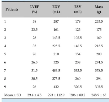
Table 2 shows the scintigraphic parameters of systolic function and basal left ventricular mass of the patients included in the study.
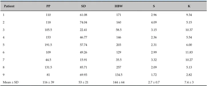
Table 3 shows the basal scintigraphic parameters of the phase analysis related to the ventricular synchrony. Two patients did not have MD, according to the scintigraphic criterion (SD > 43°), but only ED.
Table 4 shows patients’ clinical response after the cardiac resynchronizer implantation. It was observed that NYHA functional class decreased for all patients with FC > III, with two patients with NYHA IV showing a decrease to NYHA III, and only one FC III patient did not show FC improvement, with statistical significance by Fisher’s exact test. There was a statistically significant reduction in the MLHFQ scores, which, despite being subjective, showed a marked improvement in patients’ symptoms, with quality of life improvement. Regarding the 6-minute Walk Test, there was an increase in the distance covered, a decrease in the Borg index (subjective dyspnea score) and in the dyspnea assessed by the examiner, although not statistically significant.
In table 5, the findings of imaging methods in relation to desynchronization were compared. The scintigraphic values of ventricular function (LVEF, EDV, ESV and LV mass) and the values that evaluated dyssynchrony (PP, HBW, SD, S and K) were analyzed. There was a statistically significant reduction in mean systolic volume and LV mass after CRT, due to probable post-resynchronization reverse remodeling.
association between QRS duration and the presence of dyssynchrony. Figure 1 analyzes QRS duration with the SD values of the phase histogram. It was known that the higher the SD (SD > 43°), the higher the intraventricular dyssynchrony. Likewise, a QRS > 130 ms was associated with a higher probability of dyssynchrony. The association of both parameters was directly proportional. When analyzed with HBW, it was also observed that the longer the QRS duration, the greater its value. This demonstrates that HBW and SD were also directly associated, as both increased with QRS enlargement and the presence of dyssynchrony.
SD and HBW values were higher for responders than for non-responders, and the difference between HBW in both groups was statistically significant (Figure 2).
Discussion
The present study evaluated dyssynchrony at pre and post-implantation of CRT through GSPECT. CRT had a positive impacted on functional capacity, MD and ED of patients with advanced HF and LBBB and demonstrated the use of GSPECT to identify patients with a higher probability of responding to CRT.
GSPECT is a useful tool for assessing systolic function in patients submitted to perfusion studies by adding diagnostic and prognostic information without additional
exposure to radiation.15 Technological evolution has
allowed phase analysis to be employed in GSPECT studies, providing significant data regarding ventricular
synchrony.13 Trimble et al.16 used the technique of
phase analysis in myocardial perfusion scintigraphy, comparing patients with left ventricular dysfunction with patients with LBBB or right bundle branch block, patients with pacemakers and controls for the evaluation of MD. The parameters of phase analysis were able to
identify the subgroups according to the degree of ED.16
Our findings confirm, as those by Trimble et al.,16 the
feasibility of using myocardial perfusion scintigraphy Table 1 - Basal general characteristics
Characteristics n = 9
Age, years 62.4 ± 8
Body mass index, kg/m2 27.3 ± 5.5
Female gender 6
Diabetes Mellitus 5
Hypertension 7
Dyslipidemia 6
Smoking 0
Previous coronary disease 6
Previous infarction 5
CABG surgery 2
Percutaneous Coronary Intervention 1
NYHA functional class
II 2
III 5
IV 2
Beta-blocker 9
Angiotensin-converting enzyme inhibitor 3
Angiotensin-receptor blocker 5
Acetylsalicylic acid 2
Diuretics 9
Statins 3
Mineralocorticoid-receptor antagonist 6
Digoxin 4
Results expressed as number or mean ± standard deviation. NYHA: New York Heart Association.
Table 2 - Scintigraphic parameters of basal ventricular function of patients included in the study
Patients LVEF (%)
EDV (mL)
ESV (mL)
Mass (g)
1 38 287 178 233.5
2 23.5 161 123 175
3 28 143.5 102.5 169
4 35 225.5 146.5 213.5
5 26 210 154 200
6 26.5 325 238 274.5
7 31.5 483.5 333.5 378.5
8 30.5 375.5 260 294
9 26 432 320.5 302.5
Mean ± SD 29.4 ± 4.5 293 ± 112.9 206 ± 80.2 248.9 ± 65
Table 3 - Scintigraphic parameters of the pre-implantation synchronization of the resynchronizer
Patient PP SD HBW S K
1 110 61.08 171 2.96 9.34
2 118 74.04 160 4.09 5.15
3 105.5 22.41 58.5 3.15 10.37
4 153 46.77 146 2.36 5.54
5 191.5 57.74 203 2.31 6.00
6 109 49.26 129 2.99 11.83
7 44.5 15.91 35.5 3.32 10.27
8 131.5 85.71 257 2.09 5.13
9 81 69.93 134.5 1.72 2.82
Mean ± SD 116 ± 39 53 ± 21 144 ± 64 2.7 ± 0.7 7.4 ± 3
PP: peak phase; SD: standard deviation; HBW: histogram bandwidth; S: skewness; K: Kurtosis; SD: standard deviation.
Table 4 - Clinical response before and after cardiac resynchronizer implantation
Variables Pre-resynchronization Post-resynchronization p value
NYHA Functional Class
II 2 7
III-IV 7 2 0.015*
MLHFQ 63.6 ± 17.5 34.1 ± 20.5 0.006†
6-minute Walk Test
Distance covered, m 342.7 ± 82.2 376.6 ± 84.0 0.314**
Borg index 3.1 ± 1.8 1.2 ± 1.3 0.023†
Dyspnea 2.4 ± 2.0 0.89 ± 0.93 0.049†
Fisher's exact test; † paired t-test. MLHFQ: Minnesota Living with Heart Failure Questionnaire.
with phase analysis, as well as the fact that it can be used in patients with HF and CRT indication.
The pathophysiological basis for the resynchronizer implantation is the correction of a mechanical disorder secondary to an altered LV activation due to LBBB. The presence of LBBB is a sign of electrical abnormality and has been the main criterion for the selection of patients
to undergo CRT.17 However, the current criteria used to
indicate CRT are still imperfect, as a group of 20 to 40%
of patients does not respond to treatment.18,19 Bleeker et
al.20 compared the echocardiogram with QRS duration for
MD evaluation, and found that 30 to 40% of the patients with QRS duration > 120 ms did not have mechanical desynchronization, suggesting that there is an association between the findings of non-response to CRT and absence
of MD.20 MD was not necessarily associated with ED, as
evidenced by the absence of MD in patients with QRS
duration > 120 ms.20 This finding was also demonstrated
Table 5 - Statistical analysis of the pre and post-implantation resynchronizer findings between scintigraphy and echocardiography parameters, using Student’s t-test, considering p values < 0.05 as statistically significant
Scintigraphy Pre-implantation
Post-implantation p value
Ejection
fraction, % 29.4 33.89 0.32
End-diastolic
volume, mL 293.7 231.1 0.08
End-systolic
volume, mL 206.2 158 0.05
Mass, g 249 193.9 0.02
PP 116 114 0.94
SD 53.66 45.8 0.53
HBW 143.8 130.3 0.68
S 2.78 3.28 0.27
K 7.38 15. 35 0.17
PP: peak phase; SD: standard deviation; HBW: histogram bandwidth.
QRS (ms)
SD (degrees)
Correlation QRS duration x SD
y = 0.3665x - 27.378 R2 = 0.07871
Figure 1 - Correlation between pre-implantation QRS duration and pre-implantation standard deviation (SD).
T h e u s e o f i m a g i n g m e t h o d s t o i d e n t i f y
desynchronization has been validated;16 however, its
routine use as a support tool for the selection of patients
for CRT remains a topic to be studied, such as the study
of Henneman et al.,21 who evaluated patients with CRT
indication through GSPECT and observed a 29% rate of nonresponders after 6 months of therapy – comparable to the 22% observed in the present study. In the study by
Henneman et al.,21 the responders had significantly higher
dyssynchrony parameters compared to non-responders (HBW of 175° vs. 117°; and SD of 56° vs. 37°, respectively). These values are close to those found in our results (HBW of 177° vs. 76° and SD of 62° vs. 36°, respectively), confirming that the presence of MD identified at GSPECT
is a strong predictor for CRT response.21 Henneman et al.21
derived, from the sample of 42 patients, cut-off values of the scintigraphic parameters to indicate the presence of MD and to predict good response to CRT in patients
with HF: HBW > 135° and SD > 43°.21
Medical therapy decision-making should always focus on treatments that lead to changes in clinical outcomes, rather than just changes in imaging or laboratory tests. Thus, more than ventricular function improvement, the aim of this study was to select an ideal patient, who shows a reduction in morbidity and mortality after CRT. Recent studies have demonstrated that the phase analysis parameters are markers of
adverse prognosis, as observed by Al Jaroudi et al.,22
who evaluated 144 patients with chronic renal failure and had higher mortality at 2 years in those with
Figure 2 - Distribution of mean SD and HBW before implantation of the cardiac resynchronization therapy, according to the clinical response to implantation. *Chi-square test, with p = 0.03.
non-responder
responder
Henneman et al.,21 but already showing some degree
of desynchronization.
The subgroup of patients with end-stage renal disease
was also extensively studied by Aggarwal et al.,23 who
followed 828 patients with normal EF for 5 years. It was
observed that values of SD ≥ 21° or HBW ≥ 56° were
associated with worse survival in 5 years. Thus, they also demonstrated that LV desynchronization through phase analysis (GSPECT) provides prognostic value in
end-stage renal failure.23
A relatively recent study by Zafrir et al.24 had a
significant impact on desynchronization assessment and its association with cardiac mortality, by following 787 patients who underwent GSPECT in a single center
for several clinical reasons.24 These patients were
followed for 18.3 ± 6.2 months for cardiac events, and it was verified that SD had the capacity to predict cardiac mortality, and that with every 10° increment, it became an independent predictor of mortality (p = 0.04). Our study did not have data on adverse clinical outcomes in the long term, but ventricular function improvement has been used in several situations, as a valuable surrogate outcome.
Studying clinical outcomes specifically in patients
with HF, Al Jaroudi et al.22 assessed dyssynchrony in
patients with an implantable cardioverter defibrillator
(ICD) and showed that the higher the SD and the HBW, the higher the incidence of cardiopulmonary arrest or
appropriate shock by the ICD.23 The value of SD > 50°
was a predictor of death or appropriate shock by the ICD.
More recently, Zafrir et al.,24 assessing 143 patients with
HF and ICD indication, showed a higher rate of events
when they also had DM evidenced by SD > 60°.25 These
authors suggest that patients referred to a defibrillator implantation should receive associated CRT when they have SD > 60°.25
New studies have addressed the combination of GSPECT parameters to create a MD gradation, using, in addition to HBW and SD, the K and S parameters.
Aguadé-Bruix et al.26 employed a combination of these
four parameters and observed that 12% of patients with CRT indication do not have any abnormal phase
parameters26. Perhaps the study of these combined
parameters can increase the sensitivity and specificity of the technique for CRT indication.
Study limitations
The main study limitation was the small number of patients, which limited the statistical analysis. Despite the small sample size, statistical significance was observed in parameters that corroborate previous studies in the dyssynchrony area. Another study limitation was the absence of a control group with ventricular dysfunction without CRT. From the ethical point of view, it is not possible to maintain patients with CRT indication as controls, considering the impact of this treatment on mortality and its broad indication recommended in
several guidelines.6 The study had a short follow-up
period (6 months) using secondary outcomes, such as left ventricular function, rather than clinical outcomes such as death, HF progression or hospitalization.
Conclusion
The study of phase analysis by GSPECT was able to differentiate patients with isolated electrical dyssynchrony from those with associated mechanical dyssynchrony, through the intraventricular dyssynchrony parameters. The cardiac resynchronization therapy is associated with the improvement of both the mechanical desynchronization (improvement of desynchronization parameters through the phase analysis) and electrical dyssynchrony (QRS interval reduction at the electrocardiogram). Thus, because of the pre-implantation GSPECT assessment, it was possible to verify that patients with associated electrical and mechanical dyssynchrony showed better response to cardiac resynchronization therapy than those with isolated electrical dyssynchrony.
Acknowledgments
To Coordenação de Aperfeiçoamento de Pessoal de Nível Superior (CAPES) and the International Atomic Energy Agency that funded this study.
Author contributions
Conception and design of the research: Wiefels CC, Nascimento EA, Alves CR, Ribeiro FB, Ribeiro ML, Mesquita CT. Acquisition of data: Wiefels CC, Nascimento EA, Alves CR, Ribeiro FB, Fernandes FA, Ribeiro ML, Mesquita CT. Analysis and interpretation of the data: Wiefels CC, Nascimento EA, Alves CR, Ribeiro FB, Fernandes FA, Ribeiro ML, Mesquita CT. Statistical analysis: Wiefels CC, Nascimento EA, Alves CR, Mesquita CT. Obtaining financing: Ribeiro ML, Mesquita CT. Writing of the manuscript: Wiefels CC, Nascimento EA, Alves CR, Ribeiro FB, Fernandes FA, Ribeiro ML, Mesquita CT. Critical revision of the manuscript for intellectual content: Wiefels CC, Mesquita CT.
Potential Conflict of Interest
No potential conflict of interest relevant to this article was reported.
Sources of Funding
This study was partially funded by Agência Internacional de Energia Atômica.
Study Association
This article is part of the thesis of master submitted by Christiane Cigagna Wiefels, from Universidade Federal Fluminense.
Ethics approval and consent to participate
This study was approved by the Ethics Committee of the Hospital Universitário Antônio Pedro under the protocol number 884.844. All the procedures in this study were in accordance with the 1975 Helsinki Declaration, updated in 2013. Informed consent was obtained from all participants included in the study.
1. Hunt SA, Abraham WT, Chin MH, Feldman AM, Francis GS, Ganiats TG, et al. 2009 focused update incorporated into the ACC/AHA 2005 Guidelines for the Diagnosis and Management of Heart Failure in Adults: a report of the American College of Cardiology Foundation/American Heart Association Task Force on Practice Guidelines: developed in collaboration with the International Society for Heart and Lung Transplantation. Circulation. 2009;119(14):e391-479.
2. Mozaffarian D, Benjamin EJ, Go AS, Arnett DK, Blaha MJ, Cushman M, et al; American Heart Association Statistics Committee; Stroke Statistics
Subcommittee. Heart disease and stroke statistics 2016 Update: A Report From the American Heart Association. Circulation. 2016;133(4):e38-360.
3. Albuquerque DC, Souza JD, Bacal F, Rohde LE, Bernardez-Pereira S, Berwanger O, et al; Investigadores Estudo BREATHE. I Brazilian Registry of Heart Failure - clinical aspects, care quality and hospitalization outcomes. Arq Bras Cardiol. 2015;104(6):433-42.
4. Wells G, Parkash R, Healey JS, Talajic M, Arnold JM, Sullivan S, et al. Cardiac resynchronization therapy: a meta-analysis of randomized controlled trials. CMAJ. 2011;183(4):421-9.
5. Rossi A, Rossi G, Piacenti M, Startari U, Panchetti L, Morales MA. The current role of cardiac resynchronization therapy in reducing mortality and hospitalization in heart failure patients: a meta-analysis from clinical trials. Heart Vessels. 2008;23(4):217-23.
6. Ponikowski P, Voors AA, Anker SD, Bueno H, Cleland JG, Coats AJ, et al. Guidelines for the diagnosis and treatment of acute and chronic heart failure: The Task Force for the diagnosis and treatment of acute and chronic heart failure of the European Society of Cardiology (ESC) Developed with the special contribution of the Heart Failure Association (HFA) of the ESC. Eur Heart J. 2016;37(27):2129-200.
7. Cubillos-Garzon LA, Casas JP, Morillo CA, Bautista LE. Congestive heart failure in Latin America: the next epidemic. Am Heart J. 2004;147(3):412-7.
8. Abraham WT, Hayes DL. Cardiac resynchronization therapy for heart failure. Circulation. 2003;108(21):2596-603.
9. Leclercq C, Kass DA. Retiming the failing heart: principles and current clinical status of cardiac resynchronization. J Am Coll Cardiol. 2002;39(2):194-201.
10. Auricchio A, Stellbrink C, Sack S, Block M, Vogt J, Bakker P, et al. The Pacing Therapies for Congestive Heart Failure (PATH-CHF) study: rationale, design, and endpoints of a prospective randomized multicenter study. Am J Cardiol. 1999;83(5B):130D-135D.
11. Bleeker GB, Kaandorp TA, Lamb HJ, Boersma E, Steendijk P, De Roos A, et al. Effect of posterolateral scar tissue on clinical and echocardiographic improvement after cardiac resynchronization therapy. Circulation. 2006;113(7):969-76.
12. International Atomic Energy Agency (IAEA). IAEA Annual Report 2013. Vienna (Austria); 2013. [Acesso em 2017 fev 9]. Disponível em: <https:// www.iaea.org/About/Policy/GC/GC58/GC58Documents/English/ gc58-3-att1_en.pdf>.
13. Chen J, Garcia EV, Folks RD, Cooke CD, Faber TL, Tauxe EL, et al. Onset of left ventricular mechanical contraction as determined by phase analysis of ECG-gated myocardial perfusion SPECT imaging: development of a diagnostic tool for assessment of cardiac mechanical dyssynchrony. J Nucl Cardiol. 2005;12(6):687-95.
14. Reis CW, Nascimento EA, Ribeiro ML, Dias FB, Wanderley AP, Batista LA, et al. Applicability of myocardial perfusion scintigraphy in the evaluation of cardiac synchronization. Arq Bras Cardiol: Imagem cardiovasc. 2017;30(2):54-63.
15. Chen J, Garcia EV, Bax JJ, Iskandrian AE, Borges-Neto S, Soman P. SPECT myocardial perfusion imaging for the assessment of left ventricular mechanical dyssynchrony. J Nucl Cardiol. 2011;18(4):685-94.
16. Trimble MA, Borges-Neto S, Honeycutt EF, Shaw LK, Pagnanelli RJ, Chen J, et al. Evaluation of mechanical dyssynchrony and myocardial
perfusion using phase analysis of gated SPECT imaging in patients with left ventricular dysfunction. J Nucl Cardiol. 2008;15(5):663-70.
17. Poole JE, Singh JP, Birgersdotter-Green U. QRS duration or QRS morphology what really matters in cardiac resynchronization therapy. J Am Coll Cardiol. 2016;67(9):1104-17.
18. Brignole M, Auricchio A, Baron-Esquivias G, Bordachar P, Boriani G, Breithardt OA, et al; European Society of Cardiology (ESC); European Heart Rhythm Association (EHRA). 2013 ESC guidelines on cardiac pacing and cardiac resynchronization therapy: the task force on cardiac pacing and resynchronization therapy of the European Society of Cardiology (ESC). Developed in collaboration with the European Heart Rhythm Association (EHRA). Europace. 2013;15(8):1070-118.
19. Abraham WT, Hayes DL. Cardiac resynchronization therapy for heart failure. Circulation. 2003;108(21):2596-603.
20. Bleeker GB, Schalij MJ, Molhoek SG, Verwey HF, Holman ER, et al. Relationship between QRS duration and left ventricular dyssynchrony in patients with end-stage heart failure. J Cardiovasc Electrophysiol. 2004;15(5):544-9.
21. Henneman MM, Chen J, Dibbets-Schneider P, Stokkel MP, Bleeker GB, Ypenburg C, et al. Can LV dyssynchrony as assessed with phase analysis on gated myocardial perfusion SPECT predict response to CRT? J Nucl Med. 2007;48(7):1104-11.
22. AlJaroudi, W, Aggarwal, H, Venkataraman R, Heo J, Iskandrian AE, Hage FG. Impact of left ventricular dyssynchrony by phase analysis on cardiovascular outcomes in patients with end-stage renal disease. J Nucl Cardiol. 2010;17(6):1058-64.
23. Aggarwal H, AlJaroudi WA, Mehta S, Mannon R, Heo J, Iskandrian AE, et al. The prognostic value of left ventricular mechanical dyssynchrony using gated myocardial perfusion imaging in patients with end-stage renal disease. J Nucl Cardiol. 2014;21(4):739-46.
24. Zafrir N, Nevzorov R, Bental T, Strasberg B, Gutstein A, Mats I, et al. Prognostic value of left ventricular dyssynchrony by myocardial perfusion-gated SPECT in patients with normal and abnormal left ventricular functions. J Nucl Cardiol. 2014;21(3):532-40.
25. Zafrir N, Bental T, Strasberg B, Solodky A, Mats I, Gutstein A, et al. Yield of left ventricular dyssynchrony by gated SPECT MPI in patients with heart failure prior to implantable cardioverter-defibrillator or cardiac resynchronization therapy with a defibrillator: Characteristics and prediction of cardiac outcome. J Nucl Cardiol. 2017;24(1):122-9.