Research Article
Two-Dimensional PCA Highlights the Differentiated
Antitumor and Antimicrobial Activity of Methanolic and
Aqueous Extracts of
Laurus nobilis
L. from Different Origins
Maria Inês Dias,
1,2João C. M. Barreira,
1,2Ricardo C. Calhelha,
1,3Maria-João R. P. Queiroz,
3M. Beatriz P. P. Oliveira,
2Marina Sokovi
T
,
4and Isabel C. F. R. Ferreira
11Mountain Research Center (CIMO), ESA, Polytechnic Institute of Braganc¸a, Campus de Santa Apol´onia, 1172, 5301-855 Braganc¸a, Portugal
2REQUIMTE, Department of Chemical Sciences, Faculty of Pharmacy of University of Porto, Rua Jorge Viterbo Ferreira, 228, 4050-313 Porto, Portugal
3Center of Chemistry, University of Minho, Campus de Gualtar, 4710-057 Braga, Portugal
4Department of Plant Physiology, Institute for Biological Research “Siniˇsa Stankovi´c”, University of Belgrade, Bulevar Despota Stefana 142, 11000 Belgrade, Serbia
Correspondence should be addressed to Isabel C. F. R. Ferreira; [email protected]
Received 18 February 2014; Revised 24 March 2014; Accepted 27 March 2014; Published 16 April 2014
Academic Editor: Anjali Joshi
Copyright © 2014 Maria Inˆes Dias et al. This is an open access article distributed under the Creative Commons Attribution License, which permits unrestricted use, distribution, and reproduction in any medium, provided the original work is properly cited.
Natural matrices are important sources of new antitumor and antimicrobial compounds. Species such asLaurus nobilisL. (laurel) might be used for this purpose, considering its medicinal properties. Herein,in vitroactivity against human tumor cell lines, bacteria, and fungi was evaluated in enriched phenolic extracts. Specifically, methanol and aqueous extracts of wild and cultivated samples ofL. nobiliswere compared considering different phenolic groups. Principal component analysis (PCA) was applied to understand how each extract acts differentially against specific bacteria, fungi, and selected human tumor cell lines. In general, the extract type induced the highest differences in bioactivity of laurel samples. However, from the PCA biplot, it became clear that wild laurel samples were higher inhibitors of tumor cell lines (HeLa, MCF7, NCI-H460, and HCT15). HepG2 had the same response to laurel from wild and cultivated origin. It was also observed that methanolic extracts tended to have higher antimicrobial activity, except againstA. niger, A. fumigatus, andP. verrucosum. The differences in bioactivity might be related to the higher phenolic contents in methanolic extracts. These results allow selecting the extract type and/or origin with highest antibacterial, antifungal, and antitumor activity.
1. Introduction
Laurus nobilisL. (Lauraceae), commonly known as laurel or
bay leaves, is a native plant from the Southern Mediterranean
region, found in warm climate regions with high rainfall [1].
It is one of the most widely used culinary spices for seasoning of meat products, soups, and fishes but is also used as an ornamental plant, especially in Europe and USA. It is also commercially grown in Turkey, Algeria, Morocco, Portugal,
Spain, Italy, France, and Mexico [2–4]. The dry laurel and
their infusions are traditionally used to treat gastrointestinal
problems, such as epigastric, bloating, digestion, eructation, and flatulence problems. It also possesses anticonvulsive and antiepileptic activities and stimulant and narcotic properties
[2,5,6]. The ability to suppress high blood sugar and prevent
not only migraines and headaches but also bacterial and
fungal infections has also been reported [3,7].
Natural matrices, like L. nobilis, are rich sources of
bioactive compounds. In fact, nearly 60% of the antitumor and anti-infectious drugs available on the market, or under
clinical trial, are from natural origin [6, 8]. The biological
activities of plant extracts are well recognized, namely, their
2 BioMed Research International
antifungal, antimicrobial, insecticidal, and cytostatic effects. Accordingly, the bioactivity of plant extracts is often explored
in a multifactorial manner [8,9].
Nowadays, there is a worldwide concern about the use of synthetic chemical compounds as antitumor agents due to their potential negative health effects, opening ways to use plants as sources of natural compounds with similar activity
[10]. On the other hand, the indiscriminate use of antibiotics
to treat bacterial and fungal infections led to the emergence and spread of organisms resistant to broad-spectrum
antibi-otics, demanding new antimicrobial agents [11,12].
There are some reports on the antitumor potential ofL.
nobilis essential oil [13, 14], methanolic [15], ethanol, and
aqueous extracts [8]. However, most publications regard
isolated compounds [6, 16, 17]. For instance, sesquiterpene
lactones and methyl esters isolated from L. nobilis leaves
exhibited moderate-to-significant cytotoxicity towards K562
leukemia cells [16]. Likewise, there are a considerable number
of reports on the antimicrobial effects ofL. nobilisessential
oil [1,4, 9, 18–21], aqueous [11], ethanolic [12,22, 23], and
methanolic extracts [24]. The antimicrobial activity of L.
nobilisis mainly related to terpenes and phenolic compounds
[7,24–26].
Despite the previous findings, and as far as we know,
this is the first study exploring in vitro antimicrobial and
antitumor activities from cultivated and wild L. nobilis
enriched phenolic extracts. Furthermore, it was intended to compare the differentiated activity of each extract against specific bacteria, fungi, and selected human tumor cell lines, using principal component analysis.
2. Materials and Methods
2.1. Samples. Cultivated Laurus nobilis L. samples (leaves)
were purchased from Ervital (Castro Daire, Portugal), which produces Mediterranean herbs using organic farming princi-ples and methods. The wild samprinci-ples (leaves) were collected in Braganc¸a, Portugal, and further lyophilized (FreeZone 4.5, Labconco, KS, USA). Each sample was reduced to a fine dried
powder (20 mesh) and stored (7∘C) until further use.
2.2. Standards and Reagents. Fetal bovine serum (FBS),
L-glutamine, Hank’s balanced salt solution (HBSS),
trypsin-EDTA (ethylenediaminetetraacetic acid), nonessential
amino acids solution (2 mM), penicillin/streptomycin solution (100 U/mL and 100 mg/mL, resp.), RPMI-1640, and DMEM media were from HyClone (Logan, UT, USA). Acetic acid, ellipticine, sulforhodamine B (SRB), trypan blue, trichloroacetic acid (TCA), and Tris were from Sigma Chem-ical Co. (Saint Louis, USA). Mueller-Hinton agar (MH) and malt agar (MA) were obtained from the Institute of Immunol-ogy and VirolImmunol-ogy, Torlak (Belgrade, Serbia). Dimethyl sulfoxide (DMSO) (Merck KGaA, Germany) was used as a solvent. Phosphate buffered saline (PBS) was obtained from Sigma Chemical Co. (St. Louis, USA). Methanol and all other chemicals and solvents were of analytical grade and pur-chased from common sources. Water was treated in a Milli-Q water purification system (TGI Pure Water Systems, USA).
2.3. Extracts Preparation. Methanolic extracts were obtained
from cultivated and wild plant material. Each sample (≈1 g)
was extracted by stirring with 30 mL of methanol, at room temperature, 150 rpm for 1 h. The extract was filtered through Whatman number 4 paper. The residue was then reextracted with additional 30 mL of methanol. The combined extracts
were evaporated at 35∘C (rotary evaporator B¨uchi R-210,
Flawil, Switzerland) to dryness.
For aqueous extracts, plant material (≈1 g) was added to
200 mL of boiling distilled water, left to stand for 5 min out of the heating source, and then filtered under reduced pressure. The obtained extract was frozen and lyophilized.
Methanolic and aqueous extracts were redissolved in water (8 mg/mL) or 5% DMSO (10 mg/mL) for antitumor and antimicrobial activity evaluation, respectively. The final solutions were further diluted to different concentrations for bioactivity evaluation.
2.4. Antitumor Activity and Hepatotoxicity. Five human
tumor cell lines were tested: MCF7 (breast adenocarcinoma), NCI-H460 (non-small cell lung cancer), HCT15 (colon carci-noma), HeLa (cervical carcicarci-noma), and HepG2 (hepatocellu-lar carcinoma). Cells were routinely maintained as adherent cell cultures in RPMI-1640 medium containing 10% heat-inactivated FBS and 2 mM glutamine (MCF7, NCI-H460, and HCT15) or in DMEM supplemented with 10% FBS, 2 mM glutamine, 100 U/mL penicillin, and 100 mg/mL
strep-tomycin (HeLa and HepG2 cells), at 37∘C, in a humidified
air incubator containing 5% CO2. Each cell line was plated
at an appropriate density (7.5×10³cells/well for MCF-7,
NCI-H460, and HCT15 or 1.0×10⁴cells/well for HeLa and HepG2)
in 96-well plates. Sulforhodamine B assay was performed according to a procedure previously described by the authors
[27]. Ellipticine was used as positive control.
For hepatotoxicity evaluation, a cell culture (PLP2) was prepared from a freshly harvested porcine liver obtained from a local slaughter house, according to an established procedure
[27]. Cell culture was continued with direct monitoring
every 2-3 days using a phase contrast microscope. Before confluence was reached, cells were subcultured and plated in
96-well plates at a density of 1.0×10⁴cells/well and cultivated
in DMEM medium with 10% FBS, 100 U/mL penicillin, and
100𝜇g/mL streptomycin. Ellipticine was used as positive
control. The results were expressed in GI50 values (sample
concentration that inhibited 50% of the net cell growth).
2.5. Antibacterial Activity. The following gram-positive
bac-teria: Staphylococcus aureus (ATCC 6538), Bacillus cereus
(clinical isolate), Micrococcus flavus (ATCC 10240), and
Listeria monocytogenes(NCTC 7973) and gram-negative
bac-teria:Escherichia coli(ATCC 35210),Pseudomonas aeruginosa
(ATCC 27853),Salmonella typhimurium(ATCC 13311), and
Enterobacter cloacae(ATCC 35030) were used. The
BioMed Research International 3
The minimum inhibitory (MIC) and minimum bacterici-dal (MBC) concentrations were determined by the microdi-lution method. Briefly, fresh overnight culture of bacteria was
adjusted by the spectrophotometer to a concentration of 1×
105CFU/mL. The requested CFU/mL corresponded to a
bacterial suspension determined in a spectrophotometer at 625 nm (OD625). Dilutions of inocula were cultured on a solid medium to verify the absence of contamination and check the validity of the inoculum. Different solvent dilutions of methanolic extract/fractions were placed in the wells
con-taining 100𝜇L of tryptic soy broth (TSB) and afterwards 10𝜇L
of inoculum was added. The microplates were incubated for
24 h at 37∘C. The MIC of each extract was detected following
the addition of 40𝜇L of iodonitrotetrazolium chloride (INT)
(0.2 mg/mL) and incubation at 37∘C for 30 min. The lowest
concentration that produced a significant inhibition (around 50%) of the growth of the bacteria in comparison with the positive control was identified as the MIC. The minimum inhibitory concentrations (MICs) obtained from the suscep-tibility testing of various bacteria to tested extract/fraction were determined also by a colorimetric microbial viability assay based on reduction of INT color and compared with
positive control for each bacterial strain [28,29]. MBC was
determined by serial subcultivation of 10𝜇L into microplates
containing 100𝜇L of TSB. The lowest concentration not
showing growth after this subculturing was read as the MBC. Standard drugs, namely, streptomycin and ampicillin, were used as positive controls. DMSO (5%) was used as negative control.
2.6. Antifungal Activity. For the antifungal bioassays, the
following microfungi were used:Aspergillus fumigatus(1022),
Aspergillus ochraceus (ATCC 12066), Aspergillus versicolor
(ATCC 11730), Aspergillus niger (ATCC 6275), Penicillium
funiculosum(ATCC 36839),Penicillium ochrochloron(ATCC
9112),Penicillium verrucosum var. cyclopium(food isolate),
and Trichoderma viride (IAM 5061). The organisms were
obtained from the Mycological Laboratory, Department of Plant Physiology, IBRSS, Belgrade, Serbia. The micromycetes were maintained on malt agar (MA) and the cultures were
stored at 4∘C and subcultured once a month [30].
The fungal spores were washed from the surface of agar plates with sterile 0.85% saline containing 0.1% Tween 80 (v/v). The spore suspension was adjusted with sterile saline
(≈1.0×10³/𝜇L per well). The inocula were stored at 4∘C for
further use. Dilutions of the inocula were cultured on solid MA to verify the absence of contamination and to check the validity of the inoculum. MICs determination was per-formed by a serial dilution technique using 96-well microtitre plates. The extract/fractions were dissolved in 5% solution of DMSO and added to broth malt medium with fungal
inoculum. The microplates were incubated for 72 h at 28∘C.
The lowest concentrations without visible growth (at the binocular microscope) were defined as MIC. The minimum fungicidal concentrations (MFCs) were determined by serial
subcultivation of 2𝜇L in microtitre plates containing 100𝜇L
of malt broth per well and further incubation for 72 h at 28∘C.
The lowest concentration with no visible growth was defined
as the MFC, indicating 99.5% killing of the original inoculum. Bifonazole and ketoconazole were used as positive controls.
DMSO (5%) was used as negative control [31].
2.7. Statistical Analysis. For wild and cultivated plant
mate-rial, three samples were used and all the assays were carried
out in triplicate. Data were expressed as means±standard
deviations, maintaining the decimal places allowed by the magnitude of standard deviation.
An analysis of variance (ANOVA) with type III sums of squares was performed using the GLM (general linear model) procedure of the SPSS software. The dependent variables were analyzed using 2-way ANOVA with the factors “extract” (E) and “origin” (O). When a statistically significant interaction
(E×O) was detected, the two factors were evaluated
simulta-neously by the estimated marginal means plots for the two levels of each factor. Alternatively, if no statistical significant interaction was verified, means were compared using results
obtained for EB and GI that were classified using a simple𝑡
-test (after checking the equality of variances through Levene’s test), since there were fewer than three groups.
Principal components analysis (PCA) was applied as pattern recognition unsupervised classification method. The number of dimensions to keep for data analysis was assessed by the respective eigenvalues (which should be greater than one), by Cronbach’s alpha parameter (that must be positive), and also by the total percentage of variance (that should be as high as possible) explained by the number of components selected. The number of plotted dimensions was chosen in order to allow meaningful interpretations.
All statistical tests were performed at a 5% significance level using the SPSS software, version 20.0 (SPSS Inc.).
3. Results and Discussion
The interactions amongL. nobilisorigin (cultivated or wild)
and extract (methanolic or aqueous) were evaluated to verify if these factors act together to cause changes in phenolic composition and/or biological activities. Results are pre-sented as the mean value of each origin (O), comprising both extracts, as well as the mean value of each extract (E), containing samples from both origins. When the interaction
among factors (O×E) was significant (𝑃 < 0.05), acting itself
as a source of variability, the comparison of means could not be performed. In these cases, the presented conclusions were drawn from the estimated marginal means (EMM) plots obtained in each case. When the interaction was not
significant, a simple 𝑡-test (fewer than three groups) was
applied to evaluate the equality of means.
3.1. Phenolic Compound Groups Present in the Studied L. nobilis Extracts. Table 1summarizes the phenolic compound groups present inmethanolic and aqueous extracts from
cultivated and wild L. nobilis, as reported in a previous
study of our research group [32]. According to those results,
4 BioMed Research International
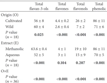
Table 1: Phenolic compounds (mg/g) of different extracts ofLaurus nobilis. The results are presented as mean±SD.
Total flavan-3-ols
Total flavones
Total flavonols
Total phenolic Origin (O)
Cultivated 56 ± 8 4.4 ± 0.2 26 ± 2 86 ± 11
Wild 60 ± 4 2.6 ± 0.4 7 ± 2 71 ± 6
𝑃value
(𝑛 = 18) 0.025 <0.001 <0.001 <0.001 Extract (E)
Methanolic 63.6 ± 0.4 4 ± 1 19 ± 10 86 ± 11
Aqueous 52 ± 5 3 ± 1 15 ± 9 70 ± 5
𝑃value
(𝑛 = 18) <0.001 0.104 0.207 <0.001 O×E
𝑃value
(𝑛 = 36) <0.001 <0.001 <0.001 <0.001
The detailed phenolic profile of all laurel samples was previously described by Dias et al. [32].
methanolic extracts had the highest flavan-3-ols contents
(e.g., (−)-epicatechin and a procyanidin trimer with an
A-type linkage). These differences were maintained after assembling samples according to their origin or extraction
type (Table 1), as previously explained. As it can be concluded,
cultivated samples had higher contents in total phenolics, especially due to their higher contents in flavonols, since the amounts in flavan-3-ols were similar for both origins. All the quantified phenolic compound groups tended to be higher in methanolic extracts, probably due to the higher temperature
used in aqueous extracts [33]. These tendencies were obtained
from the EMM plots since the interaction among factors was significant in all cases.
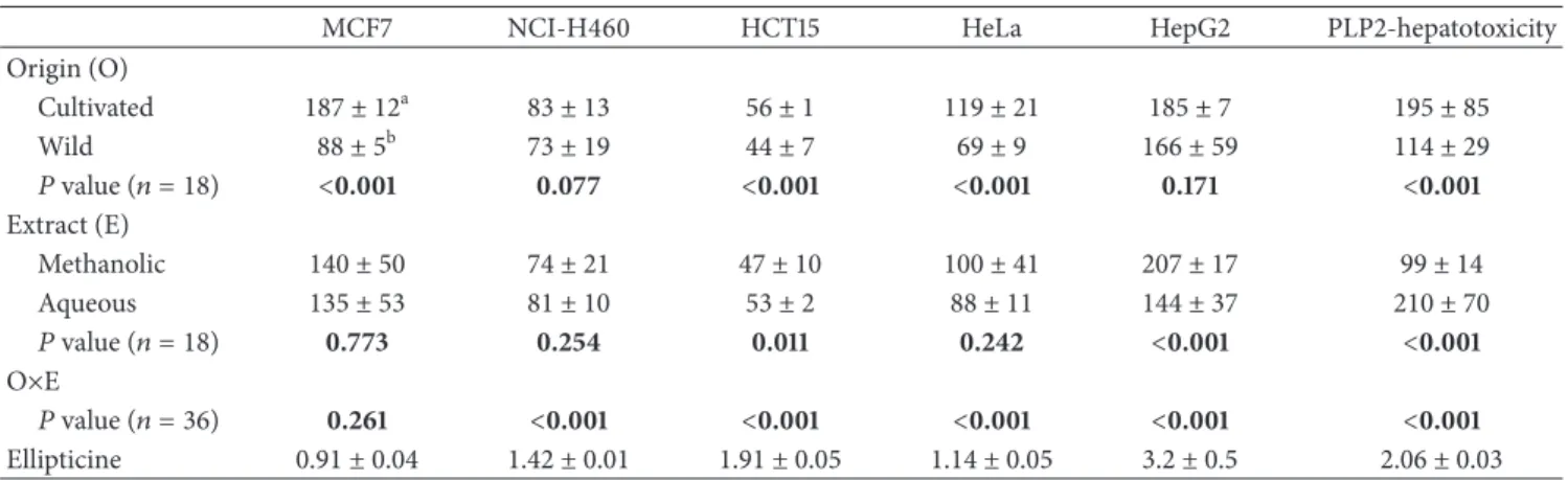
3.2. Antitumor Activity of the Studied L. nobilis Extracts.
The interaction among factors was again significant in all
cases, except MCF7 line (Table 2). Considering each factor
individually, the origin of laurel had once more higher influence, producing statistically significant differences in all
cases except HepG2. Wild laurel presented lower GI50values
than cultivated samples but also higher toxicity against
non-tumor liver primary cells (PLP2; 114𝜇g/mL). Nevertheless,
this sample might have the potential to be used for antitumor
proposes, since the GI50values for hepatotoxicity were higher
than those obtained for the tumor cell lines (except HepG2). Cultivated samples showed also antitumor activity against
NCI-H460, HCT15, and HeLa, since the corresponding GI50
values were quite lower than the toxic concentration for PLP2. Differences among aqueous and methanolic extracts
were only significant for HCT15 (47𝜇g/mL in methanolic
extracts), HepG2 (144𝜇g/mL in aqueous extracts), and PLP2
primary liver cells (99𝜇g/mL in methanolic extracts). The
results for the breast carcinoma cell line (MCF7) showed better results when compared to the essential oil of fruits and
leaves of wild L. nobilisfrom Lebanon (>400𝜇g/mL) [13],
but lower activity than aqueous extract from wild laurel from
Jordan against the same line (88.32% at 50𝜇g/mL) [8]. Kaileh
et al. [15] only reported that the methanolic extract of wild
laurel from Palestine showed no cytotoxicity.
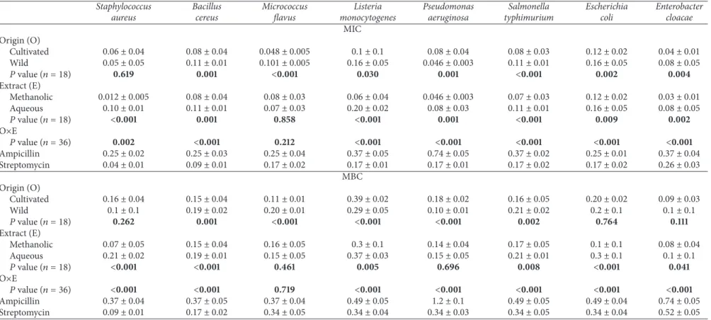
3.3. Antibacterial Activity of the Studied L. nobilis Extracts.
Extract type and origin had a significant interaction in the
antibacterial activity against all species, except M. flavus
(Table 3). Cultivated and wildL. nobiliswere active against all bacteria strains with MICs of 0.04–0.12 mg/mL and 0.046– 0.16 mg/mL, respectively. The MBCs were higher than MICs, varying from 0.09 to 0.39 mg/mL for cultivated laurel and from 0.1 to 0.29 mg/mL for wild samples. The effect of laurel
originper sewas not significant forS. aureus(MIC and MBC),
E. coli(MBC), and E. cloacae(MBC). Methanolic extracts
were better inhibitors (0.012–0.12 mg/mL) of bacterial growth than the aqueous extracts (0.07–0.20 mg/mL), except for
M. flavus (𝑃 = 0.858). In all cases, the inhibitory and
bactericidal activities were higher than those obtained for the standard ampicillin. In the case of streptomycin, the inhibitory activity of the extracts was also higher, except for
S. aureus(cultivated, wild, and aqueous samples),B. cereus
(wild and aqueous samples), andL. monocytogenes(aqueous
extract). The results were similar for bactericidal activity,
with streptomycin showing higher activity againstS. aureus
(cultivated, wild, and aqueous samples),B. cereus(wild and
aqueous samples), and L. monocytogenes (cultivated and
aqueous samples). The bacterial strains more susceptible to
cultivated and wild samples wereE. cloacaeandP. aeruginosa,
respectively. On the other hand, methanolic and aqueous
extractswere particularly active against S. aureus and M.
flavus, respectively. Regarding MBC, the results were the
same, except for aqueous extract, which proved to have
highest bactericidal effect againstE. cloacae(0.1 mg/mL).
All presented MICs were much better than those obtained
by Al-Hussaini and Mahasneh [12] on the ethanolic extracts
of L. nobilis from Jordan against S. aureus, B. cereus, E.
coli,S. typhimurium,andP. aeruginosa. The same applies to
the results obtained by El Malti and Amarouch [23] on the
ethanolic extracts of laurel from Morocco againstB. cereus,S.
aureus,L. monocytogenes,E. cloacae,E. coli,andP. aeruginosa
(>2 mg/mL). The inhibitory activity described herein is also
higher than that reported using essential oils of laurel from
Turkey (MIC values of 5 mg/mL againstE. coli,S. aureus, and
P. aeruginosa) [9]. However, better results were obtained by
Adwan and Mhanna using aqueous extracts of laurel from
Palestine againstS. aureusbacterial strain (<6.1×10⁻³mg/L),
but only when conjugated with enrofloxacin and cephalexin
antibiotics [11].
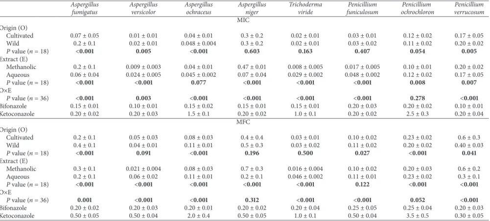
3.4. Antifungal Activity of the Studied L. nobilis Extracts.
The interaction among factors was once more significant in
almost all cases, except MIC values forP. ochrochloron(𝑃 =
0.278) and MBC values for A. niger (𝑃 = 0.312) and P.
ochrochloron(𝑃 = 0.052) (Table 4). The inhibitory activity
on fungal growth was more influenced by extract type, as it can be concluded from the statistically significant differences
verified in all cases, except A. ochraceus (𝑃 = 0.077).
BioMed Research International 5
Table 2: Antitumor activity and hepatotoxicity (GI50,𝜇g/mL) of different extracts ofLaurus nobilis. The results are presented as mean±SD1.
MCF7 NCI-H460 HCT15 HeLa HepG2 PLP2-hepatotoxicity
Origin (O)
Cultivated 187 ± 12a 83 ± 13 56 ± 1 119 ± 21 185 ± 7 195 ± 85
Wild 88 ± 5b 73 ± 19 44 ± 7 69 ± 9 166 ± 59 114 ± 29
𝑃value (𝑛 = 18) <0.001 0.077 <0.001 <0.001 0.171 <0.001
Extract (E)
Methanolic 140 ± 50 74 ± 21 47 ± 10 100 ± 41 207 ± 17 99 ± 14
Aqueous 135 ± 53 81 ± 10 53 ± 2 88 ± 11 144 ± 37 210 ± 70
𝑃value (𝑛 = 18) 0.773 0.254 0.011 0.242 <0.001 <0.001
O×E
𝑃value (𝑛 = 36) 0.261 <0.001 <0.001 <0.001 <0.001 <0.001
Ellipticine 0.91 ± 0.04 1.42 ± 0.01 1.91 ± 0.05 1.14 ± 0.05 3.2 ± 0.5 2.06 ± 0.03
1Means within a column with different letters differ significantly (𝑃 < 0.001).
Trichoderma viride, P. funiculosum, and P. ochrochloron,
while aqueous extracts were better in all remaining cases
(except, of course,A. ochraceus, which gave no differences).
Cultivated and wild samples gave MICs varying from 0.01 to 0.17 mg/mL and from 0.02 to 0.3 mg/mL, respectively. In the cases revealing statistically significant differences, cultivated laurel samples gave higher inhibitory activity.
Concerning fungicidal activity, MFCs varied from 0.03 to 0.6 mg/mL and from 0.03 to 0.5 mg/mL for cultivated and wild samples, respectively. Both origins had the same effect on
A. versicolor,A. niger,andT. viride. Comparing extract types,
MFC varied from 0.016 to 0.7 mg/mL (methanolic extracts) and from 0.046 to 0.3 mg/mL (aqueous extracts). Like it was observed for inhibitory activity, the fungicidal action was more influenced by the type of extract when compared with
laurel origin (exceptP. funiculosum).
For both origins and extracts, A. fumigatus (only
cul-tivated and aqueous samples in the case of bifonazole),
A. versicolor, A. ochraceus, T. viride, P. funiculosum, and
P. ochrochloron showed better activity than bifonazole and
ketoconazole. A. versicolor and T. viride were the most
susceptible fungal strains, whileA. niger andP. verrucosum
showed the highest resistance. Al-Hussaini and Mahasneh
[12] and Simi´c et al. [21] reported better results on ethanolic
extracts and essential oil, respectively, of laurel leaves from
Jordan and Serbia and Montenegro againstA. niger.
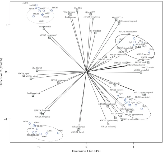
3.5. Principal Component Analysis (PCA). After analysing
individually each bioactivity indicator and the contents in phenolic compounds, PCA was applied to understand the true effect of either origin or extract type in a global manner. That is, instead of evaluating individual changes caused in each bioactivity indicator or phenolic compounds group, it was intended to obtain an integrated output dealing with all the effects at once. The plot of component loadings forextract type was designed with the first two dimensions (first:
Cronbach’s 𝛼, 0.965; eigenvalue, 17.194; second: Cronbach’s
𝛼, 0.950; eigenvalue, 13.721), which included most variance
of data (first: 40.94%; second: 32.67%). The third and fourth dimensions, despite being significant, were not plotted due to
the complexity of the corresponding output. The distribution
of objects (Figure 1) indicates a clear separation of methanolic
and aqueous extracts (black and grey ellipses). Furthermore, objects corresponding to wild and cultivated samples were clearly separated within each type of extract. The assignment of each set of objects to either wild or cultivated samples was done according to the tabled object scores (data not shown). Group corresponding to cultivated samples extracted with methanol (solid grey line ellipse) was characterized by the high amounts of bioactive compounds, specifically flavonols, flavones, and total phenolics, and its high
bioactiv-ity against bacteria (B. cereus, MIC and MBC,S. typhimurium,
MIC and MBC, E. coli, MIC, E. cloacae, MIC, and L.
monocytogenes, MIC) and fungi (A. ochraceus, MIC and
MFC,A. versicolor, MIC, andP. funiculosum, MFC).
The most distinctive features in cultivated samples extracted with water (solid black line ellipse) were the low content in flavan-3-ols, despite having high antifungal
activity againstA. fumigatusandA. niger(MIC and MFC).
A third group (dashed grey line ellipse), corresponding to wild samples extracted with methanol, was characterized
as having high antibacterial (S. aureus, MIC and MBC, L.
monocytogenes, MBC, and P. aeruginosa, MIC), antifungal
(T. viride, MIC and MFC, P. ochrochloron, MFC, and P.
funiculosum, MIC), and antitumoral activities (HCT15).
The high bioactivity of wild methanolic extracts might be related to their high content in flavan-3-ols, mainly
epicate-chin and procyanidin trimer with an A-linkage [32], which
were previously reported as having strong antibacterial
activ-ity [34,35]. Curiously, these extracts had an activity opposite
to that demonstrated by cultivated samples extracted with
water, which might indicate that the A. fumigatus and A.
flavusare poorly susceptible to flavan-3-ols.
Similarly, wild samples extracted with water (dashed black line ellipse) had the reverse behavior in comparison to cultivated samples extracted with methanol. These aqueous
extracts were mostly active against P. verrucosum, which
seemed to have low susceptibility to favonols, flavones, and total phenolics and generally high resistance against the
6
Bi
o
M
ed
R
es
earch
In
te
rn
at
io
n
al
Table 3: Antibacterial activity (MIC and MBC, mg/mL) of different extracts ofLaurus nobilis. The results are presented as mean±SD1.
Staphylococcus aureus
Bacillus cereus
Micrococcus flavus
Listeria monocytogenes
Pseudomonas aeruginosa
Salmonella typhimurium
Escherichia coli
Enterobacter cloacae MIC
Origin (O)
Cultivated 0.06 ± 0.04 0.08 ± 0.04 0.048 ± 0.005 0.1 ± 0.1 0.08 ± 0.04 0.08 ± 0.03 0.12 ± 0.02 0.04 ± 0.01
Wild 0.05 ± 0.05 0.11 ± 0.01 0.101 ± 0.005 0.16 ± 0.05 0.046 ± 0.003 0.11 ± 0.01 0.16 ± 0.05 0.08 ± 0.05
𝑃value (𝑛 = 18) 0.619 0.001 <0.001 0.030 0.001 <0.001 0.002 0.004
Extract (E)
Methanolic 0.012 ± 0.005 0.08 ± 0.04 0.08 ± 0.03 0.06 ± 0.04 0.046 ± 0.003 0.07 ± 0.03 0.12 ± 0.02 0.03 ± 0.01
Aqueous 0.10 ± 0.01 0.11 ± 0.01 0.07 ± 0.03 0.20 ± 0.02 0.08 ± 0.03 0.11 ± 0.01 0.16 ± 0.05 0.08 ± 0.05
𝑃value (𝑛 = 18) <0.001 0.001 0.858 <0.001 0.001 <0.001 0.009 0.002
O×E
𝑃value (𝑛 = 36) 0.002 <0.001 0.212 <0.001 <0.001 <0.001 <0.001 <0.001
Ampicillin 0.25 ± 0.02 0.25 ± 0.03 0.25 ± 0.04 0.37 ± 0.05 0.74 ± 0.05 0.37 ± 0.02 0.25 ± 0.01 0.37 ± 0.04
Streptomycin 0.04 ± 0.01 0.09 ± 0.01 0.17 ± 0.02 0.17 ± 0.01 0.17 ± 0.01 0.17 ± 0.02 0.17 ± 0.02 0.26 ± 0.03
MBC Origin (O)
Cultivated 0.16 ± 0.04 0.15 ± 0.04 0.11 ± 0.01 0.39 ± 0.02 0.18 ± 0.02 0.16 ± 0.05 0.20 ± 0.02 0.09 ± 0.03
Wild 0.1 ± 0.1 0.19 ± 0.02 0.20 ± 0.01 0.29 ± 0.05 0.10 ± 0.01 0.21 ± 0.02 0.2 ± 0.1 0.1 ± 0.1
𝑃value (𝑛 = 18) 0.262 0.001 <0.001 <0.001 <0.001 0.002 0.764 0.111
Extract (E)
Methanolic 0.07 ± 0.05 0.15 ± 0.04 0.16 ± 0.05 0.3 ± 0.1 0.14 ± 0.04 0.17 ± 0.05 0.1 ± 0.1 0.08 ± 0.04
Aqueous 0.21 ± 0.02 0.19 ± 0.01 0.15 ± 0.05 0.37 ± 0.03 0.15 ± 0.05 0.21 ± 0.01 0.3 ± 0.1 0.1 ± 0.1
𝑃value (𝑛 = 18) <0.001 <0.001 0.461 0.005 0.696 0.008 <0.001 0.041
O×E
𝑃value (𝑛 = 36) <0.001 <0.001 0.719 <0.001 <0.001 <0.001 <0.001 <0.001
Ampicillin 0.37 ± 0.04 0.37 ± 0.05 0.37 ± 0.04 0.49 ± 0.05 1.2 ± 0.1 0.49 ± 0.05 0.49 ± 0.04 0.74 ± 0.05
Streptomycin 0.09 ± 0.01 0.17 ± 0.02 0.34 ± 0.05 0.34 ± 0.04 0.34 ± 0.03 0.34 ± 0.05 0.34 ± 0.04 0.52 ± 0.05
Bi
o
M
ed
R
es
earch
In
te
rn
at
io
n
al
7
Table 4: Antifungal activity (MIC and MFC, mg/mL) of different extracts ofLaurus nobilis. The results are presented as mean±SD1.
Aspergillus fumigatus
Aspergillus versicolor
Aspergillus ochraceus
Aspergillus niger
Trichoderma viride
Penicillium funiculosum
Penicillium ochrochloron
Penicillium verrucosum MIC
Origin (O)
Cultivated 0.07 ± 0.05 0.01 ± 0.01 0.04 ± 0.01 0.3 ± 0.2 0.02 ± 0.01 0.03 ± 0.01 0.12 ± 0.02 0.17 ± 0.05
Wild 0.2 ± 0.1 0.02 ± 0.01 0.048 ± 0.004 0.3 ± 0.2 0.02 ± 0.01 0.03 ± 0.02 0.11 ± 0.02 0.20 ± 0.02
𝑃value (𝑛 = 18) <0.001 0.005 <0.001 0.603 0.163 0.407 0.054 0.005
Extract (E)
Methanolic 0.2 ± 0.1 0.009 ± 0.003 0.04 ± 0.01 0.47 ± 0.01 0.008 ± 0.005 0.017 ± 0.005 0.10 ± 0.01 0.20 ± 0.02
Aqueous 0.06 ± 0.04 0.024 ± 0.005 0.045 ± 0.002 0.07 ± 0.04 0.029 ± 0.002 0.048 ± 0.002 0.12 ± 0.02 0.17 ± 0.05
𝑃value (𝑛 = 18) <0.001 <0.001 0.077 <0.001 <0.001 <0.001 0.008 0.007
O×E
𝑃value (𝑛 = 36) <0.001 0.003 <0.001 <0.001 <0.001 <0.001 0.278 <0.001
Bifonazole 0.15 ± 0.01 0.10 ± 0.01 0.15 ± 0.02 0.15 ± 0.01 0.15 ± 0.01 0.20 ± 0.03 0.20 ± 0.02 0.10 ± 0.01
Ketoconazole 0.20 ± 0.02 0.20 ± 0.03 1.5 ± 0.1 0.20 ± 0.02 1.0 ± 0.1 0.20 ± 0.02 2.5 ± 0.3 0.20 ± 0.04
MFC Origin (O)
Cultivated 0.2 ± 0.1 0.05 ± 0.03 0.08 ± 0.03 0.4 ± 0.4 0.03 ± 0.01 0.10 ± 0.02 0.23 ± 0.02 0.6 ± 0.3
Wild 0.4 ± 0.1 0.04 ± 0.01 0.11 ± 0.01 0.5 ± 0.3 0.03 ± 0.02 0.11 ± 0.02 0.20 ± 0.02 0.40 ± 0.03
𝑃value (𝑛 = 18) <0.001 0.091 <0.001 0.196 0.500 0.027 <0.001 0.041
Extract (E)
Methanolic 0.3 ± 0.1 0.021 ± 0.004 0.08 ± 0.03 0.7 ± 0.3 0.016 ± 0.004 0.10 ± 0.02 0.20 ± 0.03 0.6 ± 0.2
Aqueous 0.2 ± 0.1 0.06 ± 0.02 0.11 ± 0.01 0.2 ± 0.1 0.046 ± 0.002 0.11 ± 0.01 0.23 ± 0.02 0.3 ± 0.1
𝑃value (𝑛 = 18) <0.001 <0.001 <0.001 <0.001 <0.001 0.122 <0.001 <0.001
O×E
𝑃value (𝑛 = 36) 0.001 <0.001 <0.001 0.312 <0.001 <0.001 0.052 <0.001
Bifonazole 0.20 ± 0.02 0.20 ± 0.03 0.20 ± 0.01 0.20 ± 0.02 0.20 ± 0.04 0.25 ± 0.05 0.25 ± 0.04 0.20 ± 0.03
Ketoconazole 0.50 ± 0.05 0.50 ± 0.04 2.0 ± 0.4 0.50 ± 0.05 1.0 ± 0.1 0.50 ± 0.04 3.5 ± 0.5 0.30 ± 0.05
8 BioMed Research International
MeOH MeOH
MeOH
MeOH MeOH
MeOH
MeOH
MeOH MeOH MeOH
MeOH MeOH
MeOH MeOH
MeOH
MeOH
MeOH MeOH
Total phenolics
MFC(P. verrucosum)
Total flavonols
Total flavones MBC (P. aeruginosa)
GI50-Hela
GI50-HepG2
GI50-MCF7
GI-NC-H460
MBC(L. monocytogenes)
MFC (P. ochrochloron)
GI50-HCT15
H2O
H2O
H2O
H2O
H2O
H2O
H2O
H2O
H2O
H2O
H2O H2O
H2O
H2O
H2O
H2O H2O H2O
MIC (P. Aeruginosa)
GI50-PLP1
MIC (P. ochrochloron) MBC (S. aureus)
MBC (E. cloacae)
MBC (E. coli)
MIC (E. coli)
MIC (L. monocytogenes)
MIC (A. versicolor)
MFC (P. funiculosum)
MFC (A. ochraceus) MBC (B. cereus)
MBC (S. typhimurium)
MIC (B. cereus) MIC (S. typhimurium)
MIC (E. cloacae)
MIC (S. aureus)
MIC (T. viride)
MFC (T. viride) MFC (A. versicolor) MIC (P. funiculosum))
MIC (A. ochraceus) MFC (A. fumigatus)
MFC (P. verrucosum) MFC (A. niger)
MIC (A. niger)
MIC (A. fumigatus)
MIC (M. flavus)
MBC (M. flavus)
Total flavan-3-o1
Dimen
sio
n
2
(
3
2.6
7
%)
Dimension1(40.94%)
1 1
0 0
−1 −1
Figure 1: Biplot of objects (extraction solvents) and component loadings (evaluated parameters).
4. Conclusion
The extract type induced the most marked changes in bioac-tivity of laurel samples. Furthermore, each of the assayed factors (origin and extract type) acts in a differentiated man-ner; that is, the same evaluated parameter gave sometimes statistically significant differences regarding laurel origin, but no effect at all from extract type or vice versa. From the PCA biplot, it became clear that wild laurel samples were more effective to inhibit tumor cell lines growth, especially HeLa, MCF7, NCI-H460, and HCT15. HepG2, as previously high-lighted, had the same reaction to laurel from wild and cul-tivated origin. It was also observed that methanolic extracts
tended to have higher antimicrobial activity, exceptA. niger,
A. fumigatus,andP. verrucosum.The differences in bioactivity
might be related to the higher phenolic compounds contents (mainly flavan-3-ols and flavonols) presented by methanolic extracts.
The most interesting finding in this work was the bioac-tive specificity of each laurel extract, considering its wild or cultivated origin. In fact, from the obtained results it
is possible to choose the combination extract type/origin with potentially highest effect against determined bacteria, fungi, or tumor cell line. These findings should, however, be analysed within the geographical area of study, considering eventual specific features of the used samples.
Conflict of Interests
The authors declare that they have no conflict of interests regarding the publication of this paper.
Acknowledgments
BioMed Research International 9
to Serbian Ministry of Education and Science for financial support (grant number 173032).
References
[1] H. Marzouki, A. Piras, K. B. H. Salah et al., “Essential oil composition and variability ofLaurus nobilis L. growing in Tunisia, comparison and chemometric investigation of different plant organs,”Natural Product Research, vol. 23, no. 4, pp. 343– 354, 2009.
[2] A. Barla, G. Topc¸u, S. ¨Oks¨uz, G. T¨umen, and D. G. I. Kingston, “Identification of cytotoxic sesquiterpenes fromLaurus nobilis
L,”Food Chemistry, vol. 104, no. 4, pp. 1478–1484, 2007. [3] F. Fang, S. Sang, K. Y. Chen, A. Gosslau, C.-T. Ho, and R. T.
Rosen, “Isolation and identification of cytotoxic compounds from Bay leaf (Laurus nobilis),”Food Chemistry, vol. 93, no. 3, pp. 497–501, 2005.
[4] J. Ivanoi´c, D. Miˇsin, M. Risti´c, O. Peˇsi´c, and I. ˇZiˇzovi´c, “Super-critical CO2extract and essential oil of bay (Laurus nobilisL.)— chemical composition and antibacterial activity,”Journal of the Serbian Chemical Society, vol. 75, pp. 395–404, 2010.
[5] S. Dall’Acqua, R. Cervellati, E. Speroni et al., “Phytochemical composition and antioxidant activity ofLaurus nobilisL. leaf infusion,”Journal of Medicinal Food, vol. 12, no. 4, pp. 869–876, 2009.
[6] E. Panza, M. Tersigni, M. Iorizzi et al., “Lauroside B, a megastig-mane glycoside fromLaurus nobilis(bay laurel) leaves, induces apoptosis in human melanoma cell lines by inhibiting NF-𝜅B activation,”Journal of Natural Products, vol. 74, no. 2, pp. 228– 233, 2011.
[7] C. Ramos, B. Teixeira, I. Batista et al., “Antioxidant and antibacterial activity of essential oil and extracts of bay laurel
Laurus nobilisLinnaeus (Lauraceae) from Portugal,”Natural Product Research, vol. 26, no. 6, pp. 518–529, 2012.
[8] J. Z. Al-Kalaldeh, R. Abu-Dahab, and F. U. Afifi, “Volatile oil composition and antiproliferative activity ofLaurus nobilis, Ori-ganum syriacum, OriOri-ganum vulgare, andSalvia trilobaagainst human breast adenocarcinoma cells,”Nutrition Research, vol. 30, no. 4, pp. 271–278, 2010.
[9] I. Dadalioˇglu and G. A. Evrendilek, “Chemical compositions and antibacterial effects of essential oils of Turkish oregano (Origanum minutiflorum), bay laurel (Laurus nobilis), Spanish lavender (Lavandula stoechasL.), and fennel (Foeniculum vul-gare) on common foodborne pathogens,”Journal of Agricultural and Food Chemistry, vol. 52, no. 26, pp. 8255–8260, 2004. [10] M. Carocho and I. C. F. R. Ferreira, “The role of phenolic
compounds in the fight against cancer—a review,”Anticancer Agents in Medicinal Chemistry, vol. 13, pp. 1236–1238, 2013. [11] G. Adwan and M. Mhanna, “Synergistic effects of plant extracts
and antibiotics onStaphylococcus aureusstrains isolated from clinical specimens,”Middle-East Journal of Scientific Research, vol. 3, pp. 134–139, 2008.
[12] R. Al-Hussaini and A. M. Mahasneh, “Microbial growth and quorum sensing antagonist activities of herbal plants extracts,”
Molecules, vol. 14, no. 9, pp. 3425–3435, 2009.
[13] M. R. Loizzo, R. Tundis, F. Menichini, A. M. Saab, G. A. Statti, and F. Menichini, “Cytotoxic activity of essential oils from Labiatae and Lauraceae families againstin vitrohuman tumor models,”Anticancer Research, vol. 27, no. 5, pp. 3293–3299, 2007. [14] A. M. Saab, R. Tundis, M. R. Loizzo et al., “Antioxidant and antiproliferative activity ofLaurus nobilisL. (Lauraceae)
leaves and seeds essential oils against K562 human chronic myelogenous leukaemia cells,”Natural Product Research, vol. 26, pp. 1741–1745, 2012.
[15] M. Kaileh, W. V. Berghe, E. Boone, T. Essawi, and G. Haege-man, “Screening of indigenous Palestinian medicinal plants for potential anti-inflammatory and cytotoxic activity,”Journal of Ethnopharmacology, vol. 113, no. 3, pp. 510–516, 2007.
[16] E. Julianti, K. H. Jang, S. Lee et al., “Sesquiterpenes from the leaves ofLaurus nobilisL.,”Phytochemistry, vol. 80, pp. 70–76, 2012.
[17] S. Lee, S.-C. Chung, S.-H. Lee et al., “Acylated kaempferol glycosides from Laurus nobilis leaves and their inhibitory effects on Na+/K+-adenosine triphosphatase,” Biological and Pharmaceutical Bulletin, vol. 35, no. 3, pp. 428–432, 2012. [18] U. de Corato, O. Maccioni, M. Trupo, and G. Di Sanzo,
“Use of essential oil ofLaurus nobilis obtained by means of a supercritical carbon dioxide technique against post harvest spoilage fungi,”Crop Protection, vol. 29, no. 2, pp. 142–147, 2010. [19] A. F. Millezi, D. S. Caixeta, D. F. Rossoni, M. G. Cardoso, and R. G. Piccoli, “In vitroantimicrobial properties of plant essential oilsThymus vulgaris, Cymbopogon citratusandLaurus nobilisagainst five important foodborne pathogens,”Ciˆencia e Tecnologia de Alimentos, vol. 32, pp. 167–172, 2012.
[20] S. Santoyo, R. Llor´ıa, L. Jaime, E. Iba˜nez, F. J. Se˜nor´ans, and G. Reglero, “Supercritical fluid extraction of antioxidant and antimicrobial compounds from Laurus nobilis L. Chemical and functional characterization,”European Food Research and Technology, vol. 222, no. 5-6, pp. 565–571, 2006.
[21] A. Simi´c, M. D. Sokovi´c, M. Risti´c, S. Gruji´c-Jovanovi´c, J. Vukojevi´c, and P. D. Marin, “The chemical composition of some Lauraceae essential oils and their antifungal activities,”
Phytotherapy Research, vol. 18, pp. 713–717, 2004.
[22] ¨O. Ert¨urk, “Antibacterial and antifungal activity of ethanolic extracts from eleven spice plants,”Biologia, vol. 61, no. 3, pp. 275–278, 2006.
[23] J. El Malti and H. Amarouch, “Antibacterial effect, histological impact and oxidative stress studies fromLaurus nobilisextract,”
Journal of Food Quality, vol. 32, no. 2, pp. 190–208, 2009. [24] N. Fukuyama, C. Ino, Y. Suzuki et al., “Antimicrobial
sesquiter-penoids fromLaurus nobilisL,”Natural Product Research, vol. 25, no. 14, pp. 1295–1303, 2011.
[25] M.-H. Liu, N. Otsuka, K. Noyori et al., “Synergistic effect of kaempferol glycosides purified fromLaurus nobilisand fluo-roquinolones on methicillin-resistant Staphylococcus aureus,”
Biological and Pharmaceutical Bulletin, vol. 32, no. 3, pp. 489– 492, 2009.
[26] N. Otsuka, M.-H. Liu, S. Shiota et al., “Anti-methicillin resistant
Staphylococcus aureus(MRSA) compounds isolated from Lau-rus nobilis,”Biological and Pharmaceutical Bulletin, vol. 31, no. 9, pp. 1794–1797, 2008.
[27] C. Pereira, R. C. Calhelha, L. Barros, and I. C. F. R. Ferreira, “Antioxidant properties, anti-hepatocellular carcinoma activity and hepatotoxicity of artichoke, milk thistle and borututu,”
Industrial Crops and Products, vol. 49, pp. 61–65, 2013. [28] CLSI Clinical and Laboratory Standards Institute, “Methods
for dilution antimicrobial susceptibility tests for bacteria that grow aerobically,” Approved standard, 8th ed. CLSI publication M07-A8. Clinical and Laboratory Standards Institute, Wayne, Pa, USA, 2009.
10 BioMed Research International
method for susceptibility testing against drug-resistant bacte-ria,”Journal of Microbiological Methods, vol. 90, pp. 160–166, 2012.
[30] C. Booth, “Fungal culture media,” inMethods in Microbiology, J. R. Norris and D. W. Ribbons, Eds., pp. 49–94, Academic Press, New York, NY, USA, 19711971.
[31] A. Espinel-Ingroff, “Comparison of the E-test with the NCCLS M38-P method for antifungal susceptibility testing of common and emerging pathogenic filamentous fungi,”Journal of Clinical Microbiology, vol. 39, no. 4, pp. 1360–1367, 2001.
[32] M. I. Dias, L. Barros, M. Due˜nas et al., “Nutritional and antioxidant contributions ofLaurus nobilisL. leaves: would be more suitable a wild or a cultivated sample?”Food Chemistry, vol. 156, pp. 339–346, 2014.
[33] C. Santos-Buelga, S. Gonzalez-Manzano, M. Due˜nas, and A. M. Gonzalez-Paramas, “Extraction and isolation of phenolic compounds,”Methods in Molecular Biology, vol. 864, pp. 427– 464, 2012.
[34] J. W. Betts, S. M. Kelly, and S. J. Haswell, “Antibacterial effects of theaflavin and synergy with epicatechin against clinical isolates ofAcinetobacter baumanniiandStenotrophomonas maltophilia,”
International Journal of Antimicrobial Agents, vol. 38, no. 5, pp. 421–425, 2011.
Submit your manuscripts at
http://www.hindawi.com
Hindawi Publishing Corporation
http://www.hindawi.com Volume 2014 Anatomy
Research International
Peptides
International Journal ofHindawi Publishing Corporation
http://www.hindawi.com Volume 2014
Hindawi Publishing Corporation http://www.hindawi.com
International Journal of
Volume 2014
Zoology
Hindawi Publishing Corporation
http://www.hindawi.com Volume 2014
Molecular Biology International
Hindawi Publishing Corporation http://www.hindawi.com
Genomics
International Journal ofVolume 2014
The Scientific
World Journal
Hindawi Publishing Corporation
http://www.hindawi.com Volume 2014
Hindawi Publishing Corporation
http://www.hindawi.com Volume 2014
Bioinformatics
Advances inMarine Biology
Journal of Hindawi Publishing Corporationhttp://www.hindawi.com Volume 2014
Hindawi Publishing Corporation
http://www.hindawi.com Volume 2014
Signal Transduction
Journal ofHindawi Publishing Corporation
http://www.hindawi.com Volume 2014
BioMed
Research International
Evolutionary Biology International Journal of
Hindawi Publishing Corporation
http://www.hindawi.com Volume 2014
Hindawi Publishing Corporation
http://www.hindawi.com Volume 2014 Biochemistry Research International
Archaea
Hindawi Publishing Corporation
http://www.hindawi.com Volume 2014
Hindawi Publishing Corporation
http://www.hindawi.com Volume 2014
Genetics
Research International
Hindawi Publishing Corporation
http://www.hindawi.com Volume 2014
Advances in
Virology
Hindawi Publishing Corporation http://www.hindawi.com
Nucleic Acids
Journal ofVolume 2014
Stem Cells
International
Hindawi Publishing Corporation
http://www.hindawi.com Volume 2014
Hindawi Publishing Corporation
http://www.hindawi.com Volume 2014
Enzyme
Research
Hindawi Publishing Corporation
http://www.hindawi.com Volume 2014