Rev. Bras. Frutic., v. 38, n. 3: (e-949) DOI 10.1590/0100-29452016 May/June 2016 Jaboticabal - SP ISSN 0100-2945 http://dx.doi.org/10.1590/0100-29452016949
Rev. Bras. Frutic., Jaboticabal - SP, v. 38, n. 3 : e-949 1/8
REVISION
PINK DISEASE, A REVIEW OF AN ASYMPTOMATIC
BACTERIAL DISEASE IN PINEAPPLE
VIaNey MaRíN-CeVaDa2 & LuIS eRNeStO FueNteS-RaMíRez3
ABSTRACT-Pink disease is an asymptomatic pineapple disease in the field and is evidenced with a red-dark coloration when the infectedfruit is processed to obtain products such as juice, jam, and preservatives.
Tatumella morbirosei and T. ptyseos (formerly Pantoea citrea) have been demonstrated as causal agents.
although T. morbirosei and T. ptyseos have been well studied, there are currently no cost effective control
methods in pineapple cultivation. The purpose of this review is to summarize the significant and updated research on the role of pink disease in pineapple.
Index terms: pineapple, phytopathogen,gdhB, biocontrol.
DOENçA ROSADA, uMA REVISãO DE uMA DOENçA
ASSIMTOMáTICA BACTERIANA DO ABACAxI
RESuMO - Doença rosada ou Pink disease é uma doença assintomática do abacaxi no campo, oque é evidencia com uma coloração vermelho - escura, quando a fruta infectada é processada para a obtenção de
produtos como suco, geléia, e conservantes. Tatumella morbirosei e T. ptyseos (antigo Pantoea citrea) tsão
os agentes causáis dessa doença. embora t. morbirosei e T. ptyseosforamtenham sido bem estudadas, até
o presente não existem métodos eficientes de controle da Pink disease de abacaxi. O objetivo da presente revisão é sumarizar as pesquisas significativas e atualizadas pesquisasobre o papel de Pink disease no cultivo do abacaxizeiro.
Termos para indexação: abacaxi, fitopatógeno, gdhB, biocontrole.
INTRODuCTION
Pink disease is an important global threat to pineapple production, especially for the fruit industry.
the reported causal agents are Tatumellamorbirosei
(formerly Pantoeacitrea) and Tatumella ptyseos,
both belonging to the Enterobacteriaceae (Cha
et al.,1997a; MaRIN-CeVaDa et al., 2010). Pink disease has been reported from Australia, Hawaii, Mexico, the Philippines, South Africa, and Taiwan (MaRíN-CeVaDa et al., 2006; ROhRBaCh, 1983). Cost-effective management strategies for effective control of the disease are still not available. The current schemes consist mainly of insect control, since it has been supposed that insects play a role in the transmission of the pathogen (KaDO, 2003). Additionally, plant breeding for obtaining resistant cultivars might be an alternative to overcome the problem (Kado, 2003).
Economic Status of Pineapple
The tropical fruits most cultivated in the world are bananas, mangos, avocados, papayas and pineapples (FAO 2003) with an estimated total worldwide production of about 160 million tons in 2012 (http://faostat3.fao.org/download/Q/QC/E).
About 98% of tropical-fruit production is taking place in developing countries and 80% of the world import trade for these fruits is directed to developed countries (http://fao.org/docrep/006/ y5143e/y5143e1a.htm). Pineapple had a worldwide gross production valued at $17 billion in 2012 (http:// faostat3.fao.org/download/Q/QV/E).
A large amount of the pineapple trade consists of processed products, such as canned slices and juice (BaRthOLOMew et al., 2003). Thailand, Brazil, the Philippines, Indonesia, and Costa Rica are the major producers of fresh fruit pineapples, and Costa Rica ranked as the major world pineapple exporter
1(trabalho 151-15). Recebido em: 02-06-2015. aceito em 01-09-2016.
2Vianey Marín-Cevada, Centro de Investigaciones en Ciencias Microbiológicas, ICUAP, Benemérita Universidad Autónoma de
Puebla, Mexico. email: [email protected]
3Luis Ernesto Fuentes-Ramírez, Centro de Investigaciones en Ciencias Microbiológicas, ICUAP, Benemérita Universidad Autónoma
in 2011 with 1.75 million metric tons (MMT) (http:// faostat3.fao.org/faostat-gateway/go/to/browse/T/ TP/E). The world production of canned pineapples has shown a tremendous increase over the last decade or so (http://faostat3.fao.org/faostat-gateway/go/to/ browse/Q/QC/E) (table 1). Similarly, the production of pineapple juice has showed a notable increase in the last years (http://faostat3.fao.org/faostat-gateway/ go/to/browse/Q/QC/E) (table 1).
the pineapple plant [Ananas comosus (L.)
Merr. var. comosus] is widely distributed in the New
World and shows a great adaptability to variedseveral habitats. Pineapple is one of the most ancient American crops and was domesticated and dispersed several thousand years ago by amerindian peoples in the Orinoco and Amazon basins (COLLINS, 1960). This plant is a member of the Bromeliaceae family, which contains about 58 genera and 3352 species (LutheR, 2012). “Smooth Cayenne” “Red Spanish”, “Queen”, “Abacaxi”, and “MD2” are the principal pineapple grown cultivars (Bartholomew et al.,
2003; LOeILLet et al.; 2011 uNCtaD http:// www.unctad.info/en/Infocomm/AACP-Products/ COMMODIty-PROFILe-Pineapple/).
The productivity of the pineapple depends on the control of factors such as water and nutrient supply (nitrogen, potassium, phosphorus, calcium, iron, magnesium, zinc, boron and other micronutrients), as well as management and control of pests and diseases (zhaNg et al., 1997).
Pink disease of pineapple
the pineapple plant is threatened by several phytopathogenic bacteria that are responsible for diseases like fruit collapse, marbling disease, fruit brown rot, anomalous proliferations and pink disease (table 2),(JOhNStON 1957;LIM;LOwINgS 1979;
KORReS et al. 2010;SeRRaNO 1928;DaVIS et aL.
2005;DaVIS et al. 2006; LyON 1915).
. Among them, pink disease is of great importance because of its potential impact on the postharvest processes (Cha et al., 1997b). Pasteurized fruits affected by this disease produce brownish pigments, so the commodity becomes unmarketable (ROhRBaCh, 1989; ROhRBaCh; PFeIFFeR, 1975) (Fig. 1). the challenge in the control of this disease is that infected plants remain asymptomatic in the field, making difficult to discriminate healthy from affected fruits (Pujol and Kado 1999). Unfortunately, cans of healthy fruit from the same lot of infected fruits are also commonly discarded by quality control, increasing the economic losses (Cha et al., 1997b).
Causative agents of Pink Disease
It has been suggested that this disease is caused by microorganisms of the families Acetobacteraceae
and Enterobacteriaceae (BuDDeNhageN; D u L L , 1 9 6 7 ; g O S S e L e ; S w I N g S , 1 9 8 6 ; KONtaxIS; haywaRD, 1978; ROhRBaCh;
PFeIFFeR, 1976). Almost four decades ago, an
isolate obtained from a diseased fruit, did not show perceptible symptoms when it was inoculated in fresh fruits but induced discoloration after heating the inoculated fruits (Rohrbach and Pfeiffer 1976). The authors identified that strain as belonging to
the species Enterobacter agglomerans (Pantoea
agglomerans, gaVINI, et al., 1989) (ChO, et al.,
1980; ROhRBaCh; PFeIFFeR, 1976). Afterwards,
Pantoea citrea was identified as another causative
microorganism (Cha, et al., 1997a), also in the
Family Enterobacteriaceae. A wide taxonomic
analysis of P. citrea strains causing pink disease showed their proper classification as the a new
species, Tatumella morbirosei, in a different Ggenus
of Enterobacteriaceae and not as a species of Pantoea (BRaDy et al. 2008; 2010). Furthermore, isolates obtained from pink disease affecting Mexican pineapple fruits were identified as belonging
to the species T. ptyseos (MaRíN-CeVaDa et
al. 2010). Hence, at least those two species of the
genus Tatumella are capable to cause pineapple
pink disease.
Epidemiology and other characteristics of pink disease
Seasonality is probably an important factor in the appearance of the disease, since warm and humid environments promote the appearing of the phytopathogen. In our previous research we detected the presence of T. ptyseos in 48% of pineapples in
the rainy season (MaRIN-CeVaDa, et al., 2010), and only 3% in the dry season (unpublished results of our group). Besides, the average bacterial number decreased from 106 CFU/g of fruit to 102 CFu/g of fruit in the rainy and dry season, respectively (MaRIN-CeVaDa, et al., 2010). In other geographic regions, the incidence of pink disease increases also in the rainy season, February, March and april in Hawaii and Taiwan, and August and September in the Philippines (hINe, 1976; ROhRBaCh; JOhNSON, 2003; ROhRBaCh; PFeIFFeR, 1975). Other biological or environmental factors might be required for the bacterial outbreak, since not all the wet seasons shown higher incidence of pink disease
(hINe, 1976). There is no consensus with regard to
Rev. Bras. Frutic., v. 38, n. 3: (e-949) DOI 10.1590/0100-29452016949 May/Jun 2016 Jaboticabal - SP
3
PINK DISEASE, A REVIEW OF AN ASYMPTOMATIC BACTERIAL...
with the nectar in the placental regions of flowers (ROhRBaCK; PFeIFFeR 1975). It has also been proposed that the bacteria involved in pink disease are transmitted to pineapple flowers by insects. The latter scenario is supported by the fact that spraying ofcrops with insecticide decreases the appearance of the disease (KaDO, 2003). In addition, rain splash could disperse the pathogens from infected to healthy plants (hINe, 1976).
The biochemical basis of the pink
discolouration in T. morbirosei was elucidated by
Cha et al., (1997b); PuJOL;KaDO (1999; 2000). It is noteworthy that a biochemical basis of pink disease had been suggested by Buddenhagen and Dull since 1967. The pigmentation of affected fruit is initiated by the oxidation of glucose to gluconate by glucose
dehydrogenase gdhB. gdhB is induced by glucose
in the stationary phase (PuJOL;KaDO 1999). gdhB oxidizes gluconate to 2-keto-D-gluconate, which is subsequently converted to 2,5–diketogluconate by another dehydrogenase (PuJOL; KaDO, 1999; 2000). It has been suggested that 2,5-diketogluconate can form complexes with plant compounds produced at canning temperatures, or may dimerize, producing the decolorization (Cha et al., 1997b; KaDO, 2003). Alternatively, 2-keto-D-gluconate present in infected fruits might suffer an accelerated heat oxidation
to 2,5-diketogluconate. gdhA, a gene for a second
glucose dehydrogenase in T. morbirosei, is expressed
constitutively at low levels and does not participate in the generation of pink disease pigmentation (Pujol and Kado 1999). gdhB possesses a putative binding site for pyrroloquinolinequinone (PQQ). PQQ could be synthesized endogenously in T. morbirosei since cell extracts show Gdh activity without adding an exogenous cofactor (Pujol and Kado 1999), and as a matter of fact, the T. ptyseos genome possesses at least one locus putatively implicated in PQQ biosynthesis (acc. Num. wP_025901980).
Control
The application of insecticides in pineapple fields has been suggested to decrease the incidence of pink disease (KaDO, 2003). Nevertheless, this practice could damage natural and human ecosystems and it is still unclear how insects and pink disease agents are related. Hence, it would be desirable to find alternative control methods including the production of resistant varieties and the use of biological control. In order to design suitable control and preventive measures, it is essential to get profound knowledge of all the elements of this disease. In combination with
an insecticide, the saprophytic bacterium Bacillus
gordonae have shown promising results for control
of pink disease under laboratory and field conditions
(KaDO, 2003), but its effectiveness and economic
benefits are yet to be evaluated.
Recently, our group searched for antagonists
against T. ptyseos. Among dozens of isolates from
pineapple plants, two of them that showed the greatest inhibitory activity in culture media against
T. ptyseos, where selected for determining their
activity in assays in association with plants (
MaRíN-CeVaDa et al., 2012). The antagonism assays were
carried out in double-layer plates, in liquid cultures, and in micropropagated inoculated pineapple plantlets (cv. Smooth Cayenne). The numbers of
T. ptyseos growing for 96 h in liquid co-culture
with the isolate UAPS07070 were three orders of
magnitude less thant when growing alone
(Marin-CeVaDa et al., 2012). the antagonistic bacterium was also able to abate the brownish pigmentation of culture medium produced by the pathogen in co-cultures (MaRíN-CeVaDa et al., 2012). It is still unknown if that observation was caused by the drop of T. ptyseos population itself, or by an inhibition
of the oxidation pathway. In 60 days old inoculated pineapple plantlets, the numbers of T. ptyseos were
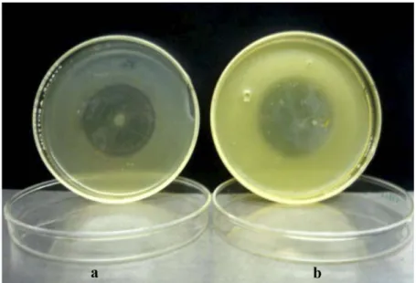
lower in co-inoculation with the isolate UAPS07070 than in lonely inoculation. Particularly, two orders of magnitude less inside the roots and three orders less in the rhizosphere (MaRIN-CeVaDa et al., 2012). Bacterial exudates produced by isolate UAPS07070 inhibited the growth of T. ptyseos
uaPS07007 in rich medium and also on rich solid medium saturated with FeCl3 (Fig. 2). Since siderophores synthesis is inhibited under high
Fe3+ concentrations, it seems unlikely that such
compounds could be responsible for the antagonism between this isolate and T. ptyseos. By comparison of partial nucleotide sequences of 16S rRNA and
recA genes, the antagonistic isolate was identified
. Bras. Frutic., v
. 38, n. 3: (e-949) DOI
10.1590/0100-29452016949
May/Jun 2016 Jaboticabal - SP
Vianey
Marín-CeV
ada
& Luis ernest
o Fuentes-raMírez
Rev. Bras. Frutic., v. 38, n. 3: (e-949) DOI 10.1590/0100-29452016949 May/Jun 2016 Jaboticabal - SP
5
PINK DISEASE, A REVIEW OF AN ASYMPTOMATIC BACTERIAL...
T
ABLE
FIGuRE 1 - Canned pineapple affected (a) compared to unaffected (b) by pink disease.
FIGuRE 2- antagonistic activity in solid media.
CONCLuSIONS
The understanding of interactions between
pineapple, Tatumella, B. gladioli, other potential
antagonists, and the environment is imperative to design preventive and control strategies for pink disease. Comparative genomic studies of the causal agents and of their antagonists might reveal genes involved in virulence and genes involved in antibiosis. Additionally, the search for new pineapple genotypes that are more resistant to pink disease could greatly reduce the economic impact of this disease in pineapple market.
ACKNOWLEDGMENTS
Work in the author’s laboratory was funded by Consejo Nacional de Ciencia y Tecnología,
México, grant CB-2009-128235-z and VIEP-BUAP.
we appreciate the valuable comments and language review by Michael Dunn (Centro de Ciencias Genómicas, uNaM, México) and Antonino Baez
(ICuaP-BuaP). We acknowledge the comments
Rev. Bras. Frutic., v. 38, n. 3: (e-949) DOI 10.1590/0100-29452016949 May/Jun 2016 Jaboticabal - SP
7
PINK DISEASE, A REVIEW OF AN ASYMPTOMATIC BACTERIAL...
REFERENCES
BaRthOLOMew, D.P.; PauL, R.e.; ROhRBaCh, K.g. The pineapple: botany, production and uses. Wallingford: CABI Publishing, 2003.
BRaDy, C.; CLeeNweRCK, I.; VeNteR, S.; VaNCaNNeyt, M.; SwINgS, J.; COutINhO, T. Phylogeny and identification of Pantoea species associated with plants, humans and the natural environment based on multilocus sequence analysis (MLSa). Systematic and Applied Microbiology, Stuttgart, v.31, n.6-8, p.460, 2008.
BRaDy, C.L.; VeNteR, S.N.; CLeeNweRCK, I.; VaNDeMeuLeBROeCKe, K.; De VOS, P.; COUTINHO, T.A. Transfer of Pantoea citrea,
Pantoea punctata and Pantoea terrea to the genus
Tatumella emend. as Tatumella citrea comb. nov.
Tatumella punctata comb. nov., and Tatumella terrea
comb. nov. (Kageyama et al., 1992) and description of Tatumella morbirosei sp.nov. International Journal Systematic Evolutionary Microbiology, Reading, v.60, n.3, p.484-494, 2010.
BuDDeNhageN, I.w.; DuLL, g.g. Pink disease of pineapple fruit caused by strains of acetic acid
bacteria. Phytopathology, St Paul, v.57, p.806, 1967.
Cha, J.S.; PuJOL, C.J.; DuCuSIN, a.R., MaCION, e.a.; huBBaRD, C.h.; KaDO, C.I. Studies on
Pantoea citrea, the causal agent of pink disease
of pineapple. Jounal of Phytopathology, utrecht, v.145, p.313-320, 1997a.
CHA, J.; PUJOL, C.; KADO, C.I. Identification and characterization of a Pantoea citrea gene encoding glucose dehydrogenase that is essential for causing pink disease of pineapple. Applied and Environmental Microbiology, washington, v.63, n.1, p.71-76, 1997b.
ChO, J.J.; haywaRD, a.C.; ROhRBaCh, K.g. Nutritional requirements and biochemical activities of pineapple pink disease bacterial strain from Hawaii. Antonie Van Leeuwenhoek, Dordrecht, v.46, n.2, p.191-204, 1980.
COLLINS, J..L. The pineapple: botany, utilization,
cultivation. London: Leonard hill, 1960.
DaVIS, R.I.; aROCha, y.; JONeS, P.; MaLau, A. First report of the association of phytoplasmas with plant diseases in the territory of Wallis and
Futuna. Australasian Plant Pathology, Clayton,
v.34, p.417-418, 2005.
DaVIS, R.I.; JONeS, P.; hOLMaN, t.J.; haLSey, K.; aMICe, R.; tuPOuNIua, S.K.; Seth, M. Phytoplasma disease surveys in Tonga, New Caledonia and Vanuatu. Australasian Plant Pathology, Clayton, v.35, n.3, p.335-340, 2006.
FaO. Medium-term prospects for agricultural
commodities. Projections to the year 2010. Rome, 2003. (technical Paper, 1)
gaVINI, J.; MeRgaeRt, J.; BeNJI, a.; MIeLCaReK, C.; IzaRD, D.; KeSteRS, K.; DE LEY, J. Transfer of Enterobacter agglomerans
(Beijerinck 1888) Ewing and Fife 1972 to Pantoea
gen. nov. as Pantoea agglomerans comb. nov.
and description of Pantoea dispersa sp.nov. International Journal of Systematic Bacteriology, ames, v.39, p.337-345,1989.
GOSSELE, F.; SWINGS, J. Identification of
Acetobacter liquefaciens as causal agent of
pink-disease of pineapple fruit. Jounal of Phytopathology, utrecht, v.116, n.3, p.167-175, 1986.
HINE, R.B. Epidemiology of pink disease of pineapple fruit. Phytopathology, St Paul, v.66, p.323-327, 1976.
JOHNSTON, A. Bacterial heart rot of the pineapple. Malaysian Agricultural Journal, Kuala Lumpur, v.40, n.1, p.2-8, 1957.
KaDO, C.I. Pink disease of pineapple. Saint Paul: the american Phytopathological Society, 2003.
Disponível em: <http://www.apsnet.org/publications/
apsnetfeatures/Pages/Pineapple.aspx>. acesso em: 27 abr. 2015.
KONtaxIS, D.g.; haywaRD, a.C. the pathogen and symptomatology of pink disease of pineapple fruit in the Philippines. Plant Disease Reporter, washington, v.62, p.446-450, 1978.
KORReS, a.M.N.; VeNtuRa, J.a.; FeRNaNDeS, P.M.B. First report of bacterium and yeasts associated with pineapple fruit collapse in Espírito Santo State, Brazil. Plant Disease, New York, v.54, n.12, p.1509, 2010.
LOEILLET, D.; DAWSON, C.; PAQUI, T. Fresh pineapple market: from the banal to the vulgar. Acta Horticulturae, the hague, v.902, p.587-594, 2011.
LutheR, h.e. An alphabetic list of bromeliad binomials. 13th ed. Sarasota: Marie Selby Botanical gardens & Bromeliad Society International, 2012.
LYON, H.L. A survey of the pineapple problems. Hawaii Plant Recreation, honolulu, v.13, p.125-139, 1915.
MaRíN-CeVaDa, V.; VaRgaS, h.V.; JuáRez, M.; LÓPez, V.g.; zagaDa, g.; heRNáNDez, S.; CRuz, a.; CaBaLLeRO-MeLLaDO, J.; LÓPez-ReyeS, L.; JIMÉNez-SaLgaDO, t.; CaRCaÑO-MONtIeL, M.; FueNteS-RaMíRez, L.e. First report of the presence of Pantoea citrea, causal agent of pink disease, in pineapple fields grown in Mexico. Plant Pathology, Oxford, v.55, n.12, p.294, 2006.
MaRíN-CeVaDa, V.; CaBaLLeRO-MeLLaDO, J.; BuStILLOS-CRIStaLeS, R.; MuÑOz-ROJaS, J.; MaSCaRÚa-eSPaRza, M.a.; CaStaÑeDa-LuCIO, M.; LÓPez-ReyeS, L.; MaRtíNez-aguILaR, L.; FueNteS-RaMíRez, L.e.
Tatumella ptyseos, an unrevealed causative agent of
pink disease in pineapple. Journal Phytopatholy, utrecht, v.158, n.2, p.93-99, 2010.
MaRíN-CeVaDa, V.; MuÑOz-ROJaS, J.; CaBaLLeRO-MeLLaDO, J.; MaSCaRÚa-e S Pa R z a , M . a . ; C a S ta Ñ MaSCaRÚa-e D a - L u C I O , M.; CaRReÑO-LÓPez, R.; eStRaDa-De LOS SaNtOS, P.; FueNteS-RaMíRez, L.e. antagonistic interactions among bacteria inhabiting
pineapple. Applied Soil Ecology, New York, v.61,
p.230-235, 2012.
PuJOL, C.; KaDO, C.I. gdhB, a gene encoding
a second quinoprotein glucose dehydrogenase in Pantoea citrae, is required for pink disease of
pineapple. Microbiology, New York, v.145, n.5,
p.1217-1226, 1999.
PuJOL, C.; KaDO, C.I. genetic and biochemical characterization of the pathway in Pantoea citrea
leading to pink disease of pineapple. Journal of Bacteriology, washigton, v.182, p.2230-2237, 2000. ROhRBaCh, K.g.; JOhNSON, M.w. Pest diseases and weeds. In: BARTHOLOMEW, D.P.; PAULL, R.e.; ROhRBaCh, K.g. (ed.). The pineapple: botany, production and uses. Wallingford: CABI Publishing, 2003. p.203-251.
ROHRBACH, K.G.; PFEIFFER, J.B. The field induction of bacterial pink disease in pineapple fruit. Phytopathology, St Paul, v.65, p.803-805, 1975.
ROhRBaCh, K.g.; PFeIFFeR, J.B. the interaction of four bacteria causing pink disease of pineapple with several pineapple cultivars. Phytopathology, St Paul, v.66, p.396-399, 1976.
ROhRBaCh, K.g. Pineapple diseases and pest and their potential for spread. In: SINGH, K.G. (Ed.). Exotic plant quarantine pests and procedures for introduction of plant materials. Serdang: aSeaN Plant Quarantine Centre and Training Institute, 1983. p.145-171.
ROHRBACH, K.G. Unusual tropical fruit diseases with extended latent periods. Plant Disease, New york, v.73, p.607-609, 1989.
SERRANO, F.B. Bacterial fruitlet brown rot of pineapple. Philippine Journal of Science, Manila, v.36, p.271-305, 1928.
SuáRez-MOReNO, z.R.; CaBaLLeRO-MeLLaDO, J.; COutINhO, B.g.; MeNDONÇa-PReVIatO, L.; JaMeS, e.K.; VeNtuRI, V. Common features of environmental and potentially beneficial plant-associated Burkholderia. Microbial Ecology, New York, v.63, p.249-266, 2012.