UNIVERSIDADE TÉCNICA DE LISBOA
Faculdade de Medicina Veterinária
EXPRESSION AND CHARACTERIZATION OF CALRETICULIN GENE ISOLATED FROM RHIPICEPHALUS ANNULATUS AFTER BABESIA BIGEMINA INFECTION.
JOANA RAMOS RAPAZ LÉRIAS
CONSTITUIÇÃO DO JÚRI
Doutora Isabel Maria Soares Pereira da Fonseca de Sampaio
Doutor Luís Manuel Madeira de Carvalho Doutor Vítor Manuel Diogo de Oliveira Alves Doutora Ana Isabel Amaro Gonçalves Domingos
ORIENTADOR
Doutora Ana Isabel Amaro Gonçalves Domingos
CO-ORIENTADOR
Doutor Luís Manuel Madeira de Carvalho
2012 LISBOA
UNIVERSIDADE TÉCNICA DE LISBOA
Faculdade de Medicina Veterinária
EXPRESSION AND CHARACTERIZATION OF CALRETICULIN GENE ISOLATED FROM RHIPICEPHALUS ANNULATUS AFTER BABESIA BIGEMINA INFECTION.
JOANA RAMOS RAPAZ LÉRIAS
DISSERTAÇÃO DE MESTRADO EM MEDICINA VETERINÁRIA.
CONSTITUIÇÃO DO JÚRI
Doutora Isabel Maria Soares Pereira da Fonseca de Sampaio
Doutor Luís Manuel Madeira de Carvalho Doutor Vítor Manuel Diogo de Oliveira Alves Doutora Ana Isabel Amaro Gonçalves Domingos
ORIENTADOR
Doutora Ana Isabel Amaro Gonçalves Domingos
CO-ORIENTADOR
Doutor Luís Manuel Madeira de Carvalho
2012 LISBOA
Acknowledgements
I would like to thank my supervisor Professor Doctor Ana Domingos, for all the help, support, patience and guidance during my Master project. Her knowledge and experience with ticks were essential to improve my work.
I express my gratitude to my co-supervisor Professor Doctor Luís Madeira de Carvalho, who encouraged my passion for parasites. My “work” in FMV/UTL - Parasitology Laboratory was essential to choose this Master theme.
I also thank Sandra Antunes, for her help at the bench, her guidance and patience.
The development of this Master project would not have been possible without the collaboration of Fábia Gomes, a dearest friend of mine and colleague at IHMT, who always showed herself available to participate in my laboratorial work.
A word of appreciation to my colleagues in IHMT and my fellow students along the 6 years of my faculty life, who always showed their sympathy, friendship and availability to help me to carrying out my tasks and duties.
I would also like to thank to João, who understood my absences while accomplishing this work and always provided me with the necessary support.
Especially to my family, mum, dad and sisters Muri and Sofia, and to my dearest dog, my four-leg best friend, Phoenix, I want to express my gratitude for all the unconditional support. Without you, I would not have become a Veterinary and certainly I would not be the same person.
Expression and characterization of calreticulin gene isolated from
Rhipicephalus (Boophilus) annulatus after Babesia bigemina
infection.
Abstract
Ticks are obligate parasites in a large variety of hosts and are considered to be, after mosquitoes, the second worldwide vector of human and animal diseases.
Bovine babesiosis causes substantial economic losses due to animal mortality, abortions and anaemia, among other effects. Calreticulin protein that has been identified in several species including ticks and previous experiments showed that calreticulin gene was up-regulated in R.
annulatus ticks infected with B. bigemina and that its knockdown by RNAi technique leads to
a reduction of both pathogen transmission and ticks weight.
This Master thesis was developed within a project on “Differential expression and functional characterization of tick (Rhipicephalus annulatus) genes in response to pathogen infection (Babesia bigemina)”, financed by the Science and Technology Foundation (FCT), with the project number of PTDC/CVT/112050/2009.
The aim of this study was the isolation of calreticulin gene, purification of calreticulin protein and its further use to produce antibodies for the purpose of immunolocalization studies and vaccination tests. In this Master thesis, calreticulin gene was amplified by PCR technique, sequenced and compared with calreticulin from the R. annulatus sequence, showing 98% identity. Afterwards, an Escherichia coli recombinant system was used in order to produce a calreticulin protein. Finally, recombinant proteins were purified using IMAC technique, due to the affinity of expressed calreticulin protein histidine tail to nickel ions. After specific elution and a final sample concentration, a unique protein was achieved in the purified sample, corresponding to recombinant calreticulin.
The results of this study were optimistic and represent one more step to improve ticks control, as we showed in this study that CRT can be produced and purified without contaminants, though further vaccination and immunolocalization studies will be the key to understand CRT future use.
Keywords: Babesia bigemina/ Rhipicephalus (Boophilus) annulatus/ Calreticulin/ Escherichia
Expressão e caracterização do gene calreticulina isolado de
Rhipicephalus (Boophilus) annulatus após infecção com Babesia
bigemina.
Resumo
As carraças são parasitas obrigatórios de uma grande variedade de hospedeiros, sendo consideradas, depois dos mosquitos, os mais importantes vectores de doenças em humanos e animais. A babesiose bovina conduz a elevados prejuízos económicos, devido ao aumento da mortalidade animal, abortos e anemia, entre outros. A calreticulina é uma proteína já identificada em várias espécies, incluindo carraças e estudos anteriores demonstraram que o gene calreticulina estava sobreexpresso em carraças R. annulatus infectadas com B. bigemina e, após o seu silenciamento através da técnica de RNAi, ocorria uma redução tanto na transmissão do agente patogénico, como no peso das carraças.
Esta tese de Mestrado foi desenvolvida em paralelo com o projecto “Expressão diferencial e caracterização de genes de carraça (Rhipicephalus annulatus) em resposta à infecção por agente patogénico (Babesia bigemina)”, financiado pela Fundação para a Ciência e Tecnologia, sendo o número do projecto PTDC/CVT/112050/2009.
A finalidade deste trabalho consistiu no isolamento do gene calreticulina e purificação da correspondente proteína, para posteriormente ser usada na produção de anticorpos destinados a estudos de imunolocalização e de vacinação. Nesta tese de mestrado, amplificou-se o gene da calreticulina pela técnica de PCR, e sequenciou-se esse gene e comparou-se com a sequência da calreticulina da carraça R. annulatus, obtendo-se uma identidade de 98%. Posteriormente, o gene foi expresso em Escherichia coli de modo a produzir-se calreticulina. Finalmente, as proteínas recombinantes foram purificadas através do método IMAC, dado a calreticulina expressa ter uma cauda de histina com afinidade para iões níquel. Após a eluição específica e a concentração das amostras finais, verificou-se que uma única proteína, correspondente à calreticulina recombinante, se encontrava presente na amostra purificada. Os resultados deste estudo foram positivos e representam mais um passo para melhorar o controlo das carraças, uma vez que este estudo demonstrou que a CRT pode ser produzida e purificada sem contaminantes, apesar de estudos posteriores de vacinação e imunolocalização serão essenciais para perceber qual o futuro da CRT.
Palavras-chave: Babesia bigemina/ Rhipicephalus (Boophilus) annulatus/ Calreticulina/
TABLE OF CONTENTS
ACKNOWLEDGEMENTS ... III ABSTRACT ... V RESUMO ... VII TABLE OF CONTENTS ... IX FIGURES ... XI TABLES ... XII ABBREVIATIONS AND SYMBOLS ... XIIICHAPTER 1. PREAMBLE ... 1
CHAPTER 2. LITERATURE REVIEW ... 3
2.1. Ticks ... 3
2.1.1. Introduction ... 3
2.1.2. Characterization, identification and morphology ... 4
2.1.3. Life cycle ... 4 2.1.4. Tick control ... 6 2.1.4.1. Introduction ... 6 2.1.4.2. Chemoprophylaxis ... 6 2.1.4.3. Biocontrol ... 7 2.1.4.4. Genetic resistance ... 7 2.1.4.5. Vaccines ... 7 2.1.4.5.1. Introduction ... 7 2.1.4.5.2. Recombinant vaccines... 8 2.1.4.5.3. Antigens selection ... 10 2.2. Babesia ... 11 2.2.1. Introduction ... 11
2.2.2. Characterization and life cycle ... 11
2.2.3. Symptoms of bovine babesiosis ... 13
2.2.4. Diagnosis... 15
2.2.5. Human babesiosis ... 16
2.2.6. Babesiosis control ... 16
2.2.6.1. Introduction ... 16
2.2.6.3. Genetic resistance ... 17
2.2.6.4. Vaccines... 17
2.3. Calreticulin... 21
2.3.1. Introduction ... 21
2.3.2. Calreticulin structure ... 22
2.3.3. Calreticulin in ticks and other parasites ... 24
CHAPTER 3. MATERIAL AND METHODS ... 27
3.1. Rhipicephalus annulatus ticks ... 27
3.2. Amplification of the calreticulin gene ... 27
3.3. Expression of recombinantcalreticulin ... 27
3.3.1. Cloning and transformation... 28
3.3.1.1. Screening of the transformed colonies ... 28
3.3.1.2. Plasmid purification for sequencing ... 28
3.3.2. Expression ... 29
3.3.3. SDS-PAGE ... 29
3.3.4. Western-Blot ... 29
3.4. Production of soluble calreticulin ... 30
3.5. Purification of recombinant calreticulin ... 31
CHAPTER 4. RESULTS ... 33
4.1. Amplification of calreticulin cDNA fragment ... 33
4.2. Cloning and transformation ... 34
4.3. Recombinant calreticulin expression ... 35
4.4. Purification of recombinant calreticulin ... 37
CHAPTER 5. DISCUSSION ... 39
CHAPTER 6. CONCLUSION ... 47
CHAPTER 7. REFERENCES ... 49
FIGURES
Figure 1 One host ticks life cycle. ... 5
Figure 2 Giemsa staining of B. bigemina (A) and B. bovis (B) infected red blood cells. ...12
Figure 3 The life cycle of Babesia bigemina in cattle and in the ixodid tick vector R. microplus. ...13
Figure 4 Cerebral form of babesiosis (A); icterus liver (B) ...14
Figure 5 Kidneys with edema and congestion (A); hematuria (“Red water”) (B) ...14
Figure 6 CRT action as a chaperone. ...22
Figure 7 Structure and functions of the CRT domains. ...24
Figure 8 PCR results. Samples were electrophoresed in a 1% Agarose/CYBRsafe gel, 1x TAE. ...33
Figure 9 Sequence of the purified sample with the amplified fragment. ...34
Figure 10 PCR results. Samples were electrophoresed in a 1% Agarose/CYBRsafe gel, 1x TAE. ...35
Figure 11 SDS-PAGE results.Protein bands from cell cultures induced by IPTG and non-induced at different time points (A) T0 to T3 and (B) T4 to T5...36
Figure 12 Western-Blot results. Protein bands from cell cultures induced by IPTG and non-induced at different time points (A) T0 to T2 and (B) T3 to T5...36
Figure 13 Western-Blot results. Protein bands from soluble and insoluble fractions. ...38
Figure 14 Western-Blot results. Protein bands from soluble and insoluble fractions. ...38
Figure 15 Western-Blot results. Protein bands from soluble and insoluble fractions. ...38
TABLES
Table 1 Prevalent ticks species in Europe ... 4
Table 2 CRT functions in parasites that cause disease in humans and animals ... 26
Table 3 Identity of calreticulin sequences from different ticks ... 34
Abbreviations and Symbols
μg Microgram
μl Microliter
µm Micrometer
A, C, G, T Adenine, cytosine, guanine, thymine
Abs Absorbance
A.D. Anno Domini
ArgE Acetylornithinase
AMA-1 Apical membrane antigen 1
BbAMA-1 Babesia bovis apical membrane antigen 1
Bbo-MIC1 Recombinant microneme protein from B. bovis
B.C. Before Christ
Bp Base pair
cDNA Complementary DNA
CRP cAMP regulatory protein
CRT Calreticulin
Cu/Zn-SODM Cu/Zn-superoxide dismutase
DDT Dichlorodiphenyltrichloroethane
DNA Deoxyribonucleic acid
EF-1a Elongation factor-1 alpha
e.g. exempli gratia
EgCRT Recombinant CRT protein from Echinococcus granulosus
ELISA Enzyme-linked immunosorbent assay
ER Endoplasmatic reticulum
F2 Second generation after cross-breeding
FPLC Fast protein liquid chromatography
GST Glutathione-S transferase
Hz Hertz
i.e. id est
IFAT Indirect immunofluorescent antibody test
IMAC Immobilized metal ion affinity chromatography
IPTG Isopropyl-β-D-thiogalactopyranoside
Kb Kilobase
kDa Kilodalton
kHz Kilohertz
LAMP Loop-mediated isothermal PCR
LB-agar Luria Bertani-agar
M Mole
mA Milliampere
ml Millilitre
min Minute
mM Millimole
MSA Merozoite surface antigen
nm Nanometer
OD Optical density
PBS Phosphate buffered saline
PCR Polymerase chain reaction
pI Isoelectric point
PVDF Polyvinylidene fluoride
rDNA Ribossomal DNA
rBmCRT Recombinant calreticulin protein from R. annulatus
rCRT Recombinant calreticulin
rHlCRT Recombinant calreticulin protein from Haemaphysalis longicornis
rHqCRT Recombinant calreticulin protein from H. qinghaiensis
RLB Reverse line blot
RNA Ribonucleic acid
RNAi RNA interference
rpm Rotations per minute
TAE Tris acetate buffer
TBE Tickborne encephalitis virus
TBV Transmission blocking vaccine
TEMED Tetramethylethylenediamine
Tris-HCl Tris hydrochloride
TTBS Tween-Tris buffer saline
s Second
SDS Sodium dodecyl sulfate
SDS-PAGE Sodium dodecyl sulfate polyacrylamide gel electrophoresis
SEL Selenoprotein W
SlyD Peptidoylproline cis-trans isomerase
SPA Soluble parasite antigens
spp. Species (plural)
S.O.C. Super-optimal catabolite repression medium
UBQ Ubiquitin
V Volt
v/v Volume per volume
w/v Weight per volume
C
C
h
h
a
a
p
p
t
t
e
e
r
r
1
1
.
.
P
P
r
r
e
e
a
a
m
m
b
b
l
l
e
e
This Master thesis was carried out in the Center for Malaria and other Tropical Diseases (CMDT), Institute of Hygiene and Tropical Medicine (IHMT) of New University of Lisbon (UNL), from September 2011 to February 2012. The main objectives of this work plan were the isolation of calreticulin (CRT) gene and the production and purification of the CRT protein in a recombinant system; this purified protein will be further used to generate antibodies to be applied in immunolocalization studies and in vaccination trials.
Among several possible themes, it was decided to study this parasite, because Babesiosis is probably one of the most relevant and expensive cattle affecting diseases in Portugal and, it may also become a public health problem, further enhancing the need to control this disease. It is already well known that the use of chemoprophylaxis to fight babesiosis is difficult and increases tick-resistance, the development of new vaccines being urgently needed.
This Master project was developed within a project on “Differential expression and functional characterization of tick (Rhipicephalus annulatus) genes in response to pathogen infection (Babesia bigemina)”, financed by the portuguese Science and Technology Foundation (FCT) and developed in IHMT in collaboration with other institutions, namely the Institute for Cinegetic Resources Research of Castilla-La Mancha University (IREC-UCLM-JCCM) in Spain and the Kimron Veterinary Institute (KVI) in Israel. The aim of this project is the functional characterization of the R. annulatus genes differentially expressed after pathogen infection, thus constituting candidate protective antigens for the development of vaccines to control ticks infestations and the transmission of tick-borne pathogens. During this project, genes differentially expressed in a R. annulatus infected with B. bigemina, but not in non-infected population, were selected by suppression-subtractive hybridization library. A total of 96 contigs were obtained and 16 candidates with putative functions in tick-pathogen interactions were further selected for expression validation by real-time PCR, CRT being one of the differentially expressed genes in B. bigemina infected ticks. Gene silencing of selected genes (including CRT), using RNA interference (RNAi) technique was performed and results revealed that CRT knockdown induced a lower B. bigemina infection levels and reduced ticks weight, when compared to controls, suggesting that CRT could contribute to the development of novel vaccines designed to reduce ticks infestations and prevent pathogens infection in
ticks and, consequently, in vertebrate hosts. So, based on these results, CRT was chosen for gene isolation and recombinant protein production, being the subject of this Master project.
C
C
h
h
a
a
p
p
t
t
e
e
r
r
2
2
.
.
L
L
i
i
t
t
e
e
r
r
a
a
t
t
u
u
r
r
e
e
R
R
e
e
v
v
i
i
e
e
w
w
2.1. Ticks
2.1.1. Introduction
Throughout history, ticks have been condemned for their activity as pathogen vectors. Dating
back to 16th Century B.C., a reference to a possible 'tick fever' was found on a papyrus scroll
(Krantz, 1978) and even in Anazasi culture (400-1300 A.D.) it was described ticks presence (Mcllwain, 1984).
Ticks are obligate ectoparasites with a worldwide distribution and a major importance in animal and human health, only matched by mosquitoes as vectors of disease (Heyman et al., 2010). Over the past two decades, ticks have become a great problem in animal production (Randolph, 2004; Pattnaik, 2006) due to their direct and indirect damage capacity and particularly their vector ability to transmit important diseases (Graf et al., 2004). Ticks parasitize terrestrial vertebrates, including amphibians, reptiles, birds and mammals (Barker & Murrel, 2004) and their prevalence differs depending on the locals, as it happens, for instance, on Europe where species on its North or South areas are different (table 1). Theobald Smith and Frederíck Kilbourne first demonstrated (1889 - 1893) that ticks were responsible for spreading diseases from their experiments on transmission by Rhipicephalus (Boophilus)
annulatus of B. bigemina in cattle (Assadian & Stanek, 2002).
Ticks transmit pathogens such as Borrelia burgdorferi (agent of Lyme disease), Anaplasma spp., Babesia spp. (agent of tick fever), Coxiella burnetti (agent of Q fever), Francisella spp.,
Rickettsia spp. (Peter, Van den Bosseche, Penzhom & Sharp, 2005), Theileria spp., Ehrlichia
spp. (Dumler et al., 2001; de la Fuente, Estrada-Pena, Venzal, Kocan & Sonenshine, 2008) and viruses such as tickborne encephalitis virus (TBE) (Peter et al., 2005), conveying some of the most important diseases transmitted by ticks.
2.1.2. Characterization, identification and morphology
Ticks belong to phylum Arthropoda, subphylum Chelicerata, class Arachnida, subclass Acari, superorder Parasitiformes, order Ixodida and superfamily Ixodoidea. There are three families: Argasidae or soft ticks with 185 species, Ixodidae or hard ticks with 713 species and Nuttalliellidae with only one species (Barker & Murrell, 2004). The Ixodidae family has a chitin cover that extends throughout all dorsal surface of the male and over a small area behind the head in case of larvae, nymphs and female ticks. The mouth pieces are anterior, there are a series of wrinkles in the cover and body, genital orifice is in ventral medial line, anus is posterior and adults have spiracles behind their fourth legs pair. In the case of Argasidae family, the difference lies in the fact that these ticks do not have a chitin cover (Urquhart, Armour, Duncan, Dunn & Jennings, 1996). As mentioned above, Nuttalliellidae comprises one single species, Nuttalliella namaqua, found in Southern Africa from Tanzania to Namibia (Aragão, 1936) and it is considered the most basal lineage of ticks (Klompen, Lekveishvili & Black, 2007; Klompen, 2010).
Table 1 Prevalent ticks species in Europe
North Europe South Europe
- Dermacentor reticulatus - Rhipicephalus annulatus - Dermacentor marginatus - Dermacentor marginatus -Haemaphysalis concinna - Haemaphysalis leachi - Hyalinella punctata - Haemaphysalis parva - Ixodes canisuga - H. punctata
- Ixodes hexagonus - Hyalomma anatolicum anatolicum - Hyalomma marginatum marginatum - I. canisuga - I. hexagonus - I. ricinus - Rhipicephalus bursa - Rhipicephalus sanguineus - Rhipicephalus turanicus
Adapted from Heyman et al. (2010).
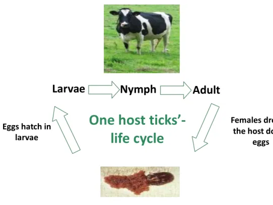
2.1.3. Life cycle
The life cycle of a tick can be classified into 4 stages. Ticks begin as eggs (stage 1) that hatch into larvae (stage 2). Larvae live and feed on animal hosts before detaching and molting
(shedding) anywhere. The larvae molt to nymph (stage 3), which feed on animals, engorge,
detach and molt. Once nymph molts, it becomes an adult tick (male or female). To distinguish larvae from nymphs and adults, the first have 6 legs and adults/nymphs has 8 legs (Kohls,
Sonenshine & Clifford, 1965). Adults climb up grass and plants to hold their prey and when a warm-blooded animal walks on the pasture, tick can crawl onto them and begin feeding. Ticks insert their mouths, attach to their prey and engorge themselves with a blood meal (stage 4). During feeding, ticks saliva can get into the host body and blood stream.
Usually Ixodes adult ticks take 4 to 10 days to get full engorgement and feed themselves by forcing their hypostome through the host skin and sucking blood and fluids that are drained from the resulting wound (Sonenshine, 1991). Ticks from Ixodidae family may have between one to three vertebral hosts. The coupling occurs in the host, where the adult female tick feeds itself for several days; then it falls into the ground, where the oviposition of millions of eggs occurs, culminating with tick’s death. The male ticks can couple many times without dying, feeding themselves intermitting. In the Argasidae family, females feed themselves frequently, coupling happens outside the host and oviposition occurs more than once in ticks life (Urquhart et al., 1996).
Figure 1 One host ticks life cycle.
Adult
Larvae
Nymph
Females drop of the host do lay
eggs Eggs hatch in
larvae
One host
ticks’-life cycle
2.1.4. Tick control
2.1.4.1. IntroductionTicks control is essential to diminish tick-borne diseases prevalence and, therefore, reduce ticks impact on livestock productivity. Nowadays, ticks control is mainly based in the application of acaricides, despite their disadvantages and limitations, although other control methods as vaccination or biocontrol agents are available (Willadsen, 2006). Probably, vaccination will be one of the most important methods for ticks control and its transmited pathogens, because several different vaccines have already been used against B. bovis and B.
bigemina in Australia (Bock & de Vos, 2001) or against Theileria annulata in Israel (Shkap &
Pipano, 2000) and China (Gu et al., 1997; Shirong, 1997) with good results.
2.1.4.2. Chemoprophylaxis
There are many acaricides that can be used against ticks: pyrethroids as flumethrin and deltamethrin; organochlorines as dichlorodiphenyltrichloroethane (DDT); organophosphates as diazinon and coumaphos; carbamates as carbaril; formamidines as amitraz; cicloamidines as clenpirin and macrocyclic lactones (avermectins and milbemycins), among others (Botana, Landoni & Martín-Jiménez, 2002). Synthetic flumethrin in pour-on protects cattle from ticks for 2 weeks and, in case of deltamethrin, for 3 weeks; this approach, in the case of babesiosis control, led to a significant decay in clinical disease (Zintl, Mulcahy, Skerrett, Taylor & Gray, 2003). The application of acaricides has some drawbacks: the appearance of residues in milk and meat products, the environmental contamination and, most important nowadays, the development of ticks’ resistance against acaricides (Botana et al., 2002). This resistance is mainly due to mutations in genes encoding detoxificating enzymes, as esterases, glutathione-S-transferases and monooxidases and due to genetic drift (Rosario-Cruz et al., 2009). Resistance to compounds such as organophosphorous, pyrethroids and amitraz has been described for R. microplus (Ortiz et al., 1995; Soberanes, Santamaría, Fragoso & García, 2002). Pesticides rotation is used in crop agriculture to minimize resistance, but its application in ticks control is not widely used yet. The association of acaricides with vaccines acts in a synergetic manner, because, in the case of R. microplus, efficacy of macrocyclic lactone acaricides is deeply enhanced in cattle vaccinated against those ticks (Kemp, McKenna, Thullner & Willadsen, 1999).
2.1.4.3. Biocontrol
Currently, an appeal is being made to biocontrol and to the use of more “environmental friendly” acaricide products. Like other parasites, ticks carry some microorganisms in their bodies, which, along with ticks, are essential for the survival of each other (endosymbiosis). Since endosymbionts are essential for ticks’ survival, elimination of those microorganisms would be deleterious for the continued existence, growth and development of ticks. Those endosymbionts could be either destroyed by a chemical approach or used to animals’ immunization, in order to interfere in ticks’ nutrition (Noda, Munderloh & Kuffi, 1997; Benson, Grawronski, Eveleigh & Benson, 2004). The use of other organisms pathogenic to ticks, like fungi of genera Beauveria and Metarhizium (Frazzon, Vaz Junior, Masuda, Scharank & Vainstein, 2000; Gindin, Samish, Zangi, Mishoutchenko & Glazer, 2002) or herbal acaricides (Khudrathulla & Jagannath, 2000; Lundh, Wiktelius & Chirico, 2005) are an alternative and important possibility of achieve the control of ticks.
2.1.4.4. Genetic resistance
The genetic resistance to ticks and tick-borne diseases is complex, but, facing other control methods and their problems, breeds resistance has become an important parameter in some regions.
Bos indicus cattle is more resistant to R. microplus ticks and babesiosis than Bos taurus, this
acquire resistance being heritable, though those animals remain susceptible to A. marginale (Bock et al., 1997, 1999). This additional resistance is possibly due to the fact that zebuine cattle breeds have more dermal mast cells than taurine breeds, as F2 crossbreed cattle has a higher developed resistance against ticks correlated with an increased number of mast cells in dermis (Engracia Filho, Bechara & Teodoro, 2006).
2.1.4.5. Vaccines 2.1.4.5.1. Introduction
Major alternatives to conventional acaricide treatments have been developed in recent years and vaccines are among the most important developments. It is now a decade since the first commercial vaccine against R. microplus based on the recombinant antigen Bm86 was released (Willadsen, Bird, Cobon & Hungerford, 1995; de la Fuente et al., 1999).
Gavac™ vaccine (Heber Biotec S.A., Havana, Cuba) and TickGARD (Hoechst Animal Health, Australia), which confirming the advantages of tick control by this method, because it is cost-effective, it reduces environmental contamination, it prevents acaricide-resistant ticks selection and it can reduce pathogens transmission, by decreasing ticks number and/or changing their vector capacity. This control scheme also has another advantage, as vaccines can protect the animal-host against both pathogens and vector, especially if antigens are conserved in both species (de la Fuente et al., 1998; de la Fuente & Kocan, 2003; de la Fuente, Kocan & Blouin, 2007).
Nevertheless, there is a problem with vaccines formulation because many of tick-protective antigens studied for propose of a future application are cytoplasmic and highly conserved, which can favour host tolerance to them and a total inefficacy of vaccine; yet this is one of the most promisor methods to fight these ectoparasites (Almazán et al., 2010).
There are two different kinds of antigens, the exposed and the concealed antigens (Willadsen, 1980). The first type is the most used in vaccines production, even though vaccines with concealed antigens may inhibit other parasites development, such as Babesia spp., by altering gut homeostasis and preventing pathogen transmission (Rachinsky, Guerrero & Scoles, 2007). Another kind of vaccines are the transmission blocking vaccines (TBV), which are supposed to block pathogen development in arthropod vectors through targeting pathogen or arthropod molecules as transmission blocking targets (Carter, 2001).
2.1.4.5.2. Recombinant vaccines
These vaccines use recombinant proteins as antigens to immunize animals and have several advantages, such as prevention or reduction of pathogens transmission (de la Fuente et al., 1998), environmental safety, low cost production (Odongo et al., 2007), prevention of drug-resistant ticks selection and inclusion of multiple antigens that could target several tick species (de la Fuente & Kocan, 2006), among others.
Canales, Almazán, Naranjo, Jongejan & de la Fuente (2009) have cloned ortholog genes (Ba86 and Bm86 genes) from R. annulatus and R. microplus, respectively, which were used in vaccine trials later on. Vaccination of cattle with Ba86 reduced, respectively for R.
annulatus and R. microplus, ticks infestations (71% and 40%), ticks weight (8% and 15%),
oviposition (22% and 5%) and eggs fertility (25% and 50%). For R. decoloratus, Odongo et al. (2007), using a Bm86 based-vaccine, found a reduction of 46% on engorged adult female ticks number, 56% on ticks weight and 61% on eggs weight after cattle immunization and de
Vos, Zeinstra, Taoufik, Willadsen & Jongejan (2001) described a reduction of 70% in R.
decoloratus reproductive capacity after feeding on vaccinated calves and 50% in engorged H. anatolicum anatolicum nymphs total weight. Despite these results, vaccine had no action
against A. variegatum and R. appendiculatus nymphs. Pipano et al. (2003) tested the efficacy of a Bm86 vaccine in protection against ticks and pathogens (B. bovis and B. bigemina) transmitted by those ectoparasites. The results showed that immunized cattle, when challenged with B. bovis-infected ticks, continued to become infected, but in the case of
B. bigemina, Bm86-immunized animals remained protected against infection, probably due to
the fact that larvae didn’t molt to nymph. Still related to the Bm86 gene, Bastos, Ueti, Knowles & Scoles (2010) studied the effect of Bm86 gene silencing on fitness of R.
microplus ticks fed in B. bovis infected cattle, showing that this procedure decreased survival
engorged ticks rate and eggs weight. A gene from R. microplus (strain A) called Bm95, homologue to Bm86, was used to immunize cattle, after it has been discovered that some R.
microplus ticks (strain A) had a moderate low susceptibly to Bm86 vaccine, possibly due to a
genetic variation in ticks (Freeman, Davey, Kappmeyer, Kammlah & Olafson, 2010). Bm95 immunized cattle showed a higher protection efficacy to both susceptible and strain A than Bm86 vaccine (García-García et al., 2000). Other authors showed that Bm95 antigen based vaccine presents better responses than Bm86 vaccine to low susceptibility ticks populations (Jittapalapong et al., 2010).
Immunization trials in cattle with recombinant subolesin, a conserved protein among vertebrates and insects, decreased R. microplus survival and reproduction rates (Merino et al., 2011), weight and oviposition (de la Fuente et al., 2011) and, moreover, protein knockdown led to degeneration of ticks parts as embryos, salivary glands and reproductive tissues (Merino et al., 2011). Further studies with this antigen showed that immunization of rats decreased vector capacity of I. scapularis nymphs for A. phagocytophilum (de la Fuente & Kocan, 2006).
The recombinant antigen 64P from R. appendiculatus was found to be involved in ticks attachment and feeding and was used to immunize guinea pigs, reducing, respectively, 48% and 70% nymph and adult infestation rates; additionally, response of animals immunized with 64P was similar to those observed during development of natural resistance in guinea pigs infested with R. appendiculatus, showing the typical local cutaneous inflammatory immune response (Trimnell, Hails & Nuttall, 2002). The recombinant protein from R. sanguineus and
I. ricinus was again tested by Trimnell et al. (2002), respectively in guinea pigs and hamsters,
demonstrating damage on ticks post-challenge. Other study indicated that there is an antigenic cross-reactivity between tick extracts of R. sanguineus, I. ricinus, A. variegatum and R.
microplus, showing the potential of this protein to develop a large-spectrum anti-tick vaccine
(Havlíková et al., 2009).
2.1.4.5.3. Antigens selection
Molecular tools are important experimental components for the study of tick gene functions. RNAi technique allows silencing of gene expression, contributing to the characterization of gene function and its phenotypic effect (Fire et al., 1998). The first report related to the use of RNAi in ticks belongs to Aljamali, Sauer & Esseberg (2002) and quickly that technique has become universally adopted for gene-silencing in ticks (de la Fuente & Kocan, 2006).
Kocan, Manzano-Roman & de la Fuente (2007) showed that subolesin knockdown in I.
scapularis, D. variabilis and A. americanum also affected oviposition, eggs embryogenesis,
larval hatching and fertility (reduction of 93% and 71% for D. variabilis and I. scapularis, respectively). Subolesin expression is activated when ticks are infected with A. marginale and
B. bigemina, suggesting a connection between tick gene expression and pathogen-infection
(Merino et al., 2011). Zivkovic et al. (2010) obtained the opposite result, showing that D.
variabilis, D. andersoni, D. reticulatus, R. sanguineus, R. microplus and R. annulatus had
equal or even lower mRNAs level than control groups.
Almazán et al. (2010) studied the knockdown effect of several R. microplus genes, such as glutathione-S transferase (GST), ubiquitin (UBQ), selenoprotein W (SEL), elongation factor-1 alpha (EF-factor-1a) and subolesin, and found out that GST and SEL genes knockdown lead to a lower ticks attachment when compared to control group, but did not influence ticks mortality or oviposition, though the other three genes results showed an increase on R. microplus mortality and reduction of oviposition.
2.2. Babesia
2.2.1. Introduction
Babesia spp. was first discovered in the 19th century in association to bovine hemoglobinuria (Babes, 1888). In 1893, Smith and Kilbourne recognized B. bigemina as the causative agent of Texas Cattle Fever (Smith & Kilbourne, 1893).
All over the world, there are some 100 Babesia species or even more (Homer, Aguilar-Delfin,
Telford 3rd, Krause & Persing, 2000), Rhipicephalus spp. being one of its main tick vector
(Urquhart et al., 1996). Bovine babesiosis are generally caused by B. bovis, B. bigemina and
B. divergens which are the most important Babesia species for cattle babesiosis; however B. major can also cause disease in cattle (Zintl et al., 2003) and in the case of human babesiosis, B. divergens and B. microti are the most dangerous species (Antunes, 2008). The different Babesia species infect a large variety of animals (Bock, Jackson, de Vos & Jorgensen, 2004;
de Vos & Geysen, 2004) and in cattle they can induce animal mortality, abortions, reduction of milk/meat production, and, sometimes, neurological symptoms (Saegerman et al., 2003).
2.2.2. Characterization and life cycle
Babesia spp. belongs to phylum Apicomplexa, class Aconoidasida, family Babesiidae and genus Babesia. In blood smears stained by Giemsa method, Babesia parasites usually appear in pairs, with a pear-shape, an elongated or a cigar form with a red nucleus and a blue cytoplasm (figure 2). Concerning to its size, Babesia species can be divided in small and big
Babesia, with 1.0-2.5 µm and 2.5-5.0 µm of length, respectively (Urquhart et al., 1996).
These morphological categorizations are usually consistent with the phylogenetic characterization based on nuclear small subunit-ribossomal RNA gene (18S rDNA) sequences, showing that small babesias are divided in two different phylogenetic clusters (Homer et al., 2000).
Taken from DPI- Queensland (EUA) http://www.dpi.qld.gov.au
Figure 2 Giemsa staining of B. bigemina (A) and B. bovis (B) infected red blood cells.
The life cycles of all Babesia parasites are very similar and all species are transmitted by infected ticks’ bites. The main difference among these life cycles is the presence of transovarial transmission in some species and not in others (Hunfeld, Hildebrandt & Gray, 2008). The Babesia spp. life cycle includes three phases, merogony, gamogony and sporogony, and two hosts, one vertebrate (mammal) and one invertebrate (tick), being R.
annulatus and R. microplus the main vectors for both B. bovis and B. bigemina (Walker et al.,
2003; Taylor, Coop & Wall, 2007). The parasite come into vertebrate hosts blood stream through infected ticks saliva, allowing sporozoites to invade erythrocytes. In these cells, parasite asexually divides itself and becomes a merozoite, with a typical pear-shape. This multiplication leads to erythrocytes lysis, with merozoites release, which infects other red blood cells, becoming trophozoites. These new-forms can divide themselves producing a new pair of merozoites, perpetuating merogony phase (1). Some merozoites turn to gametocytes and, when tick feeds in a vertebrate host, it becomes infected. In ticks’ midgut, gametocytes suffer a sexual phase involving the formation of macro and micro gametes, culminating in zygote production (gamogony). The zygote invades midgut digestive cells and then transforms into kinetes, which can access the hemolyimph in the haemocoel of tick (2). After this phase, organisms can be transferred either transtadial (between stages) or transovariac (from female to offspring via the egg). Once in larvae, kinetes migrate to salivary glands cells, where they become sporozoites and multiply themselves (sporogony (3)). When ticks feed again, they transmit the sporozoites parasites to vertebrate host (Riek, 1966; Melhorn & Schein, 1984).
Adapted from Bock et al. (2004).
Figure 3 The life cycle of Babesia bigemina in cattle and in the ixodid tick vector R. microplus.
2.2.3. Symptoms of bovine babesiosis
Most cases of Babesia infection are symptomatic. Depending on affected species, clinical signals of babesiosis are different, occasionally culminating to death in few days, as the globular volume may decrease to less than 20% (Urquhart et al., 1996; Melo, Passos, Facury-Filho, Saturnino & Ribeiro, 2001). Some of the clinical symptoms in acute disease are similar to malaria and include high fever, hemolytic anaemia, lethargy, hemoglobinuria, icterus liver (figure 3-B), kidneys with congestion (figure 4-A), abortion, weight loss, splenomegaly and decreased milk/meat production (Bock et al., 2004). Cerebral babesiosis (figure 3-A), caused
3
1
by B. bovis, is characterized by convulsions, hyperaesthesia and paralysis, due to sequestrations of parasites in the brains capillaries, resulting in low parasitaemia level (less than 1%) in circulating blood. B. bigemina infection usually leads to a less pathogenic disease, even though parasitaemia often exceeds 10% (Ristic, 1981; Bock et al., 2004). At necropsy, animal skin and mucous membranes are pale and icteric, bile is granular and dense (Urquhart et al., 1996), bladder is distended and there is hematuria (figure 4-B) (Howard, Rozza, Graça & Fighera, 2001).
Taken from Howard et al. (2001). Bovine Babesiosis.
http://www.vet.uga.edu/vpp/archives/NSEP/babesia/PORT/necropsy_findings.htm Figure 4 Cerebral form of babesiosis (A); icterus liver (B)
Taken from Howard et al. (2001). Bovine Babesiosis.
http://www.vet.uga.edu/vpp/archives/NSEP/babesia/PORT/necropsy_findings.htm Figure 5 Kidneys with edema and congestion (A); hematuria (“Red water”) (B)
A B
2.2.4. Diagnosis
Diagnosis of clinical cases of babesiosis is most frequently made by examination of blood
smears stained with Giemsa or acridine orange. Blood films from B. bovis are prepared from
capillary blood, as blood of general circulation may contain up to 20 times fewer parasites due to sequestration of infected erythrocytes in capillaries of brain and other organs (Bӧse, Jorgensen, Dalgliesh, Friedhoff & de Vos, 1995). In B. bigemina infections, parasitized cells are evenly distributed throughout blood circulation. These techniques are inexpensive and reasonably portable, though accuracy of diagnosis relies on training and skill of microscopist (Papadopoulos, Brossard & Perié, 1996). Low parasitaemias and the presence of different, but morphologically similar parasites (e.g. other Babesia spp. and also Theileria spp.) may adversely affect the proper classification of infections (Homer et al., 2000).
Another test is the cultivation in vitro, which can be used to detect infection in animals with low parasitaemia and has the advantage of being very sensitive, yet a long period of time needed for parasites to grow (Thomford, Conrad, Boyce, Holman & Jessup, 1993). Indirect immunofluorescent antibody tests (IFATs) have been used as standard diagnosis test for babesiosis; nevertheless, sensibility, specificity and subjective interpretation are the major problems with this technique. IFATs and enzyme-linked immunosorbent assay (ELISA) can reveal animals that were in contact with parasites, but do not have an active infection. However, long-term carriers are frequently sera-negative and, moreover, serological tests are often cross-reactive among different piroplasm species (Burridge, Young, Stagg, Kanhai & Kimber, 1974; Papadopoulos et al., 1996).
In cases whose diagnosis is difficult by means of blood smear or serology, or when detection of carrier animals with very low parasitaemias is required, direct recognition of parasites by
polymerase chain reaction (PCR) based assays can be used. With the evolution of more
sensitive PCR based techniques, several methods for the detection and differentiation of bovine babesiosis infections have been described, including nested PCR (Figueroa, Alvarez, Ramos, Vega & Buening, 1993), reverse line blot (RLB) hybridization (Gubbels et al., 1999), LAMP (Loop-Mediated Isothermal PCR) (Iseki et al., 2007) and real time PCR (Buling et al.,
2007). Currently, none of these methods is used globally, because some have not been
validated to worldwide use, others require complicated post-PCR detection methods to further enhance sensitivity or differentiation, or require special equipment and also some may be prone to amplicon contamination issues (Martins, 2009).
2.2.5. Human babesiosis
A risk factor for being infected with Babesia spp. sensu stricto is splenectomy (Telford 3rd &
Maguire, 2006). B. microti is recognized as a diverse species complex, parasitizing a variety of hosts, including rodents, insectivores and carnivores, but the majority of zoonotic strains utilize microtine rodents as reservoir hosts. The reservoir host for B. divergens sensu stricto, which is implicated in most cases of human babesiosis in Europe, is cattle and the vector for this parasite is I. ricinus (Duh, Petrovec & Avsic-Zupanc, 2001). In United States of America, white-footed mouse Peromyscus leucopus is the main reservoir host and I. scapularis the invertebrate vector, also known as the deer or black-legged tick. Symptoms and signs can appear one to nine weeks post infection and include hemolytic anaemia, fever, myalgia, headache, drenching sweats, malaise and chills (Hunfeld et al. 2008) or even disseminated intravascular coagulation and respiratory distress syndrome in fulminant cases (Homer et al, 2000). Diagnose involves the same techniques used for animals, namely the presence of the parasite in blood smears (Healy & Ruebush, 1980), PCR (Brandt, Healy & Welch, 1977) and serologic studies (Krause et al., 1994). In human babesiosis treatment, there are different approaches, such as the clindamycin plus quinine (Wittner et al., 1982) or the atovaquone plus azithromycin (Krause et al., 2000). Recently, randomized trials in humans infected with B.
microti showed that atovaquone plus azithromycin therapy achieved the same results of
standard quinine/clindamycin combination, but causing fewer side effects (15% versus 72%) (Hunfeld et al., 2008).
2.2.6. Babesiosis control
2.2.6.1. IntroductionBabesiosis control is essential, due to its huge implications in livestock production and its relations to public health issues (Bock et al., 2004), infecting a large variety of animals and humans. Nowadays, with the introduction of exotic breeds, babesiosis control is even more important, because those breeds usually do not have natural immunity against Babesia spp. (Graf et al., 2004). Several approaches, as ticks-vectors control (see chapter 2.1.4.), chemoprophylaxis and vaccination can be applied.
2.2.6.2. Chemoprophylaxis
One way to control this parasite is to control its vector, using acaricides, vaccines against ticks, among others (see chapter 2.1.4.). There are some drugs used against these parasites, as quinuronium sulfate, amicarbilide, diminazene and imidocarb dipropionate. The anti-babesia drugs, when used inappropriately, result in the drug-resistant Babesia strains emergence (Zintl et al., 2003).
2.2.6.3. Genetic resistance
Babesiosis control can also be achieved by introducing hosts genetically resistant to
hemoparasite infections. The nonspecific immune response against Babesia infection is attributed to age or breed related factors since, in general, young cattle is less susceptible to
Babesia spp. infections than adult cattle, possibly due to the effect of passive immunity
conferred by colostrum antibodies (Rogers et al., 2005). Bos indicus shows more resistance than Bos taurus and animals resulting from crosses between breeds are more resistant to
Babesia spp. infection and to tick infestation (Bock et al., 1997).
2.2.6.4. Vaccines
There are many types of vaccines against Babesia spp. parasites, including attenuated (calf-derived and culture-(calf-derived), recombinant and subunit vaccines.
Attenuated vaccines can be produced by parasites multiple passages in vivo in splenectomized calves, the calf-derived vaccines (Bock et al., 2004; De Waal & Combrink, 2006), or by parasites growth in vitro, the culture-derived vaccines (Jorgensen, de Vos & Dalgliesh, 1989; Echaide, de Echaide & Guglielmone, 1993; Shkap & Pipano, 2000).
Calf-derived vaccines have several associated concerns, such as the possible spread of silent pathogens, difficulties in standardizing vaccine dose, risk of virulence reversion, maintenance of carrier animals, which might serve as reservoirs for pathogens transmission, quality of vaccine production (Shkap et al., 2007), short shelf-life (Bock et al., 2004), vaccines maintenance and transportation (Shkap et al., 2007), limitations of use in animals older than 8-9 months, adverse effects (De Waal & Combrink, 2006) and potential risk of parasite transmission, since vaccinated cattle remains persistently infected for several months (Pipano, 1995).
Culture-derived vaccines do not have entail the risk of pathogens spreading, however, for
Babesia spp. culture, the main disadvantages remain in the need to fresh bovine erythrocytes
and serum from specific donors, which have to be maintained in highly strict conditions and in the fact that vaccines can lose their immunogenicity and virulence in a long-term cultivation (de Vos, 1978). Despite some attenuated vaccines disadvantages, live attenuated strains of B. bovis and B. bigemina have been used for many years, because they offer a long-lasting protection (Benavides & Sacco, 2007).
There are several studies reporting the use of these attenuated vaccines. An in vitro derived attenuated live vaccine (B. bovis-B. bigemina) was used in endemic areas to protect cattle against these parasites, conferring an effective level of protection of 93%, showing a very favorable way to protect animals against this disease (Ojeda et al., 2010).
Fish, Leiboyich, Krigel, McElwains & Shkap (2008) studied the efficacy of calf-derived B.
bovis vaccine. Immunized cattle developed a good immunity, though very susceptible animals
had fever, low parasitaemia and a decrease of hematocrit. There was a 65.3% inhibition of B.
bovis dissemination and a solid protection against babesiosis was acquired in vaccinated
animals.
Shkap et al. (2007) studied two different vaccines against B. bigemina, a culture-derived and a calf-derived. It was shown in that study that, attenuated vaccines, whether produced from splenectomized calf or from cultures, offered a total protection against clinical babesiosis upon challenge with virulent homologous parasites.
Cysteine peptidases are molecules with a huge importance to many parasites, including
Babesia spp., as specific inhibitors of these enzymes can stop B. bovis merozoites growth in vitro (Okubo, Yokoyama, Govind, Alhassan & Igarashi, 2007). Mesplet et al. (2010) showed
that gene coding to B. bovis cysteine peptidases - bovipaine-1 and bovipaine-2 - were transcripted only into the infected erythrocytes and that animals vaccinated with bovipaine-2 protein strongly reacted with the formation of antibodies. Furthermore, the anti-bovipaine-2 antibodies cross-reacted with erythrocytes infected with B. bigemina. Martins et al. (2010, 2011) also showed that cysteine proteases from B. bigemina are potential vaccine candidates. Silva et al. (2010) analyzed a recombinant microneme protein from B. bovis (Bbo-MIC1), secreted on parasite surface, demonstrating that antibodies against Bbo-MIC1 inhibit erythrocyte invasion in B. bovis in vitro cultures and this protein was recognized by antibodies in serum of B. bovis infected cattle, showing the immunogenicity of Bbo-MIC1 and its use as potential vaccine.
Carcy, Précigout, Schetters & Gorenflot (2006) recognized that recombinant merozoite surface antigens (MSA) were expressed in the merozoites and sporozoites and that antibodies
anti-MSA-1 and MSA-2c inhibit, in vitro, the invasion of erythrocytes by sporozoites, suggesting this may be used in vivo to block erythrocytes invasion. The same authors used a
B. bigemina protein, gp 45, similar to MSA-1 of B. bovis, to immunize cattle, which became
protected against the parasite.
The apical membrane antigen 1 (AMA-1), has been evaluated as a possible subunit vaccine. Antibodies against B. bovis AMA-1 (BbAMA-1) reduced the invasion efficiency in vitro; moreover, this molecule is highly similar to another AMA-1 from B. bigemina, indicating that this vaccine could, possibly, have a cross reactivity with other Babesia species (Torina et al., 2009).
2.3. Calreticulin
2.3.1. Introduction
CRT was first isolated by Ostwald and Maclennan in 1974 and further cloned by Fliegel, Burns, MacLennan, Reithmeier & Michalak in 1989 (Fliegel et al., 1989).
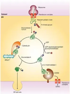
CRT has been identified in several species, but there is no CRT genes identified in yeasts and prokaryotes, whose genomes were totally sequenced (Persson, Rosenquist & Sommarin, 2002). CRT importance to cells is relevant, given that the absence of CRT gene is embryonically lethal (Mesaeli et al., 1999). Since its first detection, CRT has been identified in other cellular structures, as cytoplasm, cell membranes and extracellular matrix (Burns, Atkinson, Bleackley & Michalak, 1994); several functions have been attributed to CRT, such as acting as a chaperone to help other proteins to fold correctly (Nauseef, McCormick & Clark, 1995). This activity was demonstrated by immunoprecipitation experiments, which showed that CRT associates itself transiently to several cellular proteins immediately after their synthesis (Peterson, Ora, Van & Helenius, 1995). When a polypeptide is incorrectly folded, it bounds to one or more chaperones (including CRT) and is retained in endoplasmatic reticulum (ER), mechanism that prevents expression, aggregation and secretion of misfolded
Adapted from Mendlovic & Conconi (2011). Figure 6 CRT action as a chaperone.
Besides the essential functions that CRT performs in ER lumen, this multifaceted protein has also been implicated in many unexpected roles that occur at cells surface, cytosol, nucleus and extracellular matrix. Indeed, since discovery of CRT chaperone and calcium-regulating functions, scientists have learned that CRT has many other duties in the cell. This protein has been implicated in diverse cellular processes including signaling (Mitra & Schlaepfer, 2006), regulation of gene expression (Gardai et al., 2005), wound healing (Pallero, Elzie, Chen, Mosher & Murphy-Ullrich, 2008), removal of cancer cells (Tesniere et al., 2008) and autoimmunity (Eggleton & Llewellyn, 1999).
2.3.2. Calreticulin structure
CRT has two codifying genes, CRT-1 and CRT-2 (Persson et al., 2002), and is highly conservative with a 96% amino acid identity for CRT from human, rabbit, rat and mouse (Waser, Mesaeli, Spencer & Michalak, 1997). The CRT gene has nine exons and is localized in chromosomes 19 and 8, respectively for human and mouse genes. These sequences have
evolutionary conservation (McCauliffe, Yang, Wilson, Sontheimer & Capra, 1992). CRT gene from ticks has two exons and one intron, the last one with a conserved position, but variable size and nucleotide sequence. In R. annulatus CRT gene, intron is localized between nucleotides 88 and 412 and has 1559 base pairs (bp) (Xu, Fang, Sun, Keirans & Durden, 2005).
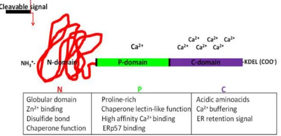
CRT proteins have a molecular weight around 46 kDa (Fliegel et al., 1989), an N-terminal cleavable amino acid signal sequence and a KDEL ER retrieval signal in the C-terminal domain. These amino acids are responsible for CRT targeting and retaining in ER lumen (Michalak et al., 1999). The protein has three cysteine residues, all located in N-domain of the protein. Two of those cysteine residues form a disulphide bridge (Matsuoka et al., 1994), which probably is responsible for correct folding of CRT N-terminal region. Structural studies show that CRT has three domains (Bedard, Szabo, Michalak & Opas, 2005), as illustrated in figure 7:
The N-terminal domain (amino acid residues 1 to 180) is the most conserved domain,
contains a binding site to monoglycosylated oligosaccharides (Schrag et al., 2001), several phosphorylation sites and is anticipated to have a folded globular structure with eight anti-parallel β-strands connected by protein loops (Michalak et al., 1999);
The P-domain (amino acid residues 181 to 290) forms an β-stranded hairpin
configuration extended curved-arm structure rich in prolin, which binds to Ca2+ with
high affinity and low capacity (Ellgaard et al., 2001) and interacts with ER chaperones (Michalak, Groenendyk, Szabo, Gold & Opas, 2009);
The C-terminal region or (C-domain) (amino acids residues 291-400) (Giraldo et al.,
2010), characterized by a helix form (Del Cid et al., 2010), mainly constituted by aspartic acid and glutamic acid amino acids (Michalak et al., 1999), shows low affinity
to Ca2+, but a high capacity, important for Ca2+ storage in ER (Castãneda-Patlán,
Razo-Paredes, Carrisoza-Gaytán, González-Mariscal & Robles-Flores, 2010). The
structure of CRT differs at C-domain considering Ca2+ level, since this domain has a
disordered structure with a low calcium concentration, but when this level rises, protein becomes more rigid and compact (Giraldo et al., 2010).
Adapted from Mendlovic & Conconi (2011). Figure 7 Structure and functions of the CRT domains.
2.3.3. Calreticulin in ticks and other parasites
CRT is a protein that exists in ticks salivary glands and saliva; probably, this protein is essential to ticks feeding and pathogen transmission, through its anti-thrombotic and complement inhibition functions. All these facts reveal the possibility of CRT being used as an antigen in a vaccine against cattle ticks (Kaewhom, Stich, Needham & Jittapalapong, 2008).
Immunization assays with CRT showed its importance to immune reaction against ticks and, consequently, its relevance as a possible global anti-ticks vaccine component. Immunization of rabbits with CRT from A. americanum, followed by rabbits’ infestation with those ticks led to necrotic lesions in ticks’ local bite, demonstrating an immune reaction capable to interfering with ticks feeding (Jaworsky et al., 1995).
Gao et al. (2008) used a recombinant CRT (rCRT) protein from H. qinghaiensis (rHqCRT) to
immunize sheep and results showed a reduced ticks weight and oviposition and a higher mortality comparing to control group.
There are various evidences of cross-reactivity between different anti-CRT antibodies, probably due to the fact that CRT is a highly conserved protein. Parizi et al. (2009) used rCRT from H. longicornis (rHlCRT) to immunize cattle and their sera reacted to both rHlCRT and rCRT from R. annulatus (rBmCRT). Besides, sera from cattle, whether immunized with rHlCRT or with rBmCRT, recognized native BmCRT.
Immunolocalization experiments using polyclonal antibodies anti-CRT from A. americanum and D. variabilis, revealed a specific protein from salivary glands homologous to CRT, which appears to be secreted during tick feeding (Jaworsky et al., 1995).
Finally, as CRT is a conserved protein, its use in a vaccine could protect animals not only from ticks, but also from tick-borne diseases. Concerning this aspect, Rachinsky et al. (2007) associated CRT with R. microplus infection by B. bovis, when they tested proteins up and down-regulated in infected and uninfected ticks and discovered that, among up-regulated proteins, there was CRT. Antunes et al. (2012) studied differentially expressed genes in R.
microplus and R. annulatus after infection by B. bigemina, showing that gene encoding CRT
was overexpressed. Knockdown of CRT in R. microplus reduced 73% the pathogen transmission as well as ticks weight.
As described previously with respect to ticks, there are several studies referring the use of CRT to protect animals from other parasites. Winter et al. (2005) used a N. americanus-CRT based vaccine to immunize mice, resulting in a significant reduction of worm number in lungs (43-49%) comparing with control group. In another study, hamsters were immunized with
Taenia solium-CRT and, besides reduction of parasite numbers, worms were unable to mature
in vaccinated animals (Mendlovic et al., 2004).
Immunolocalization studies using Haemonchus contortus-CRT antibodies showed that this protein is localized in external openings, such as the buccal cavity, vaginal tipi of female and
bursa of male worms (Suchitra & Joshi, 2005). CRT from Echinococcus granulosus (EgCRT)
is expressed in external tegument and cellular region of hydatic cysts germinal layer, possibly with the intent to inhibit classical complement pathway or to lead to an antiangiogenic effect in cysts periphery (Cabezón, Cabrera, Paredes, Ferreira & Galanti, 2008).
Debrabant, Lee, Pogue, Dwyer & Nakhasi (2002) evaluated the effect of Leishmania
donovani-CRT P-domain overexpression in transfected parasites, which resulted in reduction
of acid phosphatase-secretion and in a survival decrease in human macrophages, showing that changes in CRT expression may affect the “virulence” of the parasite.
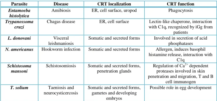
Table 2 CRT functions in parasites that cause disease in humans and animals
Parasite Disease CRT localization CRT function
Entamoeba histolytica
Amibiosis ER, cell surface, uropod Phagocytosis
Trypanossoma cruzi
Chagas disease ER, cell surface Lectin-like chaperone, interaction with C1q, recognized by iGg from
patients
L. donovani Visceral
leishmaniosis
Somatic and secreted forms Involved in secretion of acid phosphatases
N. americanus Hookworm infection Somatic and secreted forms Allergen, induces basophil histamine release, interaction with
C1q
Schistosoma mansoni
Schistosomiosis Somatic and secreted forms, penetration glands
Regulation of Ca2+ dependent proteases involved in skin penetration and migration, T and B
cell immunogen
T. solium Taeniosis and
neurocysticercosis
Somatic and secreted forms, gametes and developing
embryos
Possible role in egg development
C
C
h
h
a
a
p
p
t
t
e
e
r
r
3
3
.
.
M
M
a
a
t
t
e
e
r
r
i
i
a
a
l
l
a
a
n
n
d
d
M
M
e
e
t
t
h
h
o
o
d
d
s
s
3.1. Rhipicephalus annulatus ticks
Total RNA was extracted from R. annulatus female ticks as described in Antunes et al. (2012). Briefly, ticks were rinsed in distilled water and 75% (v/v) ethanol, dissected and the whole of the internal organs were placed in a 2 ml tube with 1 ml of Tri Reagent (Sigma-Aldrich, St. Louis, Missouri, USA). Total RNA was used in the synthesis of approximately 1 μg of cDNA using the iScript™ cDNA synthesis (Bio-Rad, Hercules, California, USA).
3.2. Amplification of the calreticulin gene
The CRT sequence was amplified by PCR technique. Primers were designed based on the CRT gene sequence of R. annulatus (accession number AY395253), ensuring full coding region coverage. The primers used were: forward 5’-CACC AT GCG GCT TCT CTG CAT TTT G -3’ and reverse 5’- CAG TTC TTC GTG CTT GTG GTC -3’.
PCR was performed using Kit GoTaq® (Promega, Madison, Wisconsin, USA) under the following conditions: one amplification round in a final volume of 25 μl, including 1 μl of template cDNA and 1 μl of each primer (95ºC for 2 min, then 40 cycles: 30 s at 94ºC, 45 s at 55ºC and 2 min at 72ºC; final extension 2 min at 72ºC). PCR assays were performed in MJ Research PTC-200 Thermo Cycler (GMI Biotech, Minnesota, USA).
The positive PCR products were purified using the illustraGFXTM PCR DNA and Gel Band
Purification Kit (GE Healthcare, Buckinghamshire, UK) according to manufacturer’s instructions. Purified samples were sequenced at Stab Vida (Almada, Portugal) and further analysed.
3.3. Expression of recombinant calreticulin
For rCRT expression in Escherichia coli system, ChampionTM pET101 directional TOPO®
expression kit uses a highly efficient 5-min cloning strategy to insert a blunt end PCR product into a vector with no requirement of post-PCR procedures or restriction enzymes (Invitrogen, 2010). The recombinant proteins produced, using this expression kit, have a histidine tail attached.
3.3.1. Cloning and transformation
The purified PCR products, previously obtained (chapter 3.2.), were initially cloned into plasmids pET101/D-TOPO vectors using the above described kit according to manufacturer’s instructions. 10 µl of pET TOPO® (Invitrogen Life Technologies, Carlsbad, California, USA) construct, previously obtained, was mixed with 50 µl of the E. coli OneShot® cells (Invitrogen Life Technologies, Carlsbad, California, USA), and then incubated in ice during 30 min, followed by incubation for 30 s at 42ºC and further incubated in ice. Afterwards, 250 µl S.O.C. medium were added to the previous sample and incubated at 37ºC, 200 rpm during 1 hour. After that period, 200 µl of the sample were used to seed 4 LB-agar/ampicillin (100 µg/ml) Petri-dishes, followed by overnight incubation at 37ºC.
3.3.1.1. Screening of the transformed colonies
Cell colonies were analyzed by PCR to confirm plasmids incorporation. Six colonies were picked up and individually suspended into 12 µl of water. Kit GoTaq® (Promega, Madison, Wisconsin, USA) was used. The PCR conditions were as followed: 94ºC for 10 min to lyse cells and inactivate nucleases, then 35 cycles: 94ºC for 3 min, 53ºC for 30 s and 72ºC for 1 min, followed by 72ºC for 10 min, in MJ Research PTC-200 Thermo Cycler (GMI Biotech, Minnesota, USA).
3.3.1.2. Plasmid purification for sequencing
For plasmids purification, illustra plasmidPrep Mini Spin Kit (GE Healthcare, Buckinghamshire, UK) was used, since it applies a simple plasmid DNA purification protocol involving a modified alkaline lysis procedure and a silica-based membrane to achieve highly efficient plasmid DNA purification (GE Healthcare, 2008).
The colonies, previously obtained (chapter 3.3.1.), were picked and suspended in 5 ml of LB/ampicillin (100 µg/ml) and incubated overnight at 37ºC and 200 rpm. Samples were then centrifuged at 16000 rpm during 10 min using a Heraeuspico 17 centrifuge (Thermo Electron