Nutritional disorder in
Pfaffia glomerata
by mercury excess in nutrient solution
Desordem nutricional em Pfaffia glomerata pelo excesso de mercúrio em solução nutritiva
Nicéia Spanholi CalgarotoI Fernando Teixeira NicolosoII Luciane Belmonte PereiraIII Denise CargneluttiIV Fabiane Goldschmidt AntesV Valderi Luiz DresslerI
ISSN 0103-8478
ABSTRACT
The mineral nutritional homeostasis in response to different concentrations of Hg (0, 25 and 50μM) was evaluated in Pfaffia glomerata plant. The exposure to the highest level of Hg (50µM) caused a decreasing in shoot and root fresh weights of 15.5% and 20%, respectively. Both shoot and root Hg concentrations increased linearly with increasing external Hg concentrations. Ca concentration decreased in shoot only at 50µM Hg, whereas shoot K and Mg concentrations decreased at both 25 and 50µM Hg, when compared to the control. A significant decrease in Cu, Zn, Fe and Mn concentrations in plants exposed to Hg was observed, but most Zn, Mn, and Cu in the roots. On the other hand, P concentration increased in both root and shoot of plants exposed at 25 and 50µM Hg, whereas Na concentration increased only in the root at 25 and 50µM Hg exposure. In general, tissue nutrient concentrations in P. glomerata plantlets exposed to Hg were significantly decreased, which indicates that the Hg may cause alteration on the mineral nutritional homeostasis of this species.
Key words: Brazilian Ginseng, nutritional homeostasis,
macronutrients, micronutrients, Hg toxicity.
RESUMO
A homeostase nutricional mineral em resposta a diferentes concentrações de Hg (0, 25 e 50μM) foram avaliadas em plantas de Pfaffia glomerata. A exposição ao mais alto nível de Hg (50µM) causou um decréscimo de 15,5% e 20%, respectivamente, na matéria fresca da parte aérea e raízes. As concentrações de Hg na parte aérea e raízes aumentaram linearmente com o aumento das concentrações de Hg. A concentração de Ca decresceu na parte aérea somente em 50µM Hg, enquanto as concentrações de K e Mg na parte aérea decresceram tanto em 25 como em 50µM Hg, quando comparado ao controle. Observou-se um
significativo decréscimo nas concentrações de Cu, Zn, Fe e Mn nas plantas expostas ao Hg, mas principalmente Zn, Mn e Cu nas raízes. Por outro lado, a concentração de P aumentou em raízes e parte aérea de plantas expostas a 25 e 50µM Hg, enquanto a concentração de Na aumentou somente nas raízes em 25 e 50µM Hg. No geral, as concentrações de nutrientes nos tecidos de P. glomerata expostas ao Hg foram significativamente diminuídas, o que indica que o Hg pode causar alterações na homeostase nutricional mineral dessa espécie.
Palavras-chave: Ginseng Brasileiro, homeostase nutricional,
macronutrientes, micronutrientes, toxicidade de Hg.
INTRODUCTION
In addition to the organic compounds produced in photosynthesis, plants require a wide variety of mineral nutrients, which are taken up mainly from the soil solution. These nutrients are required for
structural purposes and for the activity of the specific
enzymes, which regulate the cellular metabolism. Environmental variation of mineral nutrient availability is expected to result in changes in plant growth and development (EPSTEIN & BLOOM, 2005).
Plants require low levels of copper (Cu), iron (Fe), nickel (Ni), manganese (Mn), zinc (Zn), molybdenum (Mo), and chloride (Cl) as micronutrients to participate in enzyme catalyzed reactions, whereas other heavy metals as arsenic (As), cadmium (Cd),
IDepartamento de Química, Universidade Federal de Santa Maria (UFSM), Santa Maria, RS, Brasil.
IIDepartamento de Biologia, UFSM, 97105-900, Santa Maria, RS, Brasil. E-mail: [email protected]. Corresponding author. IIIInstituto Federal de Educacão, Ciência e Tecnologia de Mato Grosso, Campo Novo do Parecis, MT, Brasil.
IVUniversidade Federal da Fronteira Sul (UFFS), Erechim, RS, Brasil. VEmpresa Brasileira de Pesquisa Agropecuária, Porto Velho, RO, Brasil.
chromium (Cr), lead (Pb) and mercury (Hg) serve no know function when present in plants (EPSTEIN & BLOOM, 2005). Mercury is inadvertently added to soils in fertilizer, limestone, natural gypsum, manure (especially of marine origin), and sewage sludge and in fungicides containing Hg (PATRA et al., 2004). Mercury may inadvertently enter the food chain and pose health risks to humans and animals (PATRA et al., 2004). Propensity for plants to accumulate and translocate Hg to edible and harvested parts depends to a large extent on soil and climatic factors, plant genotype and agronomic management (PATRA et al., 2004).
The possible causal mechanisms of Hg toxicity in plants are changes in the permeability of the cell membrane, collapse of cortex cells and vascular system (RODRÍGUEZ et al., 2009; CARRASCO-GIL et al., 2013), reaction with sulphydryl (-SH) groups (GUPTA et al., 2013), alteration of cellular oxidative status (ZHOU et al., 2008; CHEN & YANG, 2012) ,
affinity for reacting with phosphate groups and active
groups of ADP or ATP, and replacement of essential ions, mainly cations (PATRA & SHARMA, 2000). It has been reported that in plants, Hg can replace some nutrients, such as Mg, Zn, and Mn, causing a reduction in chlorophyll production and inhibiting the photosynthetic electron transport chain (RUIZ et al., 2003; PATRA et al., 2004). MORENO-JIMENEZ et al. (2007) reported that Hg increased the concentration of Fe by more than 40% in roots of Marrubium vulgare, but the translocation of Fe was reduced.
The genus of Pfaffia belongs to the Amaranthaceae family and 27 species has been described in Brazil (CALGAROTO et al., 2010). SKREBSKY et al. (2008) showed that Pfaffia glomerata plantlets grown hydroponically seemed to have reasonable degree of Cd tolerance. CALGAROTO et al. (2010) reported that plants of P. glomeratagrowing in sand contaminated with Hg showed a moderate tolerance to Hg-stressed conditions by altering its antioxidant system. In line with this and taking into account the high commercial value of roots of P. glomerata to the pharmaceutical industries (NICOLOSO et al., 2001), it is important to verify if Hg can alter the concentration of macronutrients and micronutrients in the tissues of this species.
There are several reporters about the effects of Hg on the metabolism of plants (CARGNELUTTI et al., 2006; ISRAR et al., 2006; CALGAROTO et al., 2010). However, the Hg effects on the concentrations of mineral nutrients are not well known. Under this context, the present study was designed to analyze the effect of Hg on the concentration of some micronutrients
and macronutrients in both roots and shoots of P. glomerata plants, during a 9-day period of exposure to different Hg levels, grown in sand as substrate.
MATERIAL AND METHODS
Plant material and growth conditions
Pfaffia glomerata (Spreng.) Pedersen plantlets for tissue culture were obtained from the Brazilian Ginseng Germplasm Program, Universidade Federal de Santa Maria, RS, Brasil. Nodal segments (1.0cm long) without leaves were micropropagated in MS medium (MURASHIGE & SKOOG, 1962), supplemented with 30g L-1 of
sucrose, 0.1g L-1 of myo-inositol and 6g L-1 of agar
according to NICOLOSO et al. (2001). Thirty-day-old plantlets grown in vitro were transferred into pots (300mL) containing washed sand (300g). These plantlets were supplemented daily with nutrient solution containing the following composition: 1.218mM NH4Cl, 0.31mM MgSO4.7H2O, 1.5mM MgCl2.6H2O, 0.243mM KH2PO4, 2.438mM KCl, 2.438mM Ca(NO3)2.4H2O, 0.044mM CuSO4.5H2O, 0.197mM MnCl2.4H2O, 0.198mM ZnSO4.7H2O, 0.00026mM NiSO4, 0.025mM H3BO3, 0.0005mM H2MoO4.H2O and 0.048mM FeSO4.7H2O. After one month of plantlet acclimation, Hg was added to the nutrient solution as HgCl2 at concentrations
of 0 (control), 25 and 50μM. After nine days of Hg
exposure, three plantlets per replicate (each treatment consisted of three replicates) were randomly harvested and subsequently were carefully washed three times with distilled water and then divided into roots and shoot for evaluation of fresh biomass. Three independent and representative tissue samples were used for Hg determination. Both in vitro and ex vitro cultured plantlets were grown in a growth chamber at
25±1°C on a 16/8h light/dark cycle with 35μmol m-2
s-1 of irradiance by cold fluorescent lamps.
Tissue Hg concentration
To metal determination plantlets were oven-dried at 65°C to constant mass. Dried shoot and roots (10 to 200mg) were ground and digested with 5mL HNO3 and 0.2mL H2O in closed Teflon vessels, which were heated at 100°C for 3h in a digester block (Tecnal TE 007D). The samples were then diluted to 50mL with high-purity water. Hg concentrations were determined using a Varian Atomic Absorption Spectrophotometer (Spectr AA 600, Australia) equipped with a vapor generative accessory (Varian VGA-76). Concentration found
Nutrients determination
Dried plant tissues (shoot and root) were grounded and digested (using 10 to 200mg) initially with 5mL of concentrated HNO3 at 90oC during 2h.
Ca, K, Mg, P, Na, Cu, Fe, Mn, and Zn concentrations were determined by inductively coupled plasma optical emission spectrometry (ICP-OES) using a PerkinElmer Optima 4300DV (Shelton, USA) equipped with a cyclonic spray chamber and a concentric nebulizer. The emission lines used were 317.933, 324.752, 238.204, 766.490, 285.213, 259.372, 589.592, 213.617 and 213.857nm for Ca, Cu, Fe, K, Mg, Mn, Na, P and Zn, respectively. Instrumental parameters were adjusted according manufacturer recommendations. Nebulizer,
intermediate and principal gas flow rates were set to
0.65, 0.20 and 14L min-1, respectively. The content absorbed was expressed as μg g-1 dry weight.
Statistical analysis
The analyses of variance were computed
for statistically significant differences determined
based on the appropriate F-tests. The results are the means ± SD of at least three independent replicates. The mean differences were compared utilizing Tukey test at P<0.05.
RESULTS AND DISCUSSION
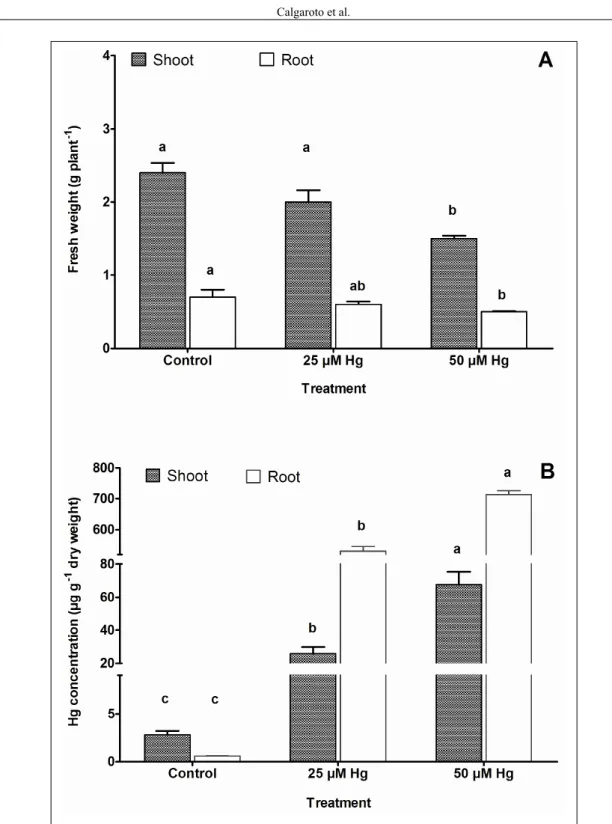
The effects of Hg on the growth of P. glomerata plants was expressed as shoot and root fresh weight (Figure 1a). The exposure to the highest level of Hg (50µM) caused a decreasing in shoot and root fresh weights of 15.5% and 20%, respectively, which can be related to the binding of Hg to SH groups of many aquaporins, present in plasma membranes obstructing the water transport from soil to plant (PATRA & SHARMA, 2000). In the present study, after nine-day of Hg exposition, P. glomerata plants showed withered leaves and darker color roots at concentration of 50µM Hg (data not shown). LOPES et al. (2013) showed that exposure to Hg reduced shoot and root growth of Hordeum vulgare, as well as stomatal conductance, carbon isotope discrimination and expression of an aquaporin transcript. These results suggested some degree of limitation to water uptake causing a moderate water stress when plants are exposed to Hg.
In the present study both shoot and root Hg concentrations increased linearly with increasing external Hg concentrations (Figure 1b).
Hg concentration in roots was 713μg g-1 dry weight
at highest level of Hg (50µM), that is about 11-fold
higher than that found in shoot at the same treatment. Some reports showed that Hg accumulation in the root can indicate that roots serve as a partial barrier to Hg transport to shoot (CALGAROTO et al., 2010; CHEN & YANG, 2012; LOPES et al., 2013). Moreover, shoot accumulated less Hg than roots, even though shoot Hg concentration has increased about 24-fold at 50µM Hg, when compared to the
control, reaching 68μg g-1 dry weight. ISRAR et
al. (2006) reported increase in Hg concentration in shoots as well as in the roots of Sesbania drummondii seedlings with increasing Hg concentrations in the growth solution. Moreover, these authors also found that the accumulation of Hg was more in roots than shoots. WANG & GREGER (2004) observed that the majority of the Hg accumulated in the root system (80%) of six clones of willow (Salix spp.) was bound in the cell wall. CARRASCO-GIL (2013) using Synchrotron X-ray Fluorescence Microprobe in cross sections of Marrubium vulgare roots observed that the most intense Hg signal in roots was found at the root external layers, and Hg was not detected in inner tissues of the root.
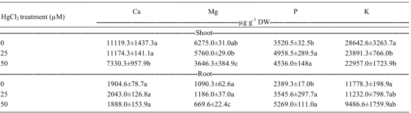
The concentration of macronutrients and micronutrients analyzed in the shoot and root tissues are shown in table 1 and 2, respectively. In general, tissue nutrient concentrations in P. glomerata
plantlets exposed to Hg were significantly decreased
when compared to the control, which indicates that the Hg may cause alteration on the nutritional homeostasis. GODBOLD & HUTTERMANN (1986) reported that exposure of Picea abies to inorganic Hg resulted in a loss of K, Mg and Mn, whereas Fe was accumulated. Such alterations might cause cellular damage leading to serious consequences for
water uptake and mainly to nutrient use efficiency.
the photosynthetic electron transport chain (RUIZ et al., 2003; PATRA et al., 2004; ISRAR et al., 2006).
Ca concentration decreased in shoot only at 50µM Hg, whereas shoot K and Mg concentrations decreased at both 25 and 50µM Hg, when compared
to the control (Table 1). SAHU et al. (2012) observed that the supply of Hg both at moderate (5µM) and high concentration (25µM) reduced the concentrations of K, Ca and Mg in Triticum aestivum. Reductions of
K, Ca and other mineral nutrients under the influence
Figure 1 - Shoot and roots fresh weight and tissues Hg concentration of Pfaffia glomerata plantlets exposed
of Hg have been reported in Marrubium vulgareand Rumex induratus (MORENO-JIMENZ et al., 2007). In contrast with our results, RODRÍGUEZ et al. (2009) reported decrease in P concentration in roots of Chilopsis linearis seedlings grown with 50 and 100µM Hg in hydroponics for 2 weeks. Interestingly, in the present study, P concentration increased in both root and shoot of P. glomerata plants exposed at 25 and 50µM Hg, when compared to the control, whereas Na concentration increased only in the root at 25 and 50µM Hg exposure. The increase of P and Na concentration might be related to the reduction in fresh weight (Figure 1a), which would lead to an increase in the concentration of cellular components. In soil, Hg dissolves as free ion or soluble complex
and is nonspecifically adsorbed by binding mainly due
to the electrostatic forces, chelated, and precipitated as sulphide, carbonate, hydroxide, and phosphate (TANGAHU et al., 2011). Possibly, the concentration of Hg in plant tissues may cause immobilization of P, similarly to what happens in the soil.
A significant decrease in Cu, Zn, Fe and
Mn concentrations in P. glomerata plants exposed to Hg was observed, but most Zn, Mn, and Cu in the roots (Table 2). In contrast to our data, MORENO-JIMENEZ et al. (2007) reported that Hg increased the concentration of Fe by more than 40% in roots of Marrubium vulgare. Interestingly, RODRÍGUES et al. (2009) reported that the concentration of Fe in roots increased in plants exposed to Hg at 50µM and decreased in roots of plants exposed to Hg at
100µM. These authors did not find any alteration
on Mn concentration in plant tissues. Therefore, the
influence of Hg on nutrient content in plants may be
related to the level o Hg in the substrate, plant species, plant organ, growth substrate, and exposure time.
Hg import into root cells is possibly through Fe, Cu, or Zn transporters/channels (PATRA & SHARMA, 2000). Taking account the Hg similarity with Cd and Zn, Hg may have inhibited some other mineral elements absorption by competition with
low affinity transporters in the plasma membrane,
leading to decreasing of shoot and root micronutrient concentrations in plants. CALGAROTO et al. (2011) observed that the addition of Zn (50µM) in the substrate
containing 50µM Hg caused a significant reduction
in the oxidative stress induced by Hg. These authors also found that upon addition of 50µM Zn in nutrient solution shoot and root Hg concentrations were 59 and 24% lower than that of plants exposed to 50µM Hg added alone. Moreover, the percentage survival,
fresh and dry weights and δ-ALA-D activity of plants
treated by 50µM Zn + 50µM Hg were greater than of that treated by Hg alone. Therefore, the data of tissue micronutrients concentrations reported in the present work suggests that excess Hg in nutrient solution may inhibit the uptake of Cu, Zn, Fe and Mn into roots of P. glomerata, with a consequent alteration in many biochemical and physiological processes, which could account for the higher oxidative stress reported by CALGAROTO et al. (2010, 2011).
Taking into account that P. glomerata has shown some degree of heavy metal tolerance, such as for Cd (SKREBSKY et al., 2008), Hg (CALGAROTO et al., 2011) and As (GUPTA et al., 2013), and considering that its roots has been used for pharmaceutical purposes and the ingestion of these metals has a great potential risk to human health, the screening for genotypes of P. glomerata that accumulate less Hg and other toxic metals mainly in the root tissues must be prioritized for purposes of cropping.
Table 1 - Macronutrient concentrations in shoot and roots of Pfaffia glomerata plantlets exposed to 0, 25 and 50µM Hg, using sand as a
substrate, for 9 days.
Ca Mg P K
HgCl2 treatment (µM)
---µg g-1 DW
---Shoot
---0 11119.3±1437.3a 6275.0±31.0ab 3520.5±32.5b 28642.6±3263.7a
25 11174.3±141.1a 5760.0±29.0b 4958.5±289.5a 23891.3±766.0b
50 7330.3±957.9b 3646.3±384.9c 4536.0±148a 22957.0±1723.9b
---Root
---0 1904.6±78.7a 1090.3±62.6a 2389.3±17.0b 11778.3±198.9a
25 2043.0±126.8a 1186.0±37.0a 3545.6±297.7a 11232.0±798.7ab
50 1888.0±153.9a 669.6±22.4c 5269.0±111.0a 9486.6±1759.9ab
CONCLUSION
The increased availability of Hg in
nutrient solution had a significant effect on the
concentration of Hg in Pfaffia glomerata and did alter the nutritional status of the plant. The growth reduction of Pfaffia glomerata plantlets might be related to a decreased in Mn, Fe, Zn, Cu, Ca, Mg, and K concentrations mainly in shoot.
ACKNOWLEGMENTS
The authors wish to thank the Conselho Nacional
de Desenvolvimento Científico e Tecnológico (CNPq),
Coordenação e Aperfeiçoamento de Pessoal de Nível Superior (CAPES) and Fundação de Amparo à Pesquisa do Estado do Rio Grande do Sul (FAPERGS).
REFERENCES
CARGNELUTTI, D. et al. Mercury toxicity induces oxidative stress in growing cucumber seedlings. Chemosphere, v.65, p.999-1006, 2006. Available from: <http://www.sciencedirect.com/ science/article/pii/S0045653506003456>. Accessed: Jul. 19, 2015. doi: 10.1016/j.chemosphere.2006.03.037.
CALGAROTO, N.S. et al. Antioxidant system activation by mercury in Pfaffia glomerata plantlets. Biometals, v.23, p.295-305, 2010. Avalaible from: <http://www.ncbi.nlm.nih.gov/ pubmed/20063044>. Accessed: Jul. 19, 2015. doi: 10.1007/ s10534-009-9287-3.
CALGAROTO, N.S. et al. Zinc alleviates mercury-induced oxidative stress in Pfaffia glomerata (Spreng.) Pedersen.
Biometals, v.24, p.959-971, 2011. Avalaible from: <http://www. ncbi.nlm.nih.gov/pubmed/21553242>. Accessed: Jul. 19, 2015. doi: 10.1007/s10534-011-9457-y.
CARRASCO-GIL, S. et al. Mercury localization and speciation in plants grown hydroponically or in a natural environmental.
Environmental Science and Tecnhology, v.47,
p.3082-3090, 2013. Avalaible from: <http://www.ncbi.nlm.nih.gov/ pubmed/23406525>. Accessed: Jul. 19, 2015. doi: 10.1021/ es303310t.
CHEN, J.; YANG, Z.M. Mercury toxicity, molecular response and tolerance in higher plants. Biometals, v.25, p.847-857, 2012. Avalaible from: <http://www.ncbi.nlm.nih.gov/ pubmed/22639189>. Accessed: Jul. 19, 2015. doi: 10.1007/ s10534-012-9560-8.
EPSTEIN, E.; BLOOM A.J. Mineral nutrition of plants: principles and perspectives. 2.ed. Sunderland: Sinauer Associates, 2005. 400p. GODBOLD, D.L.; HUTTERMANN, A. The uptake and toxicity of mercury and lead to spruce seedlings. Water, Air, and Soil Pollution, v.31, p.509-515, 1986.
GUPTA, D.K. et al. Effect of Hg, As and Pb on biomass production, photosynthetic rate, nutrients uptake and phytochelatin induction in Pfaffia glomerata. Ecotoxicology, v.22, p.1403-1412, 2013. Avalaible from: <http://www.ncbi.nlm.nih.gov/ pubmed/24068651>. Accessed: Jul. 19, 2015. doi: 10.1007/ s10646-013-1126-1.
ISRAR, M. et al. Bioaccumulation and physiological effects of mercury in Sesbania drumondii. Chemosphere, v.65, p.591-598, 2006. Avalaible from: <http://www.ncbi.nlm.nih.gov/ pubmed/16564071>. Accessed: Jul. 19, 2015. doi: 10.1016/j. chemosphere.2006.02.016.
LOPES, M.S. et al. Molecular and physiological mechanisms associated with root exposure to mercury in barley. Metallomics, v.5, p.1305-1315, 2013. Avalaible from: <http://www.ncbi. nlm.nih.gov/pubmed/23925371>. Accessed: Jul. 19, 2015. doi: 10.1039/c3mt00084b.
MORENO-JIMENEZ, E. et al. Mercury accumulation and resistance to mercury stress in Rumex induratus and Marrubium
vulgare grown in perlite. Journal of Plant Nutrition and Soil Science, v.170, p.485-494, 2007. Avalaible from: <http:// onlinelibrary.wiley.com/doi/10.1002/jpln.200625238/pdf>. Accessed: Jul. 19, 2015. doi: 10.1002/jpln.200625238.
MURASHIGE, T.; SKOOG, F. A revised medium for rapid growth and bioassays with tobacco tissue cultures. Physiologia Plantarum,
Table 2 - Micronutrient concentrations in shoot and roots of Pfaffia glomerata plantlets exposed to 0, 25 and 50µM Hg, using sand as a
substrate, for 9 days.
Fe Mn Na Zn Cu
Treatment Hg (µM)
---µg g-1 DW
---Shoot
---0 128.0±10.4a 1156.0±157.3a 2556.0±231.1a 346.6±35.8a 6.2±0.2a
25 135.0±13.2b 1255.0±44.1a 1598.3±116.2b 293.3±8.6ba 4.6±0.05b
50 106.6±7.0c 818.6±90.0b 1896.6±157.1b 209.0±23.5c 4.9±0.2b
---Root
---0 1544.0±8.0ab 712.3±43.3a 4381.3±189.7a 442.6±15.3a 9.4±0.4a
25 1273.0±36.0a 554.6±30.0b 4572.6±518.6a 337.0±14.7b 7.5±0.09b
50 973.5±4.5b 319.6±29.2c 4896.3±1033.5a 258.3±16.8c 6.1±0.7c
v.15, p.473-497, 1962. Avalaible from: <http://onlinelibrary.wiley. com/doi/10.1111/j.1399-3054.1962.tb08052.x/abstract>. Accessed: Jul. 19, 2015. doi: 10.1111/j.1399-3054.1962.tb08052.x.
NICOLOSO, F.T. et al. Micropropagação de ginseng brasileiro (Pfaffia glomerata (Spreng.) Pedersen). Brazilian Journal of Medicinal Plants, v.3, p.11-18, 2001.
PATRA, M. et al. Comparison of mercury, lead and arsenic with respect to genotoxic effects on plant systems and the development of genetic tolerance. Environmental Experimental Botany, v.52, p.199-223, 2004. Avalaible from: <http://www.sciencedirect.com/ science/article/pii/S0098847204000346>. Accessed: Jul. 19, 2015. doi: 10.1016/j.envexpbot.2004.02.009.
PATRA, M.; SHARMA, A. Mercury toxicity in plants. Botanical Review, v.66, p.379-422, 2000.
RODRÍGUEZ, E. et al. Effect of mercury and gold on growth, nutrient uptake, and anatomical changes in Chilopsis linearis.
Environmental and Experimental Botany, v.65, p.253-262, 2009. Avalaible from: <http://www.sciencedirect.com/science/ article/pii/S009884720800124X>. Accessed: Jul. 19, 2015. doi: 10.1016/j.envexpbot.2008.09.014.
RUIZ, O.N. et al. Phytoremediation of organomercurials via the chloroplast genetic engineering. Plant Physiology, v.132, p.1344-1352, 2003. Avalaible from: <http://www.ncbi.nlm.nih.gov/pmc/articles/ PMC167074/>. Accessed: Jul. 19, 2015. doi: 10. 1104/ pp. 103. 020958.
SAHU, G.K. et al. Mercury induced phytotoxicity and oxidative stress in wheat (Triticum aestivum L.) plants. Physiology and Molecular Biology of Plants, v.18, p.21-31, 2012. Avalaible from: <http://www.ncbi.nlm.nih.gov/pubmed/23573037>. Accessed: Jul. 19, 2015. doi: 10.1007/s12298-011-0090-6. SKREBSKY, E.C. et al. Effect of cadmium on growth, micronutrient
concentration, and δ-aminolevulinic acid dehydratase and acid
phosphatase activities in plants of Pfaffia glomerata. Brazilian Journal of Plant Physiology, v.20, p.285-294, 2008.
TANGAHU, B.V. et al. A review on heavy metals (As, Pb, and Hg) uptake by plants through Phytoremediation.
International Journal of Chemical Engineering, v.2011, p.1-31, 2011. Avalaible from: <http://www.hindawi.com/ journals/ijce/2011/939161/>. Accessed: Jul. 19, 2015. doi: 10.1155/2011/939161.
WANG, Y.; GREGER, M. Clonal differences in mercury tolerance, accumulation and distribution in willow. Journal of Environmental Quality, v.33, p.1779-1785, 2004. Avalaible from: <http://www.ncbi.nlm.nih.gov/pubmed/15356238>. Accessed: Jul. 19, 2015. doi: 10.2134/jeq2004.1779.