UNIVERSIDADE DA BEIRA INTERIOR
Ciências da Saúde
Desenho e produção de novos veículos para
entrega de fármacos
Paulo Filipe Brito Machado
Dissertação para obtenção do Grau de Mestre em
Ciências Biomédicas
(2º ciclo de estudos)
Orientador: Prof. Doutor Ilídio Sobreira Correia
Coorientador: Mestre Ana Sofia Matias da Silva
iv
vi
Acknowledgments
I would like to thank to my supervisor Professor Ilídio Correia for supporting the developing of my work. For all the help and stimulus to achieve the purposed goals. I learned a lot under is wing.
I also would like to thank to my co-supervisor Sofia Silva for all the teachings, help and support.
Moreover, I would like to thank to Eng. Ana Paula from the Optics centre of Universidade da Beira Interior for the help in the acquisition of the scanning electron microscopy images of the produced nanoparticles.
Thank to all my group colleges. One way or another all contributed to my work. More important they supported me and give me their friendship.
In addition I thank to my friends, especially my roommates David and Rufo, for the years passed together. I will always remember our adventures.
I could not forget my hometown friends for given me the breaks when I needed the most. Their friendship means a lot to me. Especially thank to Filipe and Nuno for being like brothers to me.
To Gabriela, for all the love and care throughout this year. Without her my life will not be the same. Thank for just being there for me.
To my family, especially my parents for the unconditional love and support on the good and especially on the bad moments. They are truly important for me, and I love them.
viii
Abstract
Nanotechnology is a science that conjugates several disciplines from engineering to chemistry, physics and biology at a molecular level. One of the areas of research with particular focus over the last few years is nanomedicine. It is expected that several improvements in medical care, from diagnosis to treatment, will occur, in the near future, due to the application of nanotechnology to medicine. Several biological and chemical compounds are being applied for the production of drug delivery systems at the nanoscale. These biocompatible systems have some interesting properties that make them capable of performing a targeted delivery of drugs and other materials. Lipossomes, solid lipid nanoparticles, dendrimers, nanoemulsions, polymeric nanoparticles and inorganic nanoparticles like carbon nanotubes, magnetic and ceramic nanoparticles are examples of drug delivery systems. Mesoporous silica nanoparticles are ceramic particles with good structural and biological properties that can be easily functionalized, allowing the bond of other compounds, like for instance gold. The objective of the present work was to produce mesoporous silica nanoparticles, to functionalize their surface and also to bond gold nanoparticles to form a core/shell nanosystem. The produced nanoparticles were characterized by Scanning Electron Microscopy, Fourier Transform Infrared spectroscopy and X-Ray Diffraction. Moreover, the loading and release profiles of the nanoparticles were also characterized using ibuprofen as model drug. In vitro studies were performed to evaluate the internalization and cytotoxicity of the produced nanocarriers. The results herein presented, suggest that the produce nanosytem are suitable for future drug delivery applications.
Keywords
x
Resumo
A nanotecnologia é uma ciência que conjuga diversas áreas científicas tais como engenharia, química, física e a biologia a nível molecular. A nanomedicina é uma das áreas de investigação mais em foco nos últimos anos. Estima-se que diversas melhorias, desde o diagnóstico até ao tratamento venham a surguir num futuro próximo devido à aplicação da nanotecnologia à medicina. Vários compostos químicos e biológicos têm sido aplicados para a produção de sistemas de entrega de drogas à nanoescala. Algumas propriedades que estes sistemas biocompatíveis possuem permitem-lhes efetuar a entrega de fármacos e outros materiais a um tecido alvo específico. Lipossomas, nanopartículas lipídicas sólidas, dendrímeros, nanoemulsões, nanopartículas poliméricas e nanopartículas inorgânicas como nanotubos de carbono e nanopartículas magnéticas e poliméricas, são exemplos de sistemas de entrega de fármacos. As nanopartículas mesoporosas de sílica são nanopartículas cerâmicas com boas propriedades estruturais e biológicas, que podem ser facilmente funcionalizadas à superfície, permitindo a ligação de outros compostos, como por exemplo ouro. O objetivo do presente trabalho foi produzir nanopartículas mesoporosas de sílica, funcionalizar a sua superfície e ligar-lhe nanopartículas de ouro para formar um nanosistema núcleo/concha. As nanopartículas produzidas foram caracterizadas por Microscopia Electrónica de Varrimento, Espectroscopia Infravermelha por Transformada de Fourier e Difracção Raio-X. Os perfis de captação e libertação de fármacos pelas nanopartículas foram também caraterizados. Para avaliar a internalização e a citotoxicidade dos nanoveículos produzidos foram também realizados estudos in vitro. Os resultados aqui apresentados sugerem que os nanosistemas produzidos podem futuramente ser aplicados em aplicações de entrega de fármacos.
Palavras-chave
Nanopartículas de Sílica; Nanopartículas núcleo/concha; Nanotecnologia; Sistemas de Entrega de Fármacos.
xii
Resumo alargado
A nanotecnologia pode ser definida como a ciência que estuda os eventos na dimensão dos nanómetros. Diversas áreas científicas tais como engenharia, química, física e a biologia a nível molecular estão enquadradas nela. Todo o processo, desde a síntese até à aplicação de um material ou dispositivo são etapas da nanotecnologia. A nanomedicina é uma das áreas de investigação mais em foco nos últimos anos. O seu desenvolvimento levará a uma melhor compreensão dos processos biológicos que ocorrem à nanoescala. Um dos seus principais objetivos é aproveitar as propriedades e características físicas dos materiais para desenvolver a prática médica em todo o seu processo. Estima-se que diversas melhorias, desde o diagnóstico até ao tratamento venham a surguir num futuro próximo devido à aplicação da nanotecnologia à medicina.
Dentro da nanomedicina encontram-se os sistemas de entrega de drogas. Prevê-se que o seu desenvolvimento venha a revolucionar o panorama farmacêutico e biotecnológico nos próximos anos. Vários compostos químicos e biológicos têm sido aplicados para a produção destes sistemas de entrega de fármacos à nanoescala. Os fármacos podem encontrar-se ligados à superfície do nanotransportador, encapsulados dentro destes ou dissolvidos na sua matriz. Estes sistemas apresentam diversas vantagens sobre os métodos tradicionais de administração de fármacos. Nomeadamente, a protecção de fármacos hidrofóbicos, a deteção do destino dos compostos no organismo, o aumento do tempo de meia-vida de um fármaco na circulação, a entrega intracelular e a possibilidade de entrega de mais do que um composto ao mesmo tempo. Para além disso, estes sistemas são biocompatíveis e possuem a capacidade de entrega de fármacos ou outros materiais a um tecido alvo específico. A capacidade de um sistema entregar especificamente o seu conteúdo, passiva ou ativamente a um alvo selecionado permite que a atuação do fármaco seja mais direccionada e desta forma, actuar ao nível das células afetadas, não interferindo com os tecidos saudáveis circundantes. A aplicação destes sistemas de entrega de fármacos ao corpo humano torna-se por isso muito atrativa. A toxicidade associada a estes sistemas, é uma propriedade fulcral, no campo das aplicações clínicas.
Lipossomas, nanopartículas lipídicas sólidas, dendrímeros, nanoemulsões, nanopartículas poliméricas e nanopartículas inorgânicas como nanotubos de carbono e nanopartículas magnéticas e poliméricas, são exemplos de sistemas de entrega de fármacos. Todos possuem vantagens e desvantagens entre eles dependendo da aplicação a que se destinam. As nanopartículas mesoporosas de sílica são nanopartículas cerâmicas inorgânicas com centenas de canais vazios (mesoporos) arranjados numa estrutura 2D em forma de favos. A aplicação destes nanosistemas em nanomedicina surge recentemente como campo de extensiva pesquisa. Elas possuem boas propriedades estruturais e biológicas que as tornam muito atrativas em termos de entrega de fármacos. Uma das suas propriedades mais relevantes é a facilidade de funcionalização à superfície, permitindo a ligação de outros compostos, como por exemplo ouro. A formação de nanopartículas constituídas por dois ou
xiii mais materiais origina um sistema designado por núcleo/concha. A junção de materiais permite aliar as propriedades de ambos para melhorar a performance do nanosistema.
O objectivo do presente trabalho foi produzir nanopartículas mesoporosas de sílica, funcionalizar a sua superfície e ligar-lhe nanopartículas de ouro para formar um nanosistema núcleo/concha.
As nanopartículas mesoporosas de sílica foram produzidas por uma adaptação do método Stöber e funcionalizadas com aminas para permitir a ligação de nanopartículas de ouro produzidas por uma adaptação do método de Frens. Várias técnicas foram utilizadas para caracterizar os nanoveículos produzidos. Microscopia Electrónica de Varrimento foi realizada para analisar a morfologia e o tamanho das partículas, a Espectroscopia Infravermelha por Transformada de Fourier efetuada permitiu fazer a analise química dos compostos e dos nanoveículos produzidos. Por fim, a Difracção Raio-X foi usada para identificação da estrutura molecular das partículas. Os perfis de captação e libertação de fármacos pelas nanopartículas foram também caracterizados para avaliar a capacidade de aplicação dos sistemas à entrega de fármacos. Para a avaliação da internalização das nanopartículas pelas células procedeu-se à obtenção de imagens de Microscopia Laser de Varrimento Confocal. A citotoxicidade dos nanoveículos produzidos foi avaliada por estudos in
vitro.
Os resultados aqui apresentados sugerem que os nanosistemas produzidos podem futuramente ser aplicados na entrega direccionada de fármacos.
xiv
Table of contents
Chapter I – Introduction ... 1 1. Introduction ... 2 1.1. Nanotechnology ... 2 1.1.1. Nanomedicine ... 31.1.2.1. Drug Delivery Systems ... 3
1.2. Targeting ... 6
1.3. Toxicity ... 7
1.4. Lipid Based Nanoparticles ... 8
1.4.1. Liposomes ... 8
1.4.2. Solid lipid nanoparticles ... 9
1.5. Dendrimers ... 10 1.6. Nanoemulsions ... 11 1.7. Polymeric nanoparticles ... 11 1.8. Inorganic nanoparticles ... 12 1.8.1. Carbon nanotubes ... 12 1.8.2. Magnetic nanoparticles ... 13 1.8.2.1. Gold Nanoparticles ... 14 1.8.3. Ceramic nanoparticles ... 15
1.8.3.1. Mesoporous Silica nanoparticles ... 15
1.9. Core/Shell nanoparticles ... 16
1.10. Objectives ... 17
Chapter II – Materials and Methods ... 19
2. Materials and Methods ... 20
2.1. Materials... 20
2.2. Methods ... 20
2.2.1. Synthesis of gold nanoparticles ... 20
xv 2.2.3. Functionalization of silica nanoparticles and preparation of silica core/ gold shell
nanoparticles ... 21
2.2.4. Scanning electron microscopy analysis ... 21
2.2.5. Fourier Transform Infrared spectroscopy ... 22
2.2.6. X-Ray Diffraction ... 22
2.2.7. Characterization of the loading profile of the vehicles ... 22
2.2.8. Characterization of the release profile of the vehicles ... 22
2.2.9. Proliferation of A549 non-small lung carcinoma cells in the presence of the various nanoparticles produced ... 23
2.2.10. In vitro transfection of cells with the nanoparticles ... 23
2.2.11. Qualitative Evaluation of in vitro transfection ... 23
2.2.12. Evaluation of the cytotoxic profile of the produced nanoparticles ... 24
2.2.13. Statistical analysis ... 24
Chapter III – Results and Discussion ... 25
3. Results and Discussion ... 26
3.1. Morphological characterization of the produced nanoparticles ... 26
3.2. Fourier Transform Infrared Spectroscopy Analysis of the produced nanoparticles ... 28
3.3. X-Ray powder diffraction analysis ... 33
3.4. Characterization of the loading profile of the vehicles ... 33
3.5. Characterization of the release profile of the vehicles ... 35
3.6. Qualitative evaluation of the in vitro transfection ... 37
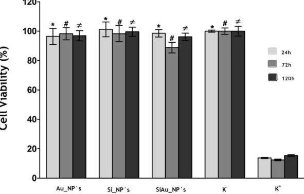
3.7. Characterization of the cytotoxic profile of the produced nanoparticles ... 39
Chapter IV – Conclusions and Future Perspectives ... 42
4. Conclusions and Future Perspectives ... 43
Chapter V – Bibliography ... 44
xvii
List of figures
Figure 1 – Nanotechnology applications in biomedical sciences. ... 2
Figure 2 – Schematic representation of some drug delivery systems ... 4
Figure 3 – Types of DDS targeting ... 7
Figure 4 - Representation of the loading and release profile of a mesoporous silica nanoparticle ... 16
Figure 5 - SEM images of Au_NP´s and Si_NP´s ... 26
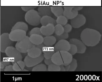
Figure 6 – SEM image of SiAu_NP´s ... 27
Figure 7 - SEM images of SiIBP_NP´s and SiAuIBP_NP´s ... 28
Figure 8 - FTIR spectra of Au_NP´s; Si_NP´s; SiAu_NP´s ... 31
Figure 9 - FTIR spectra of IBP; SiIBP_NP´s and SiAuIBP_NP´s ... 32
Figure 10 - XRD patterns of Au_NP´s; Si_NP´s and SiAu_NP´s ... 33
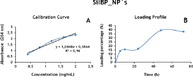
Figure 11 - Loading profile and calibration curve of SiIBP_NP´s. ... 34
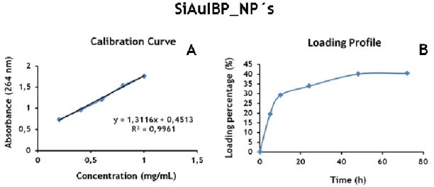
Figure 12 - Loading profile and calibration curve of SiAuIBP_NP´s ... 35
Figure 13 - Release profile and calibration curve of SiIBP_NP´s ... 36
Figure 14 - Release profile and calibration curve of SiAuIBP_NP´s ... 36
Figure 15 – CLSM images of A549 small lung cancer cells ... 38
Figure 16 – CLSM images of A549 small lung cancer cells ... 39
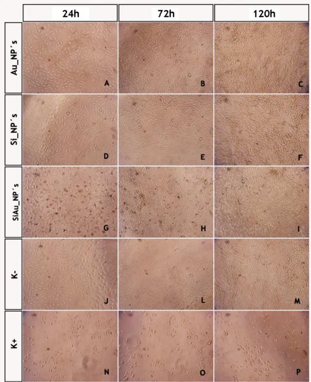
Figure 17 - Inverted Microscope Images of A549 small lung cancer cells ... 40 Figure 18 - Evaluation of the cellular viability after exposure to the produced nanoparticles 41
xix
List of Tables
xxi
List of Acronyms
μm Micrometers
APTMS (3-aminopropyl) trimethoxysilane Au_NPs Gold Nanoparticles
CLSM Confocal Laser Scaning Microscopy cm Centimeters
CNTs Carbon Nanotubes
CTAB Hexadecyltrimethylammonium Bromide DDS Drug Delivery System
DNA Deoxyribonucleic Acid EPR Enhanced Permeability EtOH Ethanol
FBS Fetal Bovine Serum
FDA Food and Drug Administration FITC Fluorescein Isothiocyanate FTIR Fourier Transform Infrared HAuCl4.3H2O Tetrachloroauric (III) Acid HCl Hydrochloric Acid
IBP Ibuprofen k+ Positive Control
K- Negative Control
MCM Mobile Crystalline Material MNPs Magnetic Nanoparticles MRI Magnetic Resonance Imaging MSNPs Mesoporous Silica Nanoparticles
MTS 3-(4,5-dimethylthiazol-2-yl)-5-(3-carboxymethoxyphenyl)-2-(4-sulfophenyl)-2H tetrazolium reagent
MWNTs Multi-walled Carbon Nanotubes NaOH Sodium Hydroxide
NIH National Institute of Health nm Nanometers
PAMAM Polyamidoamine
PBS Phosphate-buffered Saline
PBS-T Phosphate-buffered Saline - Tween 20 PEG Polyethylene Glycol
PFA Paraformaldehyde PNs Polymeric Nanoparticles
xxii PMS Phenazine Methosulfate
RES Reticulo-endothelial System RME Receptor Mediated Endocytosis SBA Santa Barbara Amorphous SiAu_NP´s Silica/gold Nanoparticles
SiAuIBP_NP´s Silica/gold Ibuprofen Nanoparticles siRNA Small Interfering Ribonucleic Acid SiIBP_NP´s Silica Ibuprofen Nanoparticles Si_NP´s Silica Nanoparticles
SEM Scaning Electron Microscopy SWNTs Single-walled Carbon Nanotubes TEOS Tetraethyl Orthosilicate
Chapter I- Introduction
2
1. Introduction
1.1. Nanotechnology
The term nanotechnology, derived from the Greek word for dwarf, “nano”, is defined as the science that studies the events in the dimension range between 1 to 100 nanometers (nm) for some authors, while others consider the maximum value of 1000 nm (Sahoo et al., 2003; Koo et al., 2005; Kingsley et al., 2006). Synthesis, characterization and application of a material, device or system production are stages to be fully characterized in the area of nanotechnology (Silva, 2004). In the last decades nanotechnology is being applied for the benefit of the human civilization (Gupta et al., 2012).
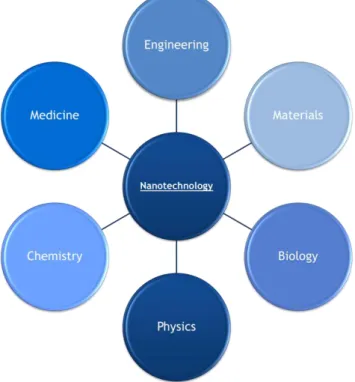
This science is not an isolated area, it aggregate a vast number of technological principles of several subjects such as engineering, electronics, chemistry, physics, biology, material science and manufacturing, at a molecular level, to produce new nanodevices (Sahoo et al., 2003). In the area of nanotechnology a huge effort has been done to develop new therapeutics to be used in nanomedicine. Nanomedicine is defined by the National Institute of Health (NIH) as a medical ramification at the molecular scale that search for new techniques of diagnosis, prevention and treatment of diseases (Park, 2007). In fact, nanomedicine is known for their large number of biomedical applications such as gene therapy, imaging and drug delivery systems (DDS) (Sahoo et al., 2003; Koo et al., 2005).
Chapter I- Introduction
3
1.1.1. Nanomedicine
Nanomedicine has the capacity to improve medical tools in all the stages of medical care, from diagnosis to treatment (Zhang et al., 2012). The metabolic disorders of various diseases take place at the molecular level, enhancing the difficulties for finding effective treatments. The main goal of nanomedicine is to take advantage of the properties and physical characteristics of nanomaterials for medical application, trying to solve these problems (Betty et al., 2010). Therefore, materials can be produced to interact with tissues and cells at a molecular level, having a high degree of specificity (Silva, 2004). Nowadays, scientists are searching and developing new nanomaterials that help transport agents through biological barriers such as ephithelia, endothelia, cell and nuclear membranes, activated monocytes and macrophages of the reticulo-endothelial system (RES). Furthermore, these materials are able to avoid enzymatic degradation and ionic or molecular efflux pumps (Ferrari, 2010). So, it is possible to say that the development of nanomedicine will ultimately lead to a better understanding of the biological processes that take place at the nanoscale level (Salata, 2004). The application of nanotechnology to medicine enhances the availability of new therapeutic weapons and improves the application of several drugs. It also allows a quicker diagnosis (Rosenholm et al., 2011). Nanotechnology in medicine has several levels of action, such as: drug and gene delivery, fluorescent biological labelling, detection of pathogens or proteins, labeling of DNA structure, tissue engineering, tumour destruction (hyperthermia), contrast agents and phagokinetic studies (Salata, 2004). An increase and expansion of this field is expected to happen, making nanomedicine an exciting area that could have a worldwide market of $70-160 billions by 2015 (Shi et al., 2010).
1.1.2.1. Drug Delivery Systems
The use of nanotechnology for drug delivery is expected to revolutionize the pharmaceutical and biotechnology panorama in a near future (Farokhzad et al., 2009). There are a large number of biological and chemical compounds that have been used for the production of drug delivery systems at the nanometer scale. These nanocarriers can be synthesised to have drugs absorbed or linked to the particle surface, encapsulated inside the carrier or dissolved within the system matrix (Bamrungsap et al., 2012). Bearing this knowledge in mind, drug targeting has become a challenging but, at the same time promising subject. By improving DDS selectivity for specific cells or sites of action, secondary effects can be diminished and the treatment effectiveness enhanced (Mattheolabakis et al., 2012). DDS presents many advantages over the traditional ways of drug administration. These advantages include:
stabilization of hydrophobic drugs reducing the use of solvents and surfactants and improving their delivery at the target site;
Chapter I- Introduction
4 biodistribution and pharmacokinetcs;
extended half-life in the circulation;
reduction of secundary effects due to the specific delivery at the target sites;
ability to provide transcytosis of drugs across tight epithelial and endothelial barriers;
possibility of internalization and release of cargo within cells; ability to co-deliver two or more drugs into a desired site;
theragnostic properties, combining therapeutics with diagnostics (Lee et al., 2011; Farokhzad et al., 2009; Bamrungsap et al., 2012; Mattheolabakis et al., 2012). In order to further improve DDS therapeutic efficacy, researchers must fully understand the interactions of carriers with the biological environment as well as with the target cells and their membrane receptors (Suri et al., 2007).
Some of the most used DDS are summarized below and depicted in figure 2. Food and Drug Administration (FDA) approved DDS are presented in table 1.
Figure 2 – Schematic representation of some drug delivery systems: (a) Liposome; (b) Polymeric micelle;
(c) Polymer-drug conjugate; (d) Dendrimer; (e) Oil nanoemulsion; (F) Mesoporous silica nanoparticle and (g) Iron oxide nanoparticle (Adapted from Hu and Zhang, 2002).
Chapter I- Introduction
5
Table 1 - FDA approved DDS available in the market (Adapted from Zhang et al., 2012).
Anti-CD30 monoclonal antibody/monomethyl auristatin E 2011
• Brentuximab Vedotin
Albumin/paclitaxel 2005
• Abraxane
Anti-CD20 monoclonal antibody/iodine-131 2003
• Bexxar
Anti-CD20 monoclonal antibody/yttrium-90 2002 • Zevalin Protein-based DDS PLGA/lanreotide 2009 • Ozurdex PLGA/dexamethasone 2007 • Somatuline PLGA/naltrexone 2006 • Vivitrol PLGA/risperidone 2003 • Risperdal Consta PLGA/leuprolide acetate 2002 • Eligard PLGA/minocycline 2001 • Arestin PLGA/triptorelin pamoatea 2000 • Trelstar
PLGA/recombinant human growth hormone 1999 • Nutropin depot PLA/doxycycline hyclate 1998 • Atridox PLGA-glucose/octreotide acetate 1998 • Sandostatin LAR Polifeprosan 20/carmustine 1996 • Gliadel PLGA/leuprolide acetate 1989 • Lupron Depot PLGA/goserelin 1989 • Zoladex Biodegradable materials PEGylated peginesatide 2012 • Omontys
PEGylated urate oxidase 2010
• Krystexxa
PEGylated tumor necrosis factor alpha inhibitor 2008
• Cimzia
PEGylated erythropoetin receptor activators 2007
• Mircera
PEGylated anti-VEGF aptamer 2004
• Macugen
PEGylated recombinant analogue of the human growth hormone 2003
• Somavert
PEGylated granulocyte colony-stimulating factor analog 2002
• Neulasta
PEGylated interferon alfa-2a 2002
• PEG-ASYS
PEGylated interferon alfa-2b 2001
• PEG-intron
PEGylated L-asparaginase 1994
• Oncaspar
PEGylated adenosine deaminase 1990
• Adagen
Polymer–drug conjugate
Liposomal morphine sulfate 2004
• DepoDur
Estradiol micellar nanoparticles 2003 • Depocyt Liposomal verteporfin 2000 • Visudyne Liposomal cytarabine 1999 • Estrasorb Liposomal amphotericin 1997 • Ambisome Liposomal daunorubicin 1996 • Daunoxome
PEGylated liposomal doxorubicin 1995
• Doxil
Liposome & Micelle
Technology Year of
approval Product name and category
Chapter I- Introduction
6
1.2. Targeting
Ultimately for a DDS be applied in the human body, it is necessary to study the interactions between these systems and the biological environment, especially the possibility of intrinsically give to the DDS the capacity to selectively target a specific cell or tissue (Zhang et al., 2012). This ability allows a specific delivery of the DDS to an area of interest, for instance deliver of a drug to cancer cells (Zhang et al., 2012). Ideally, a nanocarrier should be capable of reaching the desired cells with low, to no loss, of its content and also be capable to only deliver it to affected cells, without affecting healthy tissues (Cho et al., 2008).
Researchers generally consider two mechanisms of targeting: passive and active targeting (Koo et al., 2005; Peer et al. 2007). Passive targeting is the passage of DDS through targeting sites with leaky microvasculature, namely tumour fenestrations and inflamed tissues, into the tumour interstitium and cells by convection of passive diffusion. High molecular weight compounds only pass across pores, while low molecular weight compounds diffuse rapidly and indiscriminately through the endothelial cell layer of blood capillaries (Koo
et al., 2005; Danhier et al., 2010). Taking advantage of the unique pathophysiological characteristic of tumour vessels, the nanocarriers accumulate in this zone by the enhanced permeability and retention (EPR) effect, a characteristic phenomenon of cancer cells that occur due to the extremely disorganization and imperfection of the tumour blood vessels. Such, is caused by an extremely rapid angiogenesis and limited lymphatic drainage (Minko et
al., 2004). For effective passive targeting, the nanocarriers have to circulate in the
bloodstream for a long period of time, to allow multiple passages by the target site (Koo et
al., 2005). This constitutes a problem for the majority of the DDS available, because most of
the nanoparticles have a short circulating half-life due to the defence mechanisms of the human body, especially the reticuloendothelial system. One way to overcome this problem is by coating the nanoparticles with hydrophilic polymers such as polyethylene glycol (PEG) (Koo
et al., 2005; Torchilin, 2010). An advantage of this type of targeting is that the composition
of a DDS does not influence the EPR effect (Mattheolabakis et al., 2012).
Some diseases have, as peculiarity, the expression of epitopes or membrane receptors that can possible be used as active targeting by specific DDS. Thus, ligands that specifically bind to this epitopes or receptors must be attached on the surface of the DDS (Koo et al., 2005) This DDS/target bind and consequently uptake by cells are extremely important for the delivery of drugs that are not easily taken up by cells (Koo et al., 2005). One issue to take into consideration is that the expression of these ligands should be exclusive of the desired cells and not be present on the surface of healthy cells. In addition, an overexpression of these ligands of the target cells is also requirement (Danhier et al., 2010). Targeting ligands can be proteins (antibodies and antibody fragments), nucleic acids (aptamers) and others such as peptides, vitamins and carbohydrates (Peer et al., 2007)
Chapter I- Introduction
7
Figure 3 – Types of DDS targeting: (A) Passive targeting of nanocarriers. (1) Nanovehicles arrive to the
tumour site through the leaky vasculature. (2) Representation of the influence of size in the retention at tumour site. (B) Active targeting. Nanocarriers bond to cancer cells (1); Nanocarriers bond to angiogenic endothelial cells (2) (Adapted from Danhier et al., 2010).
1.3. Toxicity
One important property of DDS is the potential toxicity associated with the nanoparticles. So, it is important to have into consideration the hazards and safety issues on the use of this type of technology, especially when they are used for drug delivery (De Jong and Born, 2008). Nanoparticles size, chemical composition, surface structure, solubility, shape and aggregation are the main aspects that have to be taken into account in order to evaluate their biological effects in the human body. These properties are intrinsically connected with possible problems in terms of cellular uptake, protein binding, translocation from place of entry to the target site and tissue damage (Jain, 2012). The routes used for nanocarriers administration into the body are also important in order to avoid toxic effects. These routes include the gastrointestinal tract, skin, lung and systemic administration (Jain, 2012). DDS interaction with the surrounding environment, namely cells, body fluids and proteins is the key parameter to assure a successful deliver of the desired content. However, this interaction is also crucial in terms of particles toxicity (Jain, 2012). For the same therapeutic effect, the delivery of a drug within a nanoparticle requires a lower dosage,
Chapter I- Introduction
8 when compared with the delivery of free drug. Nevertheless, the particles toxicity itself must be taken into consideration as well (De Jong and Born, 2008). Another point in terms of DDS toxicity is the fate of the particles in the human body. Ideally, the non adsorbed nanoparticles should be excreted trough renal clearance or caught by the liver hepatocytes and then transport to the bile. However, the elimination route of the absorbed nanoparticles is not fully characterized and it is possible that not all nanoparticles are eliminated from the human body. These nanoparticles could be accumulated in several sites of the body. The accumulation of low concentrations of nanoparticles could not represent a high risk, but the accumulation of high doses of DDS can induce toxic secondary effects. Both chemical composition and size might contribute for the absence of nanoparticle elimination (Hagens et
al., 2007).
1.4. Lipid Based Nanoparticles
1.4.1. Liposomes
Lipossomes are the most studied nanosystem to be applied in drug delivery (Bamrungsap et al., 2012). They are artificial self-assembled vesicles obtained from amphiphilic phospholipids. Such nanodevices are constituted by one or more aqueous domains surrounded by one or more spherical bilayers of amphipathic lipid molecules with sizes ranging between 50 nm to several micrometers (µm) (Bamrungsap et al., 2012; Mattheolabakis et al., 2012). Having this characteristic structure, liposomes can incorporate both hydrophilic and hydrophobic drugs in the inner aqueous compartment and within the lipid bilayer, respectively (Mattheolabakis et al., 2012). Moreover, liposomes also have noble biological properties such as biocompatibility and biodegradability (Liu and Boyd, 2013). Their size, surface charge and functionality can be easily changed by the addition of agents to the membrane or by modifying their surface chemistry (Bamrungsap et al., 2012). There are four possible processes to load a drug into this DDS: liposome formation in an aqueous solution saturated with soluble drug; the use of organic solvents and solvent exchange mechanisms; the use of lipophilic drugs; or pH gradient methods (Malam et al., 2009). This type of nanodevices is classified accordingly to the number of bilayers presented. Therefore, liposomes can be divided into unilamellar and multilamellar vesicles (Mattheolabakis et al., 2012). Unilamellar vesicles have a mean diameter ranging between 50 to 250 nm and are liposomes with a single lipid bilayer and a large aqueous core, mostly applied to encapsulate hydrophilic drugs. On the other hand, multilamellar vesicles can be defined has liposomes with several concentric lipid bilayers in an onion-ring arrangement which allows the entrapment of lipophilic drugs due to their high lipidic content. This type of liposomes presents a mean diameter ranging from 1 to 5 μm (Mattheolabakis et al., 2012).
Chapter I- Introduction
9 However, liposomes also present some limitations such as low encapsulation efficiency, fast burst release of drugs, poor storage stability and lack of tunable triggers (Bamrungsap et al., 2012). One of the main objectives regarding the use of liposomes as DDS, is the improvement of their circulation time in the bloodstream. This has been somehow achieved by the incorporation of PEG into the phospholipids present in the liposomes, forming, therefore, a protective coating. The addition of PEG sterically stabilizes the liposome membrane against opsonisation or uptake by the reticuloendothelial system (Mattheolabakis et al., 2012). Another important goal of the research concerning this type of DDS is the development of targeted liposomes, which can be achieved through the binding of peptides or antibodies to the surface of liposomes (Mattheolabakis et al., 2012). Moreover, modified liposomes have also shown good pharmacokinetic profiles for the delivery of Deoxyribonucleic acid (DNA), antisense oligonucleotide, small interfering ribonucleic acid (siRNA), proteins and chemotherapeutic agents (Bamrungsap et al., 2012). One way used to improve the efficacy of liposomes is the use of pH-sensitive lipids. For low pH environments (usually lower than 7.4), lipossomes vesicles are destabilized and its content released to the exterior (Mattheolabakis et al., 2012).
Liposomes can deliver many types of drugs like cancer, parasite and anti-bacterial, hormones, enzymes and immunoactivators, among others. A few anti-cancer drug formulations are already available in the market, being Doxil® the one most successful to date (Liu and Boyd, 2013). The interaction between liposomes and cells can be achieved through adsorption, fusion, endocytosis or lipid transfer (Wilczewska et al., 2012).
1.4.2. Solid lipid nanoparticles
Solid lipid nanoparticles (SLNs) are constituted by a lipid matrix (solid at room and body temperatures), which can be made of highly purified triglycerides, complex glyceride mixtures or waxes, and are stabilised by one or more surfactants (Wissing et al., 2003). They have a mean diameter raging from 50 to 1000nm (Müller et al., 2000). There are several methods to produce SLNs such as high pressure homogenization, which is the more commonly used; microemulsion techniques; dispersion of a lipid in a surfactant and precipitation (Müller
et al., 2000). This type of nanoparticles has several good properties that make them suitable
for drug delivery. Among them, there are the excellent physical stability, protection of incorporated labile drugs from degradation, control of drug release profile, low toxicity, good site-specific targeting and large scale production (Wissing et al., 2003; Mehnert et al., 2012). Notwithstanding, they also have some disadvantages such as limited loading capacity due to solubility in the lipid melt, structure and polymorphic state of the lipid matrix, drug expulsion after polymorphic transition and high water content (Wissing et al., 2003; Mattheolabakis et
al., 2012).
SLNs can be administered by oral, parenteral and transdermal administration routes. SLNs can improve the immune response. Adding SLNs to a vaccine formulation allows a slower
Chapter I- Introduction
10 release rate, ensuring a prolonged exposure time to the immune system (Mattheolabakis et
al., 2012).
1.5. Dendrimers
Dendrimers are synthetic, highly branched globular macromolecules with a three dimensional structure (Gillies and Fréchet, 2005; Restani et al., 2012). The name has origin in the Greek word dendron, meaning “tree” and meros, meaning “part” (Nanjwade et al., 2009). Dendrimers consist in three different domains: a central core, constituted by a single atom or an atomic group with, at least, two similar chemical functions; several branches with origin in the core that are repeated with at least one branch junction, whose repetition is geometrically organized resulting in a series of concentric layers, called generations; and many terminal functional groups, normally located in the exterior of the macromolecule (Mattheolabakis et al., 2012). Their chemical composition and molecular weight can be precisely controlled providing excellent biocompatibility and pharmacokinetics when within a human body (Bamrungsap et al., 2012). Dendrimers constitution along with their globular structures and internal cavities, allows the encapsulation of drugs in their interior, providing also a controlled release of the drug from the inner core (Bamrungsap et al., 2012). Dendrimers can be synthesized mostly by two methods: the divergent approach and the convergent one. In the divergent technique, monomeric modules are assembled in a radial branch-upon-branch motif to the core site. On the other hand, the convergent approach consists in the reaction of several dendrons (dendrimer sections with multiple terminal groups and a single reactive function at the focal point) with a multifunctional core (Nanjwade et
al., 2009). Compared to other types of polymer architecture, dendrimers present many
advantages like low polydispersity, high functionality and a nanometer size range, between 1 to 10nm, which facilitates the passage through some biological barriers (Nanjwade et al., 2009; Bamrungsap et al., 2012). Recently, dendrimers have been described as a great improvement in many areas from gene delivery to magnetic resonance imaging. They have also been used for the development of vaccines, antivirals, antibacterials and anticancer therapeutics (Gillies and Fréchet, 2005).
Polyamidoamine (PAMAM) is one of dendrimers already in commercialization and one of the most used in biomedical applications (Gillies and Fréchet, 2005). However, PAMAM-type dendrimers have been demonstrating some drawbacks regarding their cytotoxicity (Restani et al., 2012). To overcome this limitation, Restani and co-workers have synthesised a family of water soluble blue photoluminescence biocompatible and biodegradable PURE-type dendrimers (Restani et al., 2012). Such type of dendrimers, produced using supercritical carbon dioxide (scCO2) conditions have been shown as promising tools for highly sensitive
Chapter I- Introduction
11
1.6. Nanoemulsions
Nanoemulsions are thermodynamically or kinetically stable dispersions of two immiscible liquids, belonging to the family of colloidal dispersions. In their constitution they present an oil phase and a water phase, with the addition of an interfacial film of surfactant molecules for stabilization (Devarajan and Ravichandran, 2011; Mattheolabakis et al., 2012). The dispersed phase is normally composed by small particles or droplets, with sizes ranging from 5 to 200nm and with a very low oil/water interfacial tension. Moreover, nanoemulsions are transparent, due to the small size of the droplets (Devarajan and Ravichandran, 2011). Depending on their composition, three types of nanoemulsions can be formed: oil in water nanoemulsions, which consist in a continuous aqueous phase with dispersed oil droplets; water in oil nanoemulsions, that contains water droplets dispersed in a continuous oil phase; and bi-continuous nanoemulsions, composed by interdispersed microdomains of oil and water within the system (Lovelyn and Attama, 2011; Devarajan and Ravichandran, 2011). The preparation of nanoemulsions can be achieved by high-pressure homogenization for small size particles involving a drop creation, deformation, and disruption followed by surfactant adsorption, microfluidization or phase invertion temperature technique (Shah et al., 2010; Anton et al., 2008). Nanoemulsions are applied in different areas, such as biotechnology, diagnostics, cosmetics and drug delivery. A few nanoemulsions are already commercialized (Shah et al. 2010).
1.7. Polymeric nanoparticles
Polymeric nanoparticles (PNs) are spherical structures with a maximum size of 1000 nm prepared with natural and/or synthetic polymers (Mattheolabakis et al., 2012; Pinto Reis
et al., 2006). PNs can be categorized in nanospheres and nanocapsules. Nanospheres have a
matrix type of structure in which drugs can be absorbed at the sphere or encapsulated within the particle. In the other hand, nanocapsules are vesicular systems with the drug constrained to a cavity constituted by an inner liquid core surrounded by a polymeric membrane. Drugs are usually dissolved in the inner core but may also be adsorbed to the capsule surface (Pinto Reis et al., 2006). Generally, PNs are produced by a two steps method. The first one corresponds to the preparation of an emulsified system (constituted by emulsions, miniemulsions, nanoemulsions or microemulsions). The second one is related to the formation of the nanoparticles by precipitation or gelation of a polymer or by polymerazation of monomers (Vauthier and Bouchemal, 2008). There are other less used methods that do not require the prior preparation of the emulsified system. These methods are based on the precipitation of a polymer in certain conditions to spontaneously form PN´s or self assembly of macromolecules to form nanogels from a polymer solution (Vauthier and Bouchemal, 2008). The most common PNs are made of poly-d,l-lactide-co-glycolide, polylactic acid, poly-ε-caprolactone, poly-alkyl-cyanoacrylates, chitosan and gelatine (Kumari et al., 2010). The
Chapter I- Introduction
12 specific role of a particular nanoparticle is directly associated with its morphological characteristics, surface chemistry and molecular weight. Surface modification gives the nanoparticles anti-adhesive properties, reducing their clearance by the circulating macrophages. This increases the nanoparticles life time in blood circulation and enhance the number of passages of the nanoparticles through the target sites (Kumari et al., 2010). Morevover, the surface modifications may impair a more controlled release of the drugs encapsulated within the nanoparticles (Kumari et al., 2010). Their exceptional potential in accurate drug and gene delivery makes biodegradable PNs one of the most studied types of nanodevices (Soppimath et al., 2001).
1.8. Inorganic nanoparticles
1.8.1. Carbon nanotubes
Carbon nanotubes (CNTs) are allotropes of carbon with cylindrical nanostructure that belong to the family of fullerenes (Lin et al., 2004; Lacerda et al., 2006). They are constituted exclusively by carbon atoms, in a series of condensed benzene rings arrangement, rolled-up perfectly into a tubular structure (Madani et al., 2012). They were described for the first time in the late 1950s, but their drug delivery properties were only been discovered recently (Madani et al., 2012). CNTs have several advantageous physicochemical properties such as ordered structure, low weight, high mechanical strength, high electrical and thermal conductivity, metallic or semi-metallic behaviour and high surface area (Lacerda et al., 2006). CNTs are usually produced by chemical vapour deposition. This process involves the passage of a hydrocarbon vapour through a tubular reactor with a catalyst material attached. The process is performed at temperatures between 600 and 1200º C to allow de decomposition of the hydrocarbon. After cooling, the CNTs that grow on the catalyst reactor are collected (Paradise and Goswami, 2006; Kumar and Ando, 2010). Other less used methods for CNTs synthesis involve extremely high temperature techniques such as arc discharge and laser ablation (Prasek et al., 2011; Peretz and Regev, 2012). Depending on the number of graphene layers, CNTs can be divided into single-walled carbon nanotubes (SWNTs) or multi-walled carbon nanotubes (MWNTs) (Peretz and Regev, 2012). SWTs are one-dimensional CNTs with diameters of 1-2 nm and lengths ranging from 50 nm to 1 centimetre (cm), with a different behaviour from the nanoparticles in biological environments (Kamalha et al., 2012). They normally appear in bundles due to the strong van der Waals interactions and can exhibit semi-conducting or metallic properties (Klumpp et al., 2006). Due to their flexible properties, it is possible to bend the nanotube to provide multiple binding sites of a functionalized nanotube to a specific cell. Moreover, CNTs also exhibit high surface area, which results from the great number of atoms exposed on the CNTs surface. Such fact results in an efficient loading of multiple molecules in all the nanotube sidewall (Liu et al., 2008). Conversely, MWNTs, are constituted by multiple layers of graphene with a much larger diameter, ranging
Chapter I- Introduction
13 from 10 to 100nm, with 0.34 nm between each layer (Kamalha et al., 2012). They are mainly monodispersed and have only semi-conducting properties (Klumpp et al., 2006). Their larger size enables them to deliver large biomolecules like DNA plasmids into cells, but their optical properties are worse than that of SWNTs (Liu et al., 2008).
In order to functionalize CNTs, two methods are commonly used to perform the necessary modification: oxidation by strong acids leading to a reduction of their length and consequent appearance of carboxylic groups in the same reaction, increasing their dispersibility in aqueous solutions and the binding of a large variety of active molecules like peptides, proteins, nucleic acids and other therapeutic agents (Bianco et al., 2005).
CNTs are suitable for several biomedical applications. Such applications are strongly dependent on their size, shape, structure and unique physical properties (Liu et al., 2008). Lately, CNTs have also been described as good vaccine delivery systems and protein transporters (Bianco et al., 2005).
1.8.2. Magnetic nanoparticles
Magnetic nanoparticles (MNPs) have some exciting features making them suitable to be applied in the medical field, especially in nanomedicine. MNPs can truly provide a wide range of improvements in areas like magnetic resonance imaging (MRI) (being applied as dynamic contrast agents), hyperthemic treatment for malignant cells, drug delivery under influence of an external magnetic force and manipulation of cell membranes trough mechanical stress (Berry and Curtis, 2003). The size of these particles can vary from few to tens of nanometres. This makes possible the loading of several agents with a wide range of sizes. They can also be coated with the desired biological molecules to enhance the biding potential of the particle to specific cells and tissues (Pankurst et al., 2003). MNPs are physiological well tolerated, since they present a low toxicity (Berry and Curtis, 2003).
A very interesting property of MNPs is their ability to obey to the Coulomb’s law. Such feature enables the application of an external magnetic gradient to manipulate them at distance, directing particles to a specific site, maintaining them until needed and then promote their remotion. Finally, MNPs can be produced to respond to a time-varying magnetic field, with the advantage of the transfer of energy from the exciting field to the nanoparticle whenever needed (Berry and Curtis, 2003; Pankurst et al., 2003). However, for biological applications, a biocompatle coating is needed (Tartaj et al., 2003). The fabrication method is the key to produce a valuable magnetic particle and will determine the particle size, distribution, shape, surface chemistry and magnetical properties (Tartaj et al., 2003).
One of the most researched types of magnetic nanoparticles are the iron oxide ones, which can be either paramagnetic or superparamagnetic (Gupta and Gupta, 2005). One characteristic of these nanodevices is their ability to lose their magnetism as soon as the magnetic field is removed (Berry and Curtis, 2003). Examples of such nanoparticles are magnetite or its oxidized form (maghemita), which are the most common metals in
Chapter I- Introduction
14 biomedical applications. Cobalt and nickel, for instance are normally not tested in nanomedicine due to their associated toxicity (Tartaj et al., 2003).
1.8.2.1. Gold Nanoparticles
Gold was one of the first metals discovered by humans (Dykman and Khlebtsov, 2011). The first description of colloidal gold nanoparticles (Au_NPs) was made by Michael Faraday in 1857, when he synthesized multicoloured solutions by reacting gold chloride with sodium citrate producing, without knowing, AuNPs with sizes ranging from 12 to 60 nm in diameter (Paciotti et al., 2006; Boisselier and Astruc, 2009). Since then, Au_NPs have been used in varied biological applications. In 1950s the capacity to bond proteins to the particles, without changing their activity allowed their use in immunodiagnostics and histopathology (Paciotti et
al., 2006). Recently, Au_NPs have been applied in other biological applications such as,
diagnostics and biosensors (Paciotti et al., 2006). The normal oxidation states of gold are +1 (Au [I] or aurous compounds) and +3 (Au [III] or auric compounds) (Jain et al., 2012). There are several ways of producing Au_NPs. Nevertheless, the two main methods that researchers normally use are the citrate reduction of Au [III] derivatives such as aurochloric acid (HAuCl4) in water to Au (0) and the Brust–Schiffrin method, which uses two-phase synthesis and stabilisation by thiols (Boisselier and Astruc, 2009; Jain et al., 2012). The size of AuNPs normally varies between 1 to more than 120 nm (Boisselier and Astruc, 2009). Au_NPs have some characteristic physicochemical properties that make them very exciting and interesting for biomedical applications. Such features include the surface plasmon ressoance (SPR), used for measuring the refractive index of very thin layers of material adsorbed on a metal, the abilility to bind thiol and amine groups, biocompatibility and the ability to control size and optical properties (Pattnaik, 2005; Kim and Jon, 2012; Jain et al., 2012). AuNPs also serve as practical platforms for therapeutic agents, which are related to their high surface area allowing for dense presentation of multifunctional moieties (Yeh et al., 2012). The functionalization of these nanoparticles is one of the subjects that have been widely studied by scientists for the development of biocompatible and multifunctional particles for either diagnostic or therapeutics (theragnostic) (Rana et al., 2011; Jain et al., 2012). They have been used in areas ranging from genomics, biosensors, immunoassays, clinical chemistry, detection and photothermolysis of microorganisms and cancer cells, targeted delivery of drugs, peptides, DNA, and antigens, to optical bioimaging and monitoring of cells and tissues (Dykman and Khlebtsov, 2011).
Is not yet clear the mechanism through which Au_NPs enter into cells, being the non-specific receptor mediated endocytosis (RME) the most accepted to explain their entrance into cells (Jain et al., 2012).
Chapter I- Introduction
15
1.8.3. Ceramic nanoparticles
1.8.3.1. Mesoporous Silica nanoparticles
Mesoporous silica nanoparticles (MSNPs) are solid inorganic materials with hundreds of empty channels, called mesoporos, arranged in a 2D network of honeycomb-like porous structure (Slowing et al., 2008; Vivero-Escoto et al., 2010). The most important types of MSNPs are Mobile Crystalline Material-41 (MCM-41), MCM-48 and Santa Barbara Amorphous-15 (SBA-15) (Yang at al., 2011). The synthesis of surfactant-templated mesoporous silica was first performed in 1992 (Douromis et al., 2012). However, the application in controlled release and delivery was held for the first time only in the beginning of the century (Slowing
et al., 2008). In recent years, MSNPs have been broadly studied for possible applications in
the fields of biotechnology and nanomedicine (Vivero-Escoto et al., 2010). MSNPs have interesting structural properties that make them suitable for application as DDS (Vivero-Escoto et al., 2010; Popat et al., 2011, Yang at al., 2011). The stable and rigid mesostructure gives MSNPs resistance to heat, pH, mechanical stress and hydrolysis (Slowing et al., 2008). Another property of MSNPs is that both the external surface and the internal surface (constituted by the cylindrical pores) can be functionalized (Slowing et al., 2008). Some authors consider the silica framework as a third surface domain (Wu et al., 2011). The capacity of being easily functionalized make MSNPs suitable to be connected with other inorganic particles, like gold, as well as supramolecules and proteins that can possible work as caps forming a core/shell system (Lee et al., 2011; Yang et al., 2011). Furthermore, silica has been used to enhance the biocompatibility of several DDS, like magnetic nanoparticles, polymers and micelles (Slowing et al., 2008). Moreover, the large pore, the large and uniform size of the nanoparticles and the ordered structure, besides the high surface area allow a high loading of drugs or biomolecules within these DDS (Slowing et al., 2008; Popat et al., 2011). They also enhance the therapeutic efficacy of drugs with limited clinical applicability because of the protection against the surrounding environment provided (Rosenholm et al., 2011). The well-controled size provides a good control of the particle-cell interaction allowing the deliver of a precise cargo dose (Wu et al., 2013). Another important property is their high biocompatibility, property that is fundamental for their application in the human body (Popat
et al., 2011; Wu et al., 2011)
MSNPs offer a wide range of biomedical applications. These applications include drug delivery, catalyst supports, adsorption and purification of proteins, cell imaging and labelling, enzyme adsorption and immobilisation (Popat et al., 2011). The possibility of being internalized by cells makes MSNPs suitable to be used as carriers of contrast agents. Also, they prolong the imaging time-frame trough the enhancement of their half-lives, reducing their toxicity as well (Rosenholm et al., 2011). Silica has also been used in artificial implants due to its osteogenic properties (Slowing et al., 2008). Figure 4 shows a schematic representation of the loading and release of a mesoporous silica nanoparticle.
Chapter I- Introduction
16
Figure 4 - Representation of the loading and release profile of a mesoporous silica nanoparticle
(Adapted from He and Shi, 2011).
1.9. Core/Shell nanoparticles
Nanoparticles constituted by two or more different materials are designated by core/shell nanoparticles. They are composed by an inner material (core) and an outer layer that forms a shell around the inner core (Chaudhuri and Paria, 2011). A broad range of material combinations can be applied for the production of nanocarriers. These combinations are divided in inorganic/inorganic, inorganic/organic, organic/inorganic and organic/organic, depending on the used materials. The application of the nanosystem defines the choice of the materials to be used (Chaudhuri and Paria, 2011). Core/shell systems are highly functional nanoparticles that can be easily modified. The addition of a shell to a nanoparticle provides the nanocarrier enhanced properties, such as better stability, decrease of reactivity and dispersibility, increased functionality and controlled release of materials (Chaudhuri and Paria, 2011). A great attention has been given to these core/shell nanosystems since they are suitable for applications in several fields of science, including biomedical applications. Bioimaging, controlled drug release, targeted drug delivery, cell labelling and tissue engineering are examples of these applications. The inorganic/inorganic class of core/shell nanosystems is the most important one for the production of DDS (Levin et al., 2009; Chaudhuri and Paria, 2011). The growth of a gold shell around a functional nanoparticle, like MCM-41 silica nanoparticle, improves the properties of the nanosystem. The gold shell provides a chemically inert surface layer suitable of functionalization. This shell enhances biocompatibility and preserves the properties of the core material (Levin et al., 2009).
Chapter I- Introduction
17
1.10. Objectives
In the present study different nanoparticles were produced in order to obtain a valuable nanosytem to be applied as drug delivery system. The present work plan has the following aims:
- Design and production of several drug delivery systems;
- Characterization of the properties of the different nanoparticles by Scanning Electron Microscopy (SEM), Fourier Transform Infrared (FTIR) and X-Ray Diffraction (XRD);
- Characterization of the loading and release profiles of the nanovehicles; - Evaluation of the in vitro transfection efficiency of the nanoparticles; - Determination of the cytotoxic profile of the nanoparticles.
Chapter II – Materials and Methods
20
2. Materials and Methods
2.1. Materials
A549 non-small lung carcinoma cell line was purchased from ATCC (Middlesex, UK). Sodium azide was purchased from Amresco (Solon, USA). Fetal bovine serum (FBS) was acquired from Biochrom AG (Berlin, Germany). Hexane was purchased from JMS Ltda. (Odivelas, Portugal). Tris Base and Sodium Hydroxide (Na OH) were obtained from Fisher Chemical (Loughborough, UK). Poly-d-lysine coated glass bottom dishes (35mm dishes, 10mm glass, 0mm thickness) were from by MatTek Corporation (USA). 96-well plates were purchased from Orange Scientific (Braine-L’Alleud, Belgium). Hydrochloric Acid (HCl) was acquired from Panreac (Barcelona, Spain). (3-aminopropyl) trimethoxysilane (APTMS) and Tetrachloroauric (III) acid (HAuCl4.3H2O) were bought from Alfa Aesar (Karlsruhe, Germany). Ibuprofen and Hexadecyltrimethylammonium Bromide (CTAB) were from Tokyo Chemical Industry (Tokyo, Japan). Tetraethyl orthosilicate (TEOS) was purchased from Acros Organics (Geel, Belgium). 3-(4,5-dimethylthiazol-2-yl)-5-(3-carboxymethoxyphenyl)-2-(4-sulfophenyl)-2H-tetrazolium reagent MTS and Phenazine Methosulfate (PMS) was obtained from Promega (Madison, USA). Amphotericin B, Dialysis tubing cellulose membrane, Ethanol (EtOH), Ham‘s Nutrient Mixture-F12, Penicillin G, Phosphate-buffered saline (PBS), Streptomycin, Trypsin, Triethylamine and Trisodium Citrate were from Sigma Aldrich (Sintra, Portugal).
2.2. Methods
2.2.1. Synthesis of gold nanoparticles
The gold nanoparticles (Au_NP’s) were produced through an adaptation of the Frens method was used (Frens, 1973). The process start with the addition of 0.01g of 0.01% (w/v) tetra-chloroauric[III] acid trihydrate (HAuCl4.3H2O) to 100mL of deionized water heated to boiling, under reflux conditions. After reaching boiling, 1mL of trisodium citrate hydrate 1% (w/v) was added drop by drop to the boiling solution under constant stirring. The previously yellow solution starts to turn blue after a few seconds of reaction, in a process called nucleation. Subsequently, the blue colour solution change into dark red, characteristic of the monodisperse spherical particles formation. The solution was kept under boiling conditions for 10 minutes (min) to complete the gold chloride reduction. After cooling at room temperature, 0.25g of 0.05% (w/v) sodium azide was added in order to preserve the gold nanoparticles. Finally, the resultant solution was stored at 4ºC (Frens, 1973).
Chapter II – Materials and Methods
21
2.2.2. Synthesis of silica nanoparticles
For the production of the MCM-41 silica nanoparticles (Si_NP’s), a derivation of the Stöber method was applied (Jia et al., 2012). In a round bottom flask 100 mL of Mili-Q water, 720 μL of NaOH (2M) and 0.2 g of CTAB were mixed and kept at 80ºC, under vigorous stirring. After the stabilization of the temperature, 1 mL of TEOS was added dropwise to the mixture and the reaction was left to occur. Two hours later, the reaction was stopped and cooled to room temperature, followed by centrifugation (13.500 rpm, 20 min). The produced particles were dried at room temperature overnight. To remove the surfactants, 120 mL of EtOH and 15 mL of HCl (37%) were added to each gram of particles in a round bottom flask and left at 80º C under refluxing conditions and vigorous stirring for 24 h. Finally the solution was centrifuged (13.500 rpm, 20 min) and washed with deionised water and ethanol several times to completely remove all the surfactants. After drying at room temperature, a Si_NP’s powder was obtained.
2.2.3. Functionalization of silica nanoparticles and preparation of silica
core/ gold shell nanoparticles
To obtain the silica core/ gold shell nanosystem, from now on designated by SiAu_NP´s, the previously produced Si_NP´s were amino functionalized. To do so, 100 mL of a solution of Si_NP´s in ethanol was placed in a round bottom flask and 1 mL of APTMS was added dropwise and then the mixture was left overnight, under vigorous stirring. After, the mixture was heated to 80º C for one hour, to complete the reaction. Then, the solution was centrifuged (13.500 rpm, 20 min) and washed with ethanol, 5 times, to remove the excess of APTMS. To remove the remaining ethanol, the powder was left to dry at room temperature until evaporation of the solvent. For the synthesis of the SiAu_NP´s a solution of amino functionalized Si_NP´s with ethanol was prepared and added to the Au_NP´s solution, in a ratio of 1:1, by simple contact at room temperature for several hours. After, the solution was centrifuged (13.500 rpm, 20 min) and washed with ethanol several times. A powder of SiAu_NP´s was obtained.
2.2.4. Scanning electron microscopy analysis
In order to characterize the morphology of all the nanoparticles produced, scanning electron microscopy (SEM) analysis was performed. For all the produced nanoparticles analysis, one drop of the solution was added to a 15mm cover glass and left air-dried overnight and then mounted on an aluminium board using a double-side adhesive tape and covered with gold using an Emitech K550 (London, England) sputter coater. Samples were analysed by using a Hitachi S-2700 (Tokyo, Japan) scanning electron microscope operated at an accelerating voltage of 20 kV, at several amplifications (Gaspar et al., 2011; Ribeiro et al.,
Chapter II – Materials and Methods
22 2009).
2.2.5. Fourier Transform Infrared spectroscopy
The produced nanoparticles were also analysed by FT-IR spectroscopy. In this technique an interference wave, produced by an interferometer, interacts with the sample and the resulting spectrum is analyzed (Faix, 1992). The spectrum obtained gives information about the chemical substances of the tested sample (Almeida et al., 2002). All the spectra were acquired in a Fourier transform infrared spectrophotometer Nicoletis 20 (64 scans, at a range of 4000 to 400cm−1) from Thermo Scientific (Waltham, MA, USA) equipped with a Smart
iTR auxiliary module.
2.2.6. X-Ray Diffraction
X-Ray Diffraction (XRD) is a powerful technique used to identify the crystalline phase, the structure refinement and the molecular structures of the samples as well as the grain orientation and strain of the materials (Harris et al., 2001; Tamura et al., 2002; Morgan and Gilman, 2002). To perform the XRD analysis, samples of Au_NP’s were first lyophilized and then mounted in silica supports using a double side adhesive tape. The Si_NP’s the powder was directly placed in the supports. All the experiments were performed over the range from 5º to 90º 2θ with continuous scans at a rate of 1º 2θ min-1, using a Rigaku Geigger Flex D-max
III/c diffractometer (Rigaku Americas Corporation, USA) with a copper ray tube operated at 30kV and 20mA (Gaspar et al., 2011).
2.2.7. Characterization of the loading profile of the vehicles
For ibuprofen be loaded into SiNP’s or SiAuNP´s, 20 mL of hexane and 40 mg of ibuprofen were mixed at room temperature under vigorous stirring, in a darkroom. After 30 min of homogenization, 40 mg of SiNP’s or SiAuNP´s were added to the solution. The mixtures were collected for 5, 10, 24, 48 and 72 h and then centrifuged (13.500 rpm, 10 min) in order to recover the ibuprofen loaded particles. The supernatants were stored for later quantify the amount of drug loaded in nanodevices using an UV-1700 PharmaSpec spectrophotometer from Shimadzu (Kyoto, Japan) at 264 nm and analyzed with an UVProbe Shimadzu 2.0 software (Zhang et al., 2011).
2.2.8. Characterization of the release profile of the vehicles
After ibuprofen be loaded into the nanoparticles herein produced, the release studies were performed in order to evaluate the rate of drug release. First, 500 mL of PBS was added to a goblet and placed in a darkroom, at 37º C, pH=7.4 and under constant stirring (200 rpm). The nanoparticles solution was dialysed with a cellulose membrane containing PBS. For the