Review Article
RBAFSSOCIEDADE BRASILEIRA DE ATIVIDADE FÍSICA E SAÚDE Brazilian Journal of Physical Activity and Health
RBAFS
Revista Brasileira de Atividade Física & Saúde SOCIEDADE BRASILEIRA DE ATIVIDADE FÍSICA E SAÚDE Brazilian Journal of Physical Activity and Health
RBAFS
Revista Brasileira de Atividade Física & Saúde
SOCIEDADE BRASILEIRA DE ATIVIDADE FÍSICA E SAÚDE Brazilian Journal of Physical Activity and Health Rev Bras Ativ Fis Saúde p. 667-678
DOI
http://dx.doi.org/10.12820/rbafs.v.19n6p667 1 Universidade Estadual Paulista, Department of Physical Education - Rio Claro City, São Paulo State – Brazil
2 Faculdades Estácio de Sá, Department of Physical Education– Vitória City, Espírito Santo State – Brazil
Protocols with blood flow restriction
during resistance training: a
systematic review
Protocolos com restrição do fluxo
sanguíneo durante o treino resistido: uma
revisão sistemática
Camila Bosquiero Papini1
Nuno Manuel Frade de Sousa2
Danilo Rodrigues Bertucci1
Natália de Oliveira Bertolini1
Leandro Mori Acedo1
Sebastião Gobbi1
AbstrAct
This systematic review investigated protocols used in Blood Flow Restriction (BFR) exer-cise training and evaluated the intensity of exerexer-cise and length of training for the deve-lopment of muscle hypertrophy and strength. Twelve studies were included according to criteria. The Random-effects Model was used to teste the intensity of exercise and length of training in quadriceps femoris. In general, the BFR group was associated with an increa-se in Muscle Cross-increa-sectional Area (CSA) of 3.84%; however, it was not associated with an increase in quadriceps strength compared with the control group. When the analyses were made considering the intensity of the exercise and length of training, the results showed that BFR exercise ≤ 30% of One Repetition Maximal (1RM) and the length of training ≤ 2 weeks were associated with an increase in quadriceps CSA and strength compared with the control group. The analyses provide subsidies that BFR training until 30% 1RM and a length of training until 2 weeks are the most effective to develop muscle hypertrophy and strength in lower limbs than exercise more than 30%1RM and more than 4 weeks of length training.
Keywords
Blood flow restriction; Resistance training; Strength training; Muscle strength; Hyper-trophy.
resumo
Essa revisão sistemática investigou protocolos utilizados em treinamento físico com restrição do fluxo sanguíneo (RFS) e avaliou a intensidade do exercício e período de treinamento para o desenvol-vimento de força e hipertrofia muscular. Considerando os critérios estabelecidos, 12 artigos foram incluídos. A análise de Modelos de Efeitos Aleatórios foi utilizada para testar a intensidade do exer-cício e duração do treinamento no músculo quadríceps femoral. De maneira geral, o grupo RFS foi associado com um aumento na área de secção transversa (AST) do músculo em 3,84%, no entanto, não houve associação com aumento de força no quadríceps quando comparado com o grupo controle. Quando as análises foram realizadas considerando a intensidade do exercício e tempo de treinamento, os resultados mostraram que o exercício com RFS ≤ 30% de 1 RM (Repetição Máxima) e a duração de treinamento ≤ 2 semanas foi associado com aumento da AST e força no quadríceps quando com-parado com o grupo controle. As análises fornecem subsídios de que treinamentos com RFS com uma intensidade de até 30%1RM e até 2 semanas de duração são mais efetivos para desenvolver força e hipertrofia muscular no quadríceps femoral que treinos de intensidade maior de 30% de 1RM e com duração de mais de 2 semanas.
PAlAvrAs-chAve
IntroductIon
A research focus in the physical training area is the improvement of existing methods or the development of new methods that promote better outcomes. For about 40 years, studies of physical exercise combined with blood flow restriction (BFR) have been initiated in Japan1. The technique - called Kaatsu Training - combines low intensity resistance exercise with BFR applied to the proximal ends of the limbs with a pressure cuff1-2.
Several studies have discussed the effects of BFR training on metabolic, hor-monal and mechanic responses of the muscle3-5 and on homeostatic, hemod-ynamics, inflammatory and molecular responses6-10.In addition, findings from previous studies have indicated that BFR can attenuate atrophy, increase skeletal muscle hypertrophy and strength across different age groups11-24. The possible reason for developing hypertrophy can be related to the muscle oxygen concen-tration decrease, local metabolites and insulin-like growth factor-1 (IGF1) increase and muscle cell swelling induced by blood occlusion4,7,23-26. Additionally, studies have demonstrated an acute increase in protein synthesis following BFR exer-cise, such as ribosomal S6-Kinase-1 (S6K1), promoting a key regulatory protein in the activation of the Mammalian Target of Rapamycin (mTOR) pathway6,7,9.
Abe et al.27 showed that in only 8 days (16 sessions), the low intensity re-sistance training with BFR can increase strength (9.6%) and hypertrophy (4.5%). Similarly, Yasuda et al.6,26 reported an increase in strength (14%) and hypertrophy (7.8%) in lower limbs and an increase in strength (8%) and hy-pertrophy (16%) in upper limbs over 2 weeks of training. Although the BFR exercise training is widely used in research, there is no standard protocol, specifically with reference to the most effective intensity, volume, length of training and pressure cuff to develop muscle hypertrophy and strength.
The development of a low intensity intervention to increase strength and hypertrophy is clinically relevant, because there are some conditions that the traditional methods (high intensity) are not indicate, e.g. patients with com-promised muscle-tendon integrity or neurological conditions that result in the inability to activate their muscles voluntarily25,28,29. The use of a non-standard technique can lead to misunderstanding in the inference of its effectiveness.
In this sense, this review summarizes the recent studies demonstrating the effect of BFR training in hypertrophy and strength adaptations. Thus, the ob-jectives of this study are (i) to discuss the different protocols used during BFR training to increase muscle hypertrophy and strength and (ii) to evaluate the intensity of exercise and length of training for the most effective development of muscle hypertrophy and strength through BFR training.
methods
The systematic review was conducted by an electronic search in MEDLINE/ PubMed. The descriptors utilized to identify the studies were divided into 2 groups: a) Blood flow restriction, Blood flow occlusion, Restriction of muscular venous blood flow, Hypoxia, Kaatsu; b) Strength training, Strength exercise, Muscle train-ing, Muscle exercise, Resistance exercise, Resistance training. We refer to Boolean operators: AND to combine the groups, OR for the words at the same group and QUOTATION MARKS in each descriptor.
The inclusion criteria adopted to select the studies were: a) original stud-ies published from 2005 to August 2013; b) using controlled and randomized sample; c) using resistance training or resistance exercise with BFR located in limbs; d) showing results about hypertrophy and strength effects; e) using 1RM (One Repetition Maximum), ultrasonography, biopsy, anthropometric measure and magnetic resonance imaging as assessment techniques; f) written in English language.
The terms “resistance training” and “resistance exercise” were defined as strength exercise performed with load, located by muscle groups. Case studies, environmental hypoxia (altitude), aerobic exercise, strength exercise without load/burden or performed with corporal weight, acute sections or metabolic and molecular responses were not included.
Initially, 173 scientific studies were identified. The selection was performed in the study title and 101 were excluded. After analysis of the abstracts and the methodology, considering the inclusions criteria, 09 studies were selected to include in this review. It was performed a hand searching in the reference list and 03 studies were included. (Figure 1).
FIgure 1 – Flow diagram for systematic review, 2005 – 2013.
The studies of upper limbs21,26,30 were not included in the statistical anal-yses due to the low number of studies reviewed. The study20 about a limb suspension was also not included in the statistical analyses because it com-pares the CSA and strength gain in a situation of muscle atrophy. Therefore, a descriptive analyses for upper limbs and suspension studies were performed. The statistical analyses were applied to the lower limbs studies5-7,19,22-24,27. The study of Laurentino et al. (2008)23 was divided in 2 once they have utilized 2 BFR Groups. A total of 9 studies were included in the statistical analyses.
The Random-effects Model was used to test the intensity of exercise and length of training of the protocols discussed. The variables were dichoto-mized into ≤ 30% of 1RM and > 30% of 1RM for intensity of exercise and into ≤ 2 weeks and >4 weeks for length of training. The absolute changes in mid-thigh Cross Sectional Area (CSA) and quadriceps strength (1RM) were
reported as percentage of change for the control group and BFR group after interventions. Pooled-effect estimates were obtained by comparing the least squares mean percentage change from baseline to the end of the intervention for each group, and were expressed as the weighted mean difference between groups. The comparison was made between the BFR groups and the control groups. The control group performed the identical exercise than BFR Group, however, without BFR.
An alpha value of 0.05 was considered statistically significant. Statisti-cal heterogeneity of the treatment effect among studies was assessed using Cochran Q test, a threshold p-value of 0.01 was considered statistically sig-nificant, and the inconsistency I2 test in which values greater than 50% were considered indicative of high heterogeneity. The analyses were conducted us-ing Review Manager 5.2 software (Copenhagen: The Nordic Cochrane Cen-tre, The Cochrane Collaboration, 2012).
results
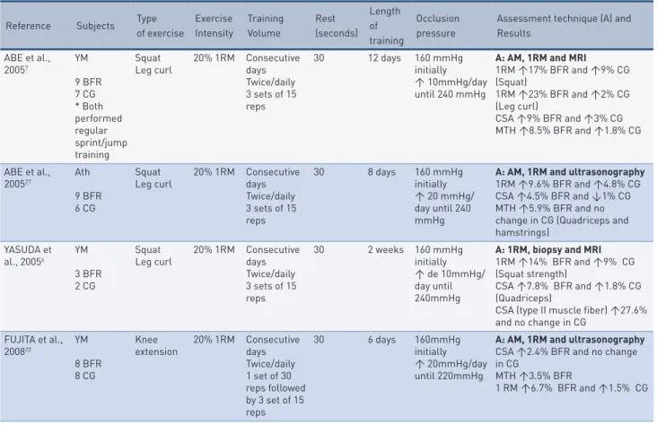
Table 1 presents the summary of the studies using BFR to increase muscle hypertrophy and strength, evidencing their methodology and main results. tAble 1 – Reviewed studies using blood flow restriction, demonstrating the training protocol, assessment technique and results.
Reference Subjects Type of exercise Exercise Intensity Training Volume Rest (seconds) Length of training Occlusion pressure
Assessment technique (A) and Results ABE et al., 20057 YM 9 BFR 7 CG * Both performed regular sprint/jump training Squat
Leg curl 20% 1RM Consecutive days Twice/daily 3 sets of 15 reps 30 12 days 160 mmHg initially ↑ 10mmHg/day until 240 mmHg
A: AM, 1RM and MRI
1RM ↑17% BFR and ↑9% CG (Squat) 1RM ↑23% BFR and ↑2% CG (Leg curl) CSA ↑9% BFR and ↑3% CG MTH ↑8.5% BFR and ↑1.8% CG ABE et al., 200527 Ath 9 BFR 6 CG Squat
Leg curl 20% 1RM Consecutive days Twice/daily 3 sets of 15 reps 30 8 days 160 mmHg initially ↑ 20 mmHg/ day until 240 mmHg
A: AM, 1RM and ultrasonography 1RM ↑9.6% BFR and ↑4.8% CG CSA ↑4.5% BFR and ↓1% CG MTH ↑5.9% BFR and no change in CG (Quadriceps and hamstrings) YASUDA et al., 20056 YM 3 BFR 2 CG Squat
Leg curl 20% 1RM Consecutive days Twice/daily 3 sets of 15 reps 30 2 weeks 160 mmHg initially ↑ de 10mmHg/ day until 240mmHg
A: 1RM, biopsy and MRI 1RM ↑14% BFR and ↑9% CG (Squat strength)
CSA ↑7.8% BFR and ↑1.8% CG (Quadriceps)
CSA (type II muscle fiber) ↑27.6% and no change in CG FUJITA et al., 200822 YM 8 BFR 8 CG Knee
extension 20% 1RM Consecutive days Twice/daily 1 set of 30 reps followed by 3 set of 15 reps 30 6 days 160mmHg initially ↑ 20mmHg/day until 220mmHg
A: AM, 1RM and ultrasonography CSA ↑2.4% BFR and no change in CG
MTH ↑3.5% BFR
Reference Subjects Type of exercise Exercise Intensity Training Volume Rest (seconds) Length of training Occlusion pressure
Assessment technique (A) and Results LAURENTINO et al., 200823 YM 8 BFR 8 HLE *Right leg with BFR and left leg trained as a control Knee extension BFR: 60% 1RM HLE: 80% 1RM 2 days/week 1ª - 3ª week: 3 sets 4ª - 5ª week 4 sets 6ª - 8ª week: 5 sets BFR: 12 reps HLE: 6 reps 120 8 weeks BFR: 131± 12 mmHg HLE: 125 ± 15 mmHg A: 1RM and MRI
CSA ↑4.6% BFR (right and left legs)
and ↑5.3% HLE (right and left legs)
1RM ↑36% (right leg) BFR, ↑38% (left leg) BFR and ↑35% HLE (right and left legs)
MARADAME et al., 200824 YM 8 BFR 7 CG Knee extension Knee flexion Dumbbell curl Leg: 30% 1RM Arm: 50% 1RM 2 days/week 3 sets of 15 reps 2 days/week 3 sets of 10 reps 30 180 10 weeks 160mmHg initially ↑ de 20mmHg in each 2 weeks A: 1RM and MRI 1RM ↑19% BFR and ↑10% CG (Knee extension) 1RM ↑18% BFR and ↑9% CG (Knee flexion) CSA ↑5.7% BFR (Leg) 1RM ↑20% BFR and ↑20% CG (Elbow flexion) CSA↑10% BFR (Arm) COOK et al., 201020 YA 8 BFR 8 NT Knee extension * 30 days of unilateral lower limb suspension 20% MVC 3 days/ week 3 sets until fatigue
90 4 weeks + 1,3 of SBP A: MRI and dynamometry (MVC) CSA ↓1.2% BFR and ↓7.4% NT MVC ↓2% BFR and ↓21% NT CREUDER et al., 201021 YA 12 BFR *One arm were randomly selected as CG Handgrip 60% MVC 3 days/week 20 minutes/ day 15 grips/ minute
No rest 4 weeks 80mmHg A: Dynamometry (MVC), AM and ultrasonography MVC ↑16.2% BFR and ↑16.2% CG Forearm circumference ↑2.42% BFR and ↑1.62% CG KARABULUT et al., 20105 EM 13 BFR 13 HLE 11 NT Leg press Leg extension BFR: 20% 1RM HLE: 80% 1RM – 3 sets of 8 reps 3 days/ week 1 set of 30 reps followed by 2 sets of 15 reps 60 6 weeks 160 mmHg initially ↑ de 20mmHg according to Perceived Exertion Scale (BORG = 16) A: 1RM 1RM ↑19.3% BFR and 20.4% HLE (Leg press) 1RM ↑19.1% BFR and ↑31.2% HLE (Leg extension)
YASUDA et al., 201026 YM
5 BFR 5 CG
Bench
press 30% 1RM 6 days/weekTwice/daily 1 set of 30 reps followed by 3 sets of 30 reps 30 2 weeks 30 mmHg initially ↑ 10 mmHg/ day until 160mmHg A: 1RM and ultrasonography CSA ↑8% BFR and ↓1% CG (Triceps) CSA ↑16% BFR and ↑2% CG (Pectoralis Major) 1RM ↑6% BFR and ↓2% CG (Bench press) CLARK et al., 201119 YA 9 BFR 7 HLE Knee extension BFR: 30% 1RM HLE: 80% 1 RM 3 days/week 3sets of 15 reps 90 4 weeks +1,3 of SBP A: Dynamometry (MVC) ↑8% BFE and ↑13% HLE
YASUDA et al., 201130 YM 10 BFR 10 HLE 10 NT Bench press BFR: 30% 1RM HLE: 75% 1 RM 3 days/week 1 set of 30 reps followed by 3 sets of 15 reps 30 6 weeks 100 mmHg initially ↑ de 10mmHg/day until 160mmHg A: AM, MVC and 1RM 1RM ↑8.7% BFR and ↑19.9% HLE (Bench press)
CSA ↑4.9% BFR, ↑8.6% HLE and ↓1.1% NT (Triceps) CSA ↑8.3% BFR and 7.6% HLE (Pectoralis Major)
Legend: AM = Anthropometric Measure; Ath= Athletes; BFR= Blood Flow Restriction Group; CG= Control Group; CSA= Muscle Cross-sectional Area; EM= Elderly Men; HLE= High Load Exercise Group; MRI= Magnetic Resonance Imaging; MTH= Mid-thigh Muscle Thickness; MVC= Maximum Voluntary Contraction; NT= No Training Group; Rep = repetitions; SBP = Systolic Blood Pressure; YA= Young Adults; YM= Young Men; 1RM= One Repetition Maximum.
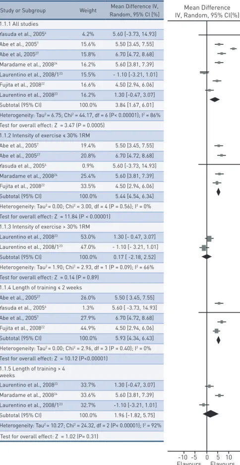
Figure 2 shows the BFR training effects on mid-thigh CSA. Seven studies (99 subjects) demonstrated that BFR group was associated (p < 0.001) with an increase in mid-thigh CSA of 3.84% (95% CI, 1.67 to 6.01); I2, 86%; p for heterogeneity < 0.001 as compared with control group.
When the intensity of the exercise was ≤ 30% of 1RM, BFR group was associated (p < 0.001) with an increase in quadriceps CSA of 5.44% (95% CI, 4.54 to 6.34); I2, 0%; p for heterogeneity = 0.56 as compared with the control group at the same intensity of exercise. Conversely, intensity of exercise > 30% of 1RM with BFR group was not associated (p = 0.89) with an increase in mid-thigh CSA [0.17% (95% CI, -2.18 to 2.52); I2, 66%; p for heterogeneity = 0.09] as compared with the control group at the same intensity of exercise. The length of training ≤ 2 weeks was also associated (p < 0.001) with an in-crease in quadriceps CSA for the BFR group [5.39% (95% CI, 4.34 to 6.43); I2, 0%; p for heterogeneity = 0.40], however, the increase was not demonstrat-ed (p = 0.31) for lengths of training > 4 weeks [1.96% (95% CI, -1.82 to 5.57); I2, 92%; p for heterogeneity < 0.001] as compared with the control group.
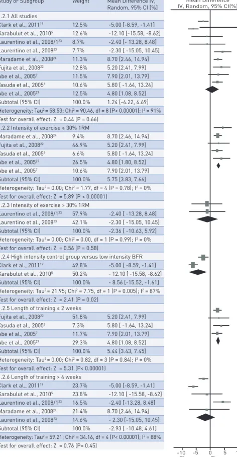
Figure 3 shows the BFR effects on quadriceps strength. Nine studies (115 subjects) demonstrated that BFR group was not associated (p = 0.66) with an increase in quadriceps strength [1.24% (95% CI, -4.22 to 6.69); I2, 91%; p for heterogeneity < 0.001] as compared with the control group.
The intensity of exercise ≤ 30% of 1RM was associated (p < 0.001) with an increase in quadriceps strength for BFR group [5.75% (95% CI, 3.83 to 7.66); I2, 0%; p for heterogeneity = 0.78] as compared with the control group at the same intensity of exercise. There was no association (p = 0.58) between BFR group and intensity of exercise > 30% of 1RM [-2.36% (95% CI, -10.63 to 5.92); I2, 0%; p for heterogeneity = 0.99]. The high intensity control group was associated (p = 0.02) with an increase in quadriceps strength of 8.56% (95% CI, 1.61 to 15.52); I2, 87%; p for heterogeneity = 0.005 as compared with the low intensity BFR group. The length of training ≤ 2 weeks was also associated (p < 0.001) with an increase in quadriceps strength for the BFR group [5.44% (95% CI, 3.43 to 7.45); I2, 0%; p for heterogeneity = 0.84], however, the increase was not demon-strated (p = 0.45) for lengths of training > 4 weeks [-2.93% (95% CI, -10.48 to 4.61); I2, 88%; p for heterogeneity < 0.001] as compared with the control group.
dIscussIon
The BFR training studies have been demonstrating effective outcomes in both acute and chronic training to develop hypertrophy and muscle strength. However, the problem with most previously reported studies is the fact that there is no stan-dard protocol. The pressure cuff, training volume, intensity and length of training during interventions are different. Thereby, this study aimed to discuss the BFR physical exercise protocols related with muscle hypertrophy and strength.
In general, the statistical analyses evidenced that the BFR training was associ-ated with an increase in mid-thigh CSA of 3.84% (p < 0.001) compared with the control group at the same intensity of exercise. Some possibilities are presented for this results, such as an increase in muscle protein synthesis and muscle ac-tivity; increase in endogenous anabolic hormones, like GH and IGF-17,31,32 and increase in S6K1 phosphorylation and hypertrophy pathway7,914,26,31,32. Although most studies have evaluated in an acute form, these are the expected responses
Study or Subgroup Weight Mean Difference IV, Random, 95% CI [%] 1.1.1 All studies Yasuda et al., 20056 4.2% 5.60 [-3.73, 14.93] Abe et al., 20057 15.6% 5.50 [3.45, 7.55] Abe et al, 200527 15.8% 6.70 [4.72, 8.68] Maradame et al., 200824 16.2% 5.60 [3.81, 7.39] Laurentino et al., 2008/123 15.5% - 1.10 [-3.21, 1.01] Fujita et al., 200822 16.6% 4.50 [2.94, 6.06] Laurentino et al., 200823 16.2% 1.30 [-0.47, 3.07] Subtotal (95% CI) 100.0% 3.84 [1.67, 6.01] Heterogeneity: Tau2 = 6.75; Chi2 = 44.17, df = 6 (P< 0.00001); I2 = 86% Test for overall effect: Z = 3.47 (P = 0.0005)
1.1.2 Intensity of exercise ≤ 30% 1RM Abe et al., 20057 19.4% 5.50 [3.45, 7.55] Abe et al., 200527 20.8% 6.70 [4.72, 8.68] Yasuda et al., 20056 0.9% 5.60 [-3.73, 14.93] Maradame et al., 200824 25.4% 5.60 [3.81, 7.39] Fujita et al., 200822 33.5% 4.50 [2.94, 6.06] Subtotal (95% CI) 100.0% 5.44 [4.54, 6.34] Heterogeneity: Tau2 = 0.00; Chi2 = 3.00, df = 4 (P = 0.56); I2 = 0% Test for overall effect: Z = 11.84 (P < 0.00001)
1.1.3 Intensity of exercise > 30% 1RM
Laurentino et al., 200823 53.0% 1.30 [- 0.47, 3.07] Laurentino et al., 2008/123 47.0% - 1.10 [- 3.21, 1.01] Subtotal (95% CI) 100.0% 0.17 [ -2.18, 2.52] Heterogeneity: Tau2 = 1.90; Chi2 = 2.93, df = 1 (P = 0.09); I2 = 66% Test for overall effect: Z = 0.14 (P = 0.89)
1.1.4 Length of training ≤ 2 weeks
Abe et al., 200527 26.0% 5.50 [ 3.45, 7.55] Yasuda et al., 20056 1.3% 5.60 [ -3.73, 14.93] Abe et al., 20057 27.9% 6.70 [4.72, 8.68] Fujita et al., 200822 44.9% 4.50 [2.94, 6.06] Subtotal (95% CI) 100.0% 5.93 [4.34, 6.43] Heterogeneity: Tau2 = 0.00; Chi2 = 2.96, df = 3 (P = 0.40); I2 = 0% Test for overall effect: Z = 10.12 (P<0.00001)
1.1.5 Length of training > 4 weeks Laurentino et al., 200823 33.7% 1.30 [-0.47, 3.07] Maradame et al., 200824 33.6% 5.60 [3.81, 7.39] Laurentino et al., 2008/123 32.7% -1.10 [-3.21, 1.01] Subtotal (95% CI) 100.0% 1.96 [-1.82, 5.75] Heterogeneity: Tau2 = 10.27; Chi2 = 24.32, df = 2 (P< 0.00001); I2 = 92%
Test for overall effect: Z = 1.02 (P= 0.31)
FIgure 2 – Effects of BFR exercise training on mid-thigh Cross Sectional Area compared with control group. After a global analysis, the studies were divided and evaluated separately by inten-sity (≤ 30% or > 30% of 1RM) and by length (≤ 2 weeks or > 2 weeks).
Study or Subgroup Weight Mean Difference IV, Random, 95% CI [%] 1.2.1 All studies Clark et al., 201119 12.5% -5.00 [-8.59, -1.41] Karabulut et al., 20105 12.6% -12.10 [-15.58, -8.62] Laurentino et al., 2008/123 8.7% -2.40 [- 13.28, 8.48] Laurentino et al., 200823 7.7% -2.30 [ -15.05, 10.45] Maradame et al., 200824 11.3% 8.70 [2.46, 14.94] Fujita et al., 200822 12.8% 5.20 [2.41, 7.99] Abe et al., 20057 11.5% 7.90 [2.01, 13.79] Yasuda et al., 20056 10.6% 5.80 [ -1.64, 13.24] Abe et al., 200527 12.5% 4.80 [1.08, 8.52] Subtotal (95% CI) 100.0% 1.24 [-4.22, 6.69] Heterogeneity: Tau2 = 58.53; Chi2 = 90.46, df = 8 (P< 0.00001); I2 = 91% Test for overall effect: Z = 0.44 (P = 0.66)
1.2.2 Intensity of exercise ≤ 30% 1RM Maradame et al., 200824 9.4% 8.70 [2.46, 14.94] Fujita et al., 200822 46.9% 5.20 [2.41, 7.99] Yasuda et al., 20056 6.6% 5.80 [ -1.64, 13.24] Abe et al., 200527 26.5% 4.80 [1.80, 8.52] Abe et al., 20057 10.6% 7.90 [2.01, 13.79] Subtotal (95% CI) 100.0% 5.75 [3.83, 7.66] Heterogeneity: Tau2 = 0.00; Chi2 = 1.77, df = 4 (P = 0.78); I2 = 0% Test for overall effect: Z = 5.89 (P < 0.00001)
1.2.3 Intensity of exercise > 30% 1RM
Laurentino et al., 2008/123 57.9% -2.40 [ -13.28, 8.48] Laurentino et al., 200823 42.1% -2.30 [ -15.05, 10.45] Subtotal (95% CI) 100.0% -2.36 [ -10.63, 5.92] Heterogeneity: Tau2 = 0.00; Chi2 = 0.00, df = 1 (P = 0.99); I2 = 0% Test for overall effect: Z = 0.56 (P = 0.58)
1.2.4 High intensity control group versus low intensity BFR Clark et al., 201119 49.8% -5.00 [ -8.59, -1.41] Karabulut et al., 20105 50.2% - 12.10 [ -15.58, -8.62] Subtotal (95% CI) 100.0% - 8.56 [-15.52, -1.61] Heterogeneity: Tau2 = 21.95; Chi2 = 7.75, df = 1 (P = 0.005); I2 = 87% Test for overall effect: Z = 2.41 (P = 0.02)
1.2.5 Length of training ≤ 2 weeks
Fujita et al., 200822 51.8% 5.20 [2.41, 7.99] Yasuda et al., 20056 7.3% 5.80 [ -1.64, 13.24] Abe et al., 20057 11.7% 7.90 [2.01, 13.79] Abe et al., 200527 29.3% 4.80 [1.08, 8.52] Subtotal (95% CI) 100.0% 5.44 [3.43, 7.45] Heterogeneity: Tau2 = 0.00; Chi2 = 0.82, df = 3 (P = 0.84); I2 = 0% Test for overall effect: Z = 5.31 (P< 0.00001)
1.2.6 Length of training > 4 weeks
Clark et al., 201119 23.7% -5.00 [-8.59, -1.41] Karabulut et al., 20105 23.8% -12.10 [ -15.58, -8.62] Laurentino et al., 2008/123 16.5% -2.40 [-13.28, 8.48] Maradame et al., 200824 21.4% 8.70 [2.46, 14.94] Laurentino et al., 200823 14.6% - 2.30 [-15.05, 10.45] Subtotal (95% CI) 100.0% -2.93 [ -10.48, 4.61] Heterogeneity: Tau2 = 59.21; Chi2 = 34.16, df = 4 (P< 0.00001); I2 = 88% Test for overall effect: Z = 0.76 (P= 0.45)
FIgure 3 – Effects of BFR exercise training on quadriceps strength compared with control group. After a global analysis, the studies were divided and evaluated separately by intensity (≤ 30% or > 30% of 1RM) and by length (≤ 2 weeks or > 2 weeks). The comparison between high intensity control group and low intensity with BFR were studied.
for the process of hypertrophy. The increase of these variables in each training session influences the chronic adaptation during physical training in long term.
The BFR training was not associated with an increase in quadriceps strength as compared with the control group at the same intensity of exercise. Previous studies have reported that relative strength (i.e. the maximal strength per unit of muscle size) did not change significantly between pre and post training following BFR exercise2,26,33. Additionally, the results showed that the high intensity control group were associated with an increase in quadriceps strength of 8.56% (p = 0.02) as compared with the low intensity BFR group. The increase of strength is dependent of the fiber recruitment34. In the high intensities (i.e. 60%1RM) exercises, the depolarization of muscle fiber type IIa and IIb is responsible for the increase of strength.
In general, the protocols were applied from 6 days and up to 10 weeks of training. The most studies utilized a training volume of 3 days per week5,19,20,30 using 3 sets of 15 repetitions6,7,19,24,27 or 1 set of 30 repetitions following 3 sets of 15 repetitions5,22,26,30 and a rest of 30 seconds6,7,22,24,26,27,30. Some studies applied a twice-daily exercise session6,7,22,27. It is believed that the low-intensity BFR training does not require a long recovery time between training sessions and in this condition occurs minimal muscle damage27. Thus, it is possible to prescribe a larger frequency of exercise sessions with a BFR training protocol. The pro-tocols lasting between 4-10 weeks stipulated 2-3 sessions per week5,19,20,21,23,24,30.
In this review the analyses found that length of training ≤ 2 weeks was as-sociated with an increase in quadriceps CSA of 5.39% (p < 0.001) and quadri-ceps strength of 5.44% (p < 0.001) for the BFR groups. However, the in-crease in quadriceps CSA and quadriceps strength were not demonstrated for lengths of training > 4 weeks as compared with the control group at the same intensity of exercise. These results show that BFR exercise protocols with a short length of training are better than one long protocol, because they result in positive effects in hypertrophy and strength muscle in lower limbs.
In shorter periods of training without BFR (eg. 2 weeks) no increase in muscle mass or hypertrophy happens, but a brief increase in muscle strength can hap-pen due to the neural adaptations and the inter and intra-muscular coordination. However, these outcomes are intensified due to metabolic stress that the BFR training exercise promotes in two weeks. In more long training periods with BFR (eg. 4 weeks), a response continues to happen due to the metabolic stress, howev-er, an adaptation of the organism occurs requiring a new overload. Furthermore, traditional training without BFR in <4 weeks have sharp gains in strength.
The practical application of BFR exercise training up to 2 weeks may in-crease the efficacy of an intervention, e.g. the rehabilitation of an injured limb. According to Abe et al.23, exercise training with BFR for 2 weeks in high-level athletes is beneficial in gaining of strength and hypertrophy with minimal muscle damage and less recovery time required. Thus, regular training in the competition season can be performed in combination with a low intensity training with RFS without loss of performance.
The cuff pressure in lower limbs is always bigger than the pressure applied in upper limbs because the blood flows between them are different at rest. The sys-tolic blood flow is lesser in upper limbs and it requires small pressure35. In the most studies reviewed, the pressure applied in the lower limbs was initially 160 mmHg, gradually increasing (10 mmHg or 20 mmHg per day/week or session) up to 240
mmHg5-7,22,24,27. In the upper limbs the initial pressure ranged from 30 mmHg to 100 mmHg gradually increasing up to 160 mmHg26,30. One study5 used the subjective perceived exertion for controlling the gradual increase in cuff pressure. Thus, when perceived exertion was below 16 on the BORG scale, the cuff pressure was increased (20 mmHg) in the next training session. The researchers did not substantiate the in-tensity of occlusion pressure and its gradual increase. The compressive pressure var-ies between studvar-ies, but typically, the cuff is inflated to a pressure greater than brachial diastolic blood pressure and upward of pressures exceeding systolic blood pressure28.
In the lower limbs BFR training, the most widely used intensity by re-searchers was 20% 1RM5-7,20,22,27. We examined the relationship between BFR exercise intensity with quadriceps CSA and strength. When the intensity of the exercise was ≤ 30% of 1RM, the BFR group was associated with an in-crease in quadriceps CSA of 5.44% (p < 0.001) as compared with the control group at the same intensity of exercise. Conversely, intensity of exercise > 30% of 1RM with BFR was not associated with an increase in mid-thigh CSA as compared with the control groups at the same intensity of exercise. In the same way, the intensity of exercise ≤ 30% of 1RM was associated with an in-crease in quadriceps strength for the BFR group of 5.75% (p < 0.001) as com-pared with the control groups at the same intensity of exercise. An association with BFR and intensity of exercise > 30% of 1RM was not shown.
Our results confirm that BFR training should be performed at intensities below 30% of 1RM to develop hypertrophy and muscle strength. During exercise, the BFR induces an effect of local hypoxia. The BFR low intensity exercise results in a decrease in oxygen and pH levels in muscle tissue and an increase in systemic blood lactate levels, resulting in a shift toward anaerobic metabolism3,9,28 while in low intensity exercise without BFR the aerobic me-tabolism is predominant. The high intensity exercise (e.g. >301RM) induces a mechanical occlusion during muscle contraction, for this reason the increase of strength and hypertrophy were not different between high intensity group compared with BFR group at the same intensity of exercise. In other words, there is no advantage in BFR training at high load intensities23.
There is no agreement in the intensity in the protocols applied in upper limbs. The BFR exercise load ranged from 30% of 1RM26,30 to 60% of MCV21. Com-paring the results of bench press protocols with BFR (100 a 160 mmHg) at 30% of 1RM, it is possible to verify that a bigger training volume in a short length of training (12 sessions/week, in 2 weeks, 120 repetitions/session)26 promoted better outcomes in hypertrophy when compared to a smaller training volume but long length of training (3 sessions/week, in 6 weeks, 75 repetitions/session)30. This con-clusion is consistent with the results found in the lower limbs statistical analyses. Interestingly, one study20 simulated the disuse atrophy (unilateral lower limb suspension for 30 days) and evaluated the effectiveness of BFR exercise to attenuate muscle loss and weakness. The results show that the muscle loss and weakness was respectively 1.2% and 2% in the BFR group; while the control group (non-exercised) had a loss of 7.4% and 21% respectively. The loss of strength of the plantar flexors (muscles not exercised) was similar in both groups. Thus, BFR exercise is also important for situations of disuse as in periods of joint immobilization, bed rest or limb suspension.
In this sense, intervention protocols using low mechanical stress in order to develop muscle strength and hypertrophy have considerable clinical
signif-icance. There are conditions when high mechanical load is not recommended or when it is not possible to develop exercise training with high load intensity, e.g. spaceflight missions9,14,15. Thus, the BFR training with low load can be considered a useful method to promote good outcomes.
Although Kaatsu Training is an efficient method, the BFR in local limbs can produce adverse responses in cardiovascular systems. Some collateral effects are subcutaneous hemorrhage, cerebral anemia, venous thrombosis, pain, pul-monary embolism, and increased blood pressure. However, these effects have a low incidence regarding the amount of people that have used the training method and it can be considered relatively safe29.
The major finding of this study was that BFR training until 30% of 1RM and a length of training until 2 weeks are the most effective to develop muscle hypertrophy and strength in lower limbs than exercise more than 30%1RM and more than 4 weeks of length training.
Although the initial cuff pressure of 160 mmHg with a gradual increase up to 240 mmHg and 3 sets of 15 repetitions are the most used procedures, it was not possible to conclude the most appropriate pressure and volume training for greater outcomes in muscle hypertrophy and strength.
Authorship credit
CBP led the manuscript writing. CBP, NOB e LMA delineated the study, per-formed the literature searches and the data extraction; participated in the writing of the manuscript and approved the final version to be published. NMFS e DRB performed analyses and interpretation of data; participated in the writing of the manuscript and approved the final version to be published. SG revised it critically for important intellectual content and approved the final version to be published.
reFerences
1. Sato Y. The history and future of KAATSU Training. Int J KAATSU Train Res. 2005;1(1):1–5.
2. Takarada Y, Sato Y, Ishii N. Effects of resistance exercise combined with vascular occlusion on muscle function in athletes. Eur J Appl Physiol. 2002;86(4):308–14.
3. Gundermann DM, Fry CS, Dickinson JM, et al. Reactive hyperemia is not responsible for stimulating muscle protein synthesis following blood flow restriction exercise. J Appl Physiol. 2012;112(9):1520–28.
4. Hardie DG, Hawley SA, Scott JW. AMP-activated protein kinase--development of the energy sensor concept. J Physiol. 2006;574(Pt 1):7–15.
5. Pierce JR, Clark BC, Ploutz-Snyder LL, Kanaley J a. Growth hormone and muscle function responses to skeletal muscle ischemia. J Appl Physiol. 2006;101(6):1588–95.
6. Fry CS, Glynn EL, Drummond MJ, et al. Blood flow restriction exercise stimulates mTORC1 signaling and muscle protein synthesis in older men. J Appl Physiol. 2010;108(5):1199–209.
7. Fujita S, Abe T, Drummond MJ, et al. Blood flow restriction during low-intensity resistance exercise increases S6K1 phosphorylation and muscle protein synthesis. J Appl Physiol. 2007;103(3):903–10
8. Madarame H, Kurano M, Fukumura K, Fukuda T, Nakajima T. Haemostatic and inflammatory responses to blood flow-restricted exercise in patients with ischaemic heart disease: a pilot study. Clin Physiol Funct Imaging. 2013;33(1):11–7.
9. Manini TM, Vincent KR, Leeuwenburgh CL, et al. Myogenic and proteolytic mRNA expression following blood flow restricted exercise. Acta Physiol (Oxf). 2011;201(2):255–63.
10. Vieira PJC, Chiappa GR, Umpierre D, Stein R, Ribeiro JP. Hemodynamic responses to resistance exercise with restricted blood flow in young and older men. J Strength Cond Res. 2013;27(8):2288–94.
11. Abe T, Sakamaki M, Fujita S, et al. Effects of low-intensity walk training with restricted leg blood flow on muscle strength and aerobic capacity in older adults. J Geriatr Phys Ther. 2010;33(1):34–40.
CoRResPonDIng AuthoR
CAMIlA B. PAPInI
Avenida 3A, n 1007, Bela Vista, PostCode 13506-790, Rio Claro City, SP, Brazil (+55) 19 99211 5627 e-mail: [email protected] ReCeIVeD 06/06/2014 ReVIseD 12/08/2014 APPRoVeD 24/09/2014
12. Beekley MD, Sato Y, Abe T. KAATSU-walk training increases serum bone-specific alkaline phosphatase in young men. Int J KAATSU Train Res. 2005;1(2):77–81.
13. Karabulut M, Abe T, Sato Y, Bemben MG. The effects of low-intensity resistance training with vascular restriction on leg muscle strength in older men. Eur J Appl Physiol. 2010;108(1):147–55.
14. Yasuda T, Abe T, Sato Y, et al. Muscle fiber cross-sectional area is increased after two weeks of twice daily KAATSU-resistance training. Int J KAATSU Train Res. 2005;1(2):65–70.
15. Abe T, Loenneke JP, Fahs C a, Rossow LM, Thiebaud RS, Bemben MG. Exercise intensity and muscle hypertrophy in blood flow-restricted limbs and non-restricted muscles: a brief review. Clin Physiol Funct Imaging. 2012;32(4):247–52.
16. Clark BC, Manini TM, Hoffman RL, et al. Relative safety of 4 weeks of blood flow-restricted resistance exercise in young, healthy adults. Scand J Med Sci Sports. 2011;21(5):653–62.
17. Cook SB, Brown KA, Deruisseau K, Kanaley JA, Ploutz-Snyder LL. Skeletal muscle adaptations following blood flow-restricted training during 30 days of muscular unloading. J Appl Physiol. 2010;109(2):341–49.
18. Credeur DP, Hollis BC, Welsch MA. Effects of handgrip training with venous restriction on brachial artery vasodilation. Med Sci Sports Exerc. 2010;42(7):1296–302.
19. Fujita T, Brechue WF, Kurita K, Sato Y, Abe T. Increased muscle volume and strength following six days of low-intensity resistance training with restricted muscle blood flow. Int J KAATSU Train Res. 2008;4(1):1–8.
20. Laurentino G, Ugrinowitsch C, Aihara a Y, et al. Effects of strength training and vascular occlusion. Int J Sport Med. 2008;29(8):664–7.
21. Madarame H, Neya M, Ochi E, Nakazato K, Sato Y, Ishii N. Cross-transfer effects of resistance training with blood flow restriction. Med Sci Sports Exerc. 2008;40(2):258–63.
22. Wernbom M, Augustsson J, Raastad T. Ischemic strength training: a low-load alternative to heavy resistance exercise? Scand J Med Sci Sports. 2008;18(4):401–16.
23. Abe T, Yasuda T, Midorikawa T, et al. Skeletal muscle size and circulating IGF-1 are increased after two weeks of twice daily “KAATSU” resistance training. Int J KAATSU Train Res. 2005;1(1):6–12.
24. Yasuda T, Fujita S, Ogasawara R, Sato Y, Abe T. Effects of low-intensity bench press training with restricted arm muscle blood flow on chest muscle hypertrophy: a pilot study. Clin Physiol Funct Imaging. 2010;30(5):338–43.
25. Clanton TL. Hypoxia-induced reactive oxygen species formation in skeletal muscle. J Appl Physiol. 2007;102(6):2379–88.
26. Takano H, Morita T, Iida H, et al. Hemodynamic and hormonal responses to a short-term low-intensity resistance exercise with the reduction of muscle blood flow. Eur J Appl Physiol; 2005:65–73.
27. Abe T, Kawamoto K, Yasuda T, Kearns CF, Midorikawa T, Sato Y. Eight days KAATSU-resistance training improved sprint but not jump performance in collegiate male track and field athletes. Int J KAATSU Train Res. 2005;1(1):19–23.
28. Manini TM, Clark BC. Blood flow restricted exercise and skeletal muscle health. Exerc Sport Sci Rev. 2009;37(2):78–85.
29. Nakajima T, Kurano M, Iida H, et al. Use and safety of KAATSU training:Results of a national survey. Int J KAATSU Train Res. 2006;2(1):5–13.
30. Yasuda T, Ogasawara R, Sakamaki M, Ozaki H, Sato Y, Abe T. Combined effects of low-intensity blood flow restriction training and high-intensity resistance training on muscle strength and size. Eur J Appl Physiol. 2011;111(10):2525–33.
31. Takarada Y, Nakamura Y, Aruga S, Onda T, Miyazaki S, Ishii N. Rapid increase in plasma growth hormone after low-intensity resistance exercise with vascular occlusion.; 2000:61–65.
32. Reeves G V, Kraemer RR, Hollander DB, et al. Comparison of hormone responses following light resistance exercise with partial vascular occlusion and moderately difficult resistance exercise without occlusion. J Appl Physiol. 2006;101(6):1616–22.
33. Takarada Y, Takazawa H, Sato Y, Takebayashi S, Tanaka Y, Ishii N. Effects of resistance exercise combined with moderate vascular occlusion on muscular function in humans. J Appl Physiol. 2000;88(6):2097–106.
34. Peterson MD, Rhea MR, Alvar BA. Applications of the dose-response for muscular strength development: a review of meta-analytic efficacy and reliability for designing training prescription. J Strength Cond Res. 2005;19(4):950–58.
35. Sato Y, Yoshitomi a., Abe T. Acute growth hormone response to low-intensity KAATSU resistance exercise: Comparison between arm and leg. Int J KAATSU Train Res. 2005;1(2):45–50.