Regulate Anterior-Posterior Axis Polarization by
Guiding Cell Migration in Mouse Visceral Endoderm
Chiharu Kimura-Yoshida,1,5Hiroshi Nakano,1,5Daiji Okamura,6Kazuki Nakao,3 Shigenobu Yonemura,4Jose A. Belo,7,8 Shinichi Aizawa,2,3Yasuhisa Matsui,6 and Isao Matsuo1,5,*
1Head Organizer Project, Vertebrate Body Plan Group 2Vertebrate Body Plan Group
3Laboratory for Animal Resources and Genetics Engineering Team
4Laboratory for Cellular Morphogenesis RIKEN Center for Developmental Biology (CDB) 2-2-3 Minatojima-minami-cho
Chuou-ku Kobe Hyogo 650-0047 Japan
5Department of Molecular Embryology Osaka Medical Center and Research Institute
for Maternal and Child Health 840 Murodo-cho
Izumi
Osaka 594-1101 Japan
6Cell Resource Center for Biomedical Research Institute of Development, Aging, and Cancer Tohoku University
Seiryo-machi 4-1 Sendai
Miyagi 980-8575 Japan
7Instituto Gulbenkian de Ciencia Rua da Quinta Grande
6. Apartado 14 2781-901 Oeiras Portugal
8Centro de Biomedicina Molecular e Estrutural Universidade do Algarve
Campus de Gambelas 8005-139 Faro Portugal
Summary
The mouse embryonic axis is initially formed with a proximal-distal orientation followed by subsequent conversion to a prospective anterior-posterior (A-P) po-larity with directional migration of visceral endoderm cells. Importantly, Otx2, a homeobox gene, is essential to this developmental process. However, the genetic regulatory mechanism governing axis conversion is poorly understood. Here, defective axis conversion due to Otx2 deficiency can be rescued by expression of Dkk1, a Wnt antagonist, or following removal of one copy of the b-catenin gene. Misexpression of a canon-ical Wnt ligand can also inhibit correct A-P axis rota-tion. Moreover, asymmetrical distribution of-catenin localization is impaired in the Otx2-deficient and Wnt-*Correspondence: [email protected]
misexpressing visceral endoderm. Concurrently, ca-nonical Wnt and Dkk1 function as repulsive and attractive guidance cues, respectively, in the migra-tion of visceral endoderm cells. We propose that Wnt/ -catenin signaling mediates A-P axis polarization by guiding cell migration toward the prospective anterior in the pregastrula mouse embryo.
Introduction
By embryonic day 5.5 (E5.5), the mouse embryonic axis is initially generated in a proximal-distal (P-D) orienta-tion; subsequently, prior to gastrulation, this axis is converted to the anterior-posterior (A-P) direction ( Bed-dington and Robertson, 1999). During the “axis rota-tion” process, a distinct population of visceral endo-derm cells marked by Hex expression is located at the distal tip of the egg cylinder; these cells migrate proxi-mally to the prospective anterior side of the embryo (Srinivas et al., 2004; Thomas et al., 1998). Coinciden-tally, in the proximal ectoderm region, several genes, Cripto, Nodal, and Wnt(s), are expressed; furthermore, expression shifts to the posterior side, where the pros-pective primitive streak forms (Kimura et al., 2000, 2001; Thomas et al., 1998). Axis conversion involves the coordination of both anterior migration of distal visceral endoderm (DVE) cells and the posterior shift of proxi-mal markers, which transforms the P-D orientation to the definitive A-P polarity.
A-P axis formation has been shown to be regulated by the TGF-β/Nodal signaling pathway. Notably, TGF-β/ Nodal signaling promotes DVE formation; Nodal in the epiblast induces Nodal in the visceral endoderm as well as other target genes such as Otx2 and Nodal antago-nists, e.g., Cerl and Lefty1 (Brennan et al., 2001). Addi-tionally, the Nodal antagonists Cerl and Lefty1 also par-ticipate in the formation of anterior visceral endoderm (AVE) by controlling cell proliferation in the visceral en-doderm layer (Yamamoto et al., 2004).
Moreover, the axis conversion process requires the function of Otx2, a paired-type homeobox gene. The null mutation of the Otx2 gene demonstrated axis rota-tion failure, resulting in a headless phenotype in the mouse embryo (Kimura et al., 2000; Perea-Gomez et al., 2001). Although Nodal antagonists Cerl and Lefty1 were present in DVE of Otx2−/−embryos (Kimura et al.,
2000; Perea-Gomez et al., 2001), dickkopf1 (Dkk1), a Wnt antagonist, was absent (Kimura et al., 2001; Perea-Gomez et al., 2001; Zakin et al., 2000). These findings suggest that Wnt or other signaling pathways may be involved in axis rotation, in addition to Nodal signaling. However, the genetic mechanism by which Otx2 con-trols A-P axis specification in terms of Wnt signaling remains unknown.
During axis specification in Xenopus embryos, ca-nonical Wnt signaling is mediated byβ-catenin (Logan and Nusse, 2004). In the absence of the Wnt ligand, β-catenin is constituitively phosphorylated by the ser-ine threonser-ine kinase, GSK3; subsequently,
phosphory-latedβ-catenin is thought to undergo degradation. Upon inhibition of GSK3 activity by Wnt signaling, dephos-phorylatedβ-catenin translocates into the nucleus as an active form, and, consequently,β-catenin activates expression of specific target genes. A secreted Wnt an-tagonist, Dkk1, can bind Wnt coreceptors LRP5/6 and degradeβ-catenin indirectly (Glinka et al., 1998; Mao et al., 2002).
The current investigation provided evidence corre-sponding to a link between Otx2 and Wnt/β-catenin sig-naling with respect to A-P axis polarization. Data sug-gested that Dkk1, which is a downstream target of Otx2, functions as an attractive guidance cue controlling di-rectional migration of visceral endoderm cells and can inhibit the nuclear localization ofβ-catenin in visceral endoderm. Moreover, this study also demonstrated that defective DVE migration consequent to Otx2 deficiency can be rescued by expression of Dkk1 alone or hetero-zygosity of the b-catenin mutation. In conclusion, we propose that Otx2 attenuatesβ-catenin activity in the visceral endoderm via Wnt antagonists including Dkk1, and that this serves as a mechanism to control Dkk1/ Wnt-mediated guidance of DVE cell migration toward the future anterior side during A-P axis polarization. Results
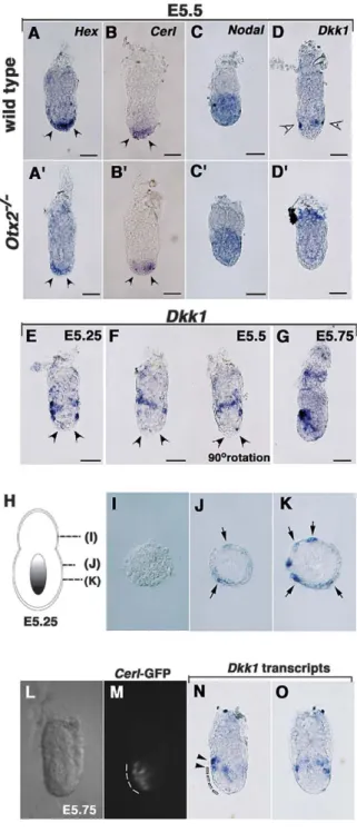
Expression of the Dkk1 Gene during DVE Migration We previously demonstrated that Otx2−/−embryos fail to form the A-P axis correctly (Kimura et al., 2000). Pre-cise expression analysis of molecular markers during DVE migration involving whole-mount in situ hybridiza-tion revealed apparent normal formahybridiza-tion of DVE in Otx2−/−embryos at E5.5, although migration to one side proximally was not possible (Figures 1A, 1B, 1A#, and 1B#). Expression of the DVE markers Hex, Cerl, and Lefty1 was detected in the distal portion of visceral en-doderm at E5.5 in Otx2−/−embryos, and this expression was similar to that of wild-type (Figures 1A, 1B, 1A#, and 1B# and data not shown). Moreover, Nodal expres-sion was normal in the epiblast of mutant embryos ( Fig-ures 1C and 1C#). At E6.5, expression of these markers remained in the distal portion of visceral endoderm and the proximal epiblast (Kimura et al., 2001, 2000; Perea-Gomez et al., 2001). These data clearly demonstrated that Otx2−/−embryos can form DVE normally around E5.5, whereas mutant DVE fails to migrate anteriorly even at E6.5.
In order to elucidate the role of Dkk1 during DVE mi-gration, Dkk1 expression was examined in detail ( Fig-ures 1E–1K). Around E5.5, prior to migration, Dkk1 ex-pression was initially detected in the proximal portion of DVE in a circular pattern (Figures 1D–1F and 1H–1K). Importantly, Dkk1 expression was negative in the most distal tip of the visceral endoderm, which is evidenced by the thickened morphology (Kimura et al., 2000); in con-trast, Hex and Cerl expression was positive throughout the entire DVE (Figures 1A and 1B). Dkk1 expression in Otx2−/−mutants at E5.5 was assessed to determine whether Otx2 is necessary for induction of Dkk1 ex-pression from the initial phase or simply for mainte-nance at subsequent stages. Consequently, Dkk1 ex-pression was not observed in mutant embryos, which
Figure 1. Marker Analysis of Wild-Type and Otx2 Mutant Embryos during A-P Axis Development
(A–D#) Whole-mount in situ hybridization in (A–D) wild-type and (A#– D#) Otx2−/−embryos at E5.5. (A and A#) Hex, (B and B#) Cerl, (C and C#) Nodal, and (D and D#) Dkk1.
(E–G) Dkk1 expression at (E) E5.25, (F) E5.5, and (G) E5.75. (H–K) Dkk1 expression at E5.25 in cross-sections at the level indi-cated in (H). Arrows indicate Dkk1 expression.
(L–O) Comparative expression analysis of Cerl and Dkk1 at E5.75. The GFP signal of the Cerl-GFP transgenic embryo was recorded, and, subsequently, Dkk1 mRNA expression was examined by in situ hybridization. (L) Bright and (M) dark field views of the Cerl-GFP embryo. (N) Lateral and (O) frontal views of Dkk1 expression of the
Cerl-GFP embryo. (M and N) Dotted lines indicate the Cerl-positive
domain. (N) Arrowheads indicate the Dkk1-positive domain. Scale bars indicate 50m.
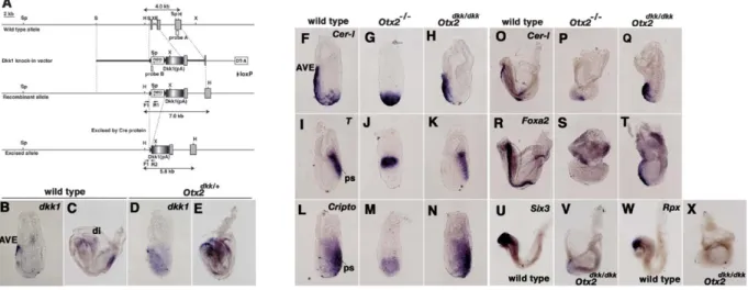
Figure 2. Dkk1 Expression Rescues Axis Rotation Defects Caused by Otx2 Mutation
(A) Generation of the Dkk1 knockin mutation in the Otx2 locus. Diagrammatic representations of the wild-type Otx2 allele, knockin vector, recombinant allele, and excised allele. Probes A and B are those employed for Southern blotting to identify the knockin mutation.
(B–E) Whole-mount in situ hybridization with the Dkk1 probe in (B and C) wild-type and (D and E) Otx2dkk/+embryos. (B and D) E6.5 and (C
and E) E7.8.
(F–X) Whole-mount in situ hybridization in (F, I, L, O, R, U, and W) wild-type, (G, J, M, P, and S) Otx2−/−, and (H, K, N, Q, T, V, and X)
Otx2dkk/dkkembryos. (F–H and O–Q) Cerl, (I–K) T, (L–N) Cripto, (R–T) Foxa2, (U and V) Six3, and (W and X) Rpx. (F–N) E6.5, (O–T) E7.8, and
(U–X) E8.0.
AVE, anterior visceral endoderm; di, diencephalic region; ps, primitive streak.
indicated that Otx2 is essential for induction of Dkk1 expression (Figure 1D#).
When DVE cells began to migrate to the anterior side by E5.75, Dkk1 expression was observed in the future anterior side of the visceral endoderm; however, Dkk1 expression was not evident in the posterior side (Figure 1G). Additional comparative expression analysis with Cerl and Dkk1 revealed that Dkk1-positive visceral en-doderm cells are apparently located in the foremost as-pect of Cerl-positive migrating cells (Figures 1L–1O). These data suggest the possibility that asymmetrical Dkk1 expression in the visceral endoderm might play a role in directional migration of DVE cells.
Dkk1 Expression Rescues Defective Axis Conversion Caused by Otx2 Deficiency
In order to determine whether Dkk1 participates in axis rotation as a downstream target of Otx2, mutant mice were generated in which the Dkk1 cDNA was inserted into the Otx2 locus (Otx2dkk/+) (Figure 2). In the targeting vector, the Otx2 gene was disrupted via insertion of Dkk1 cDNA and the PGKneo cassette flanked by two loxP sites (Figure 2A; Figure S1; see the Supplemen-tal Data available with this article online). Chimeras (Otx2dkk-neo/+) were mated with b-actin cre transgenic mice in order to remove the PGKneo cassette. Nor-mally, Dkk1 is expressed in AVE at the gastrulation stage and in the presumptive diencephalic region at the subsequent head fold stage (Figures 2B and 2C) (Glinka et al., 1998). However, in Otx2dkk/+embryos, in addition to endogenous Dkk1 expression domains, Dkk1 ex-pression was detected in the epiblast at E6.5 and in the entire rostral brain region at E7.8 (Figures 2D and 2E). Further RT-PCR analysis confirmed the absence of
Otx2 mRNA in Otx2dkk/dkk embryos (Figure S1). These findings clearly indicated that the knockin Dkk1 cDNA is expressed correctly in lieu of the Otx2 gene.
To determine whether axis rotation defects caused by the Otx2 mutation can be restored upon replace-ment of Dkk1 cDNA, several molecular markers were analyzed in Otx2dkk/dkk embryos (Figures 2F–2X). In Otx2−/−embryos, expression of the AVE markers Cerl, Hex, and Lefty1 occur in the distal tip even at E6.5 ( Fig-ures 1A#, 1B#, and 2G; data not shown). Coincidentally, T expression is present in the proximal side, and Cripto expression is apparent in the entire epiblast of Otx2−/− embryos (Figures 2J and 2M). Importantly, in Otx2dkk/dkk embryos, expression of AVE markers Cerl and Hex was detected in AVE in a manner similar to that of wild-type embryos (Figure 2H; data not shown). Concurrently, T and Cripto expression were restricted normally to the posterior side of Otx2dkk/dkk embryos (Figures 2K and 2N). Restoration of axis conversion was observed fre-quently in Otx2dkk/dkkembryos (n = 20/50). These find-ings demonstrate that axis rotation defects due to the Otx2 mutation can be partially rescued by Dkk1 expres-sion alone. In addition, Otx2 regulates Dkk1 expresexpres-sion in the AVE directly through Otx2 binding sites in the Dkk1 promoter (Figures S2 and S3). As a result, the aforementioned data suggest that Dkk1 can mediate axis conversion from the P-D to the A-P orientation as a crucial downstream target of Otx2.
The anterior neuroectoderm markers Rpx/Hesx1 and Six3 were examined to determine whether later fore-brain abnormalities of Otx2−/− mutant embryos could be rescued by Dkk1 cDNA (Figures 2U–2X). However, neither marker was induced in Otx2dkk/dkk embryos at E8.0 (Figures 2V and 2X; Rpx/Hesx1, n = 0/5; Six3, n =
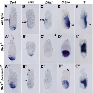
Figure 3. Rescue of A-P Axis Patterning Defects in Otx2−/−; b-catenin+/−Embryos
(A–E$) Whole-mount in situ hybridization analysis of (A, A#, and A$)
Cerl, (B, B#, and B$) Hex, (C, C#, and C$) Dkk1, (D, D#, and D$) Cripto, and (E, E#, and E$) T expression at E6.5; (A–E) wild-type,
(A#–E#) Otx2−/−, and (A$–E$) Otx2−/−;b-catenin+/−. AVE, anterior vis-ceral endoderm; ps, primitive streak.
0/17). In order to assess whether failure in forebrain in-duction of Otx2dkk/dkk embryos is a consequence of anterior mesendoderm defects, expression of the mo-lecular markers Cerl and Foxa2 were analyzed (Figures 2O–2T). As expected, expression of these markers was severely affected in Otx2dkk/dkk embryos (Figures 2Q and 2T). This finding indicates that Otx2dkk/dkkembryos are unable to form the anterior mesendoderm properly. These results in concert suggest that failure of AVE mi-gration may be primarily attributable to the absence of the Wnt antagonist, Dkk1, in Otx2−/−embryos; more-over, additional signaling molecules may be necessary for subsequent forebrain induction as previously pro-posed (Stern, 2001).
Rescue of Axis Rotation Defects in Otx2−/−Embryos Lacking One Copy of
Dkk1 inhibits canonical Wnt signaling at the extracellu-lar level via direct interaction with LRP5/6 and Kremen, which ultimately downregulatesβ-catenin activity in the nucleus (Mao et al., 2002). In order to examine genetic interaction between Otx2 and Wnt/β-catenin signaling, Otx2+/− mutants were mated with mice carrying the b-catenin null allele; subsequently, double-mutant phe-notypes were examined (Brault et al., 2001) (Figure 3). Otx2+/−;b-catenin+/− mutants were viable and fertile (data not shown); as a result, Otx2−/−;b-catenin+/−mice were obtained by crossing Otx2+/−;b-catenin+/−mutants with Otx2+/− mutants. Molecular marker analysis was performed at E6.5 to analyze axis development in Otx2−/−;b-catenin+/−embryos. Cerl, an AVE marker, ex-pression in the DVE of Otx2−/−embryos was restricted
to one side of the Otx2−/−;b-catenin+/−visceral endo-derm (Figures 3A, 3A#, and 3A$; n = 3/10). Similarly, proper expression of another AVE marker, Hex, was ob-served in the anterior side of the visceral endoderm in Otx2−/−;b-catenin+/− embryos (Figure 3B$; n = 2/6). These findings indicate that the anterior migration de-fect of DVE cells in Otx2 null mutants is partially res-cued after removal of one copy of the b-catenin gene.
To establish whether restoration of Otx2 null mutant defects is a consequence of ectopic induction of Dkk1 expression due to heterozygosity of b-catenin, Dkk1 ex-pression was examined in these mutant embryos. How-ever, Dkk1 expression was never induced in Otx2−/−; b-catenin+/−embryos (n = 0/6; Figure 3C$). Moreover, expression of posterior markers Cripto and T appeared to be restricted to one side of Otx2−/−;b-catenin+/− em-bryos (n = 2/7, 2/4;Figures 4D$ and 4E$, respectively). These results, in concert, demonstrate that defective axis rotation in Otx2−/−embryos could be partially res-cued after removal of one copy of the b-catenin gene. The aforementioned genetic evidence clearly supports Otx2 participation in specification of A-P polarity via attenuation of Wnt/β-catenin signaling.
Misexpression of Wnt8, a Canonical Wnt Ligand, Prevents Axis Conversion from the P-D
to the A-P Orientation
The aforementioned findings suggest the possibility that the attenuation of Wnt/β-catenin signaling can initi-ate A-P axis conversion in the mouse embryo. In order to determine whether attenuation of canonical Wnt sig-naling is essential for correct A-P axis formation during normal development, a transgenic mouse misexpress-ing the canonical Wnt ligand, mouse Wnt8A gene (mWnt8A), was generated; subsequently, phenotypes of these animals were analyzed (Figure S4andFigure 4). mWnt8A is a mouse cognate of Xenopus Wnt8 and chicken Wnt8c, both of which possess strong double axis-inducing activity in frog and mouse, respectively (Popperl et al., 1997; Logan and Nusse, 2004). The Cre/ loxP system was employed in order to examine precise phenotypes of transgenic embryos reliably during A-P axis development (Figure S4). The lacZ gene flanked by two loxP sites was inserted between the CAG promoter and mWnt8A cDNA. To remove the blockade of the lacZ stop codon, chimeras were mated with b-actin-cre transgenic mice, which ubiquitously express Cre pro-tein (Lewandoski et al., 1997). mWnt8A expression was evaluated in the resultant Tg(CAG-mWnt8A) embryos to identify mWnt8A misexpression. Normally, mWnt8A expression occurs in the proximal epiblast prior to E6.0; subsequently, by E6.5, it is restricted to the posterior side (Figure S4andFigure 4A). In contrast, hemizygous Tg(CAG-mWnt8A) embryos exhibited ectopic expres-sion of the mWnt8A gene throughout the epiblast ( Fig-ure 4A#).
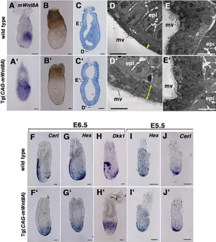
Morphological analysis of Tg(CAG-mWnt8A) embryos revealed failure to specify the embryonic region of the visceral endoderm appropriately at E6.5 (Figures 4B– 4E and 4B#–4E#). Wild-type embryos lost their symmet-rically cylindrical shape; additionally, distinct curva-tures were apparent on the opposite side of the primi-tive steak at this stage (Figure 4B). In contrast, the
Figure 4. Morphological and Molecular Marker Analyses of Tg(CAG-mWnt8A) Em-bryos
(A–J#) Whole-mount in situ hybridization in (A and F–J) wild-type and (A# and F#–J#)
Tg(CAG-mWnt8A) embryos; (A and A#) mWnt8A, (F,
F#, J, and J#) Cerl, (G, G#, I, and I#) Hex, and (H and H#) Dkk1 at (I, I#, J, and J#) E5.5 and (A, A#, F–H, F#–H#) E6.5. Morphological an-alysis of (B–E) wild-type and (B#–E#) trans-genic embryos at E6.5. Fine structure of em-bryos by (C and C#) semithin sections and corresponding (D, D#, D, and E#) electron mi-crographs. Scale bars are 50m in (A)–(C), (A#)–(C#), (F)–(J), and (F#)–(J#) and are 10m in (D), (D#), (E), and (E#). epi, epiblast; mv, microvilli.
external morphology of Tg(CAG-mWnt8A) embryos was nearly symmetrical, and curvature was not evident at E6.5 (Figure 4B#). The size of Tg(CAG-mWnt8A) em-bryos was nearly identical to that of wild-type emem-bryos; consequently, morphological abnormalities are not at-tributable to developmental retardation.
Subsequently, fine structure was examined via utility of semithin and ultrathin sections to assess the visceral endoderm structure more precisely. These experiments revealed that embryonic visceral endoderm is improp-erly regionalized in these embryos (Figures 4C–4E and 4C#–4E#). In wild-type embryos, visceral endoderm cells of the extraembryonic region displayed a tall, co-lumnar appearance and a high degree of vacuolization in association with dense microvilli, whereas the em-bryonic region consisted of squamous cells with lower numbers of microvilli (Figures 4D and 4E) (Batten and Haar, 1979). DVE cells of Tg(CAG-mWnt8A) embryos possessed a tall, columnar shape, but not a squamous shape, and many microvilli, characteristics identical to those of visceral endoderm cells at the level of the ex-traembryonic region (Figures 4D# and 4E#). However, morphological features of epiblast cells were un-changed in the Tg(CAG-mWnt8A) embryos (Figures 4D and 4D#). These findings suggest that misexpression of mWnt8A leads to developmental failure of visceral endoderm, but not of epiblasts.
To further assess A-P axis phenotypes in transgenic embryos, several marker genes were examined at the gastrulation stage (Figures 4F–4J and 4F#–4J#).
Expres-sion of AVE markers Cerl, Hex, and Lhx1 was observed in the DVE of Tg(CAG-mWnt8A) embryos; expression was not shifted to one side at E6.5 (Figures 4F# and 4G#; data not shown). Circular, symmetrical Dkk1 ex-pression remained in the proximal portion of DVE; how-ever, it was not restricted to the anterior side (Figure 4H#). Notably, expression of Cerl and Hex occurred nor-mally in DVE of the transgenic embryos at E5.5, sug-gesting that DVE is formed appropriately in Tg(CAG-mWnt8A) embryos (Figures 4I# and 4J#). Concurrent with these findings, in terms of posterior markers, prox-imal T expression remained, and Cripto and Nodal ex-pressions were apparent throughout the entire epiblast of Tg(CAG-mWnt8A) embryos at E6.5 (Figure S5). The aforementioned data demonstrate that misexpression of mWnt8A leads to failure of axis conversion with re-spect to the P-D to the A-P orientation.
Asymmetrical Distribution of-Catenin Localization in the Visceral Endoderm Is Impaired in Otx2-Deficient and Tg(CAG-mWnt8A) Embryos
These lines of evidence suggest the possibility that, during normal A-P axis development,β-catenin activity is attenuated in AVE. In order to test this point directly, expression and cellular localization of the active form of β-catenin, which is dephosphorylated at residues Ser37 and Thr41 (Staal et al., 2002), was analyzed via confocal immunofluorescence. Findings indicate that nuclear and cytoplasmicβ-catenin is specifically diminished in AVE of wild-type embryos (Figures 5A and 5B).
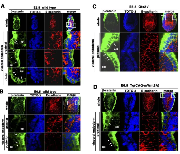
Figure 5. Expression and Cellular Localization of the Active Form of theβ-Catenin Protein during A-P Axis Specification
(A–D) Fluorescent images of (A and B) wild-type, (C) Otx2−/−, and (D) Tg(CAG-mWnt8A) embryos at (A) E5.5 and (B–D) E6.5. (A–D) Anti-active-β-catenin (green), TOTO-3 (nuclei, blue), E-cadherin (adherens junctions, red), and the merged image. The areas shown in the magnified views are marked in the upper merged images (white squares). (A) Active-β-catenin protein is strongly detected in the cytoplasm of the entire visceral endoderm as well as in the nucleus, where the TOTO-3 image is merged (white arrows). (B) Nuclear and cytoplasmicβ-catenin expression is upregulated in the posterior visceral endoderm (white arrows). (C) Localization of nuclearβ-catenin is evident in both proximal aspects of the visceral endoderm (white arrows). (D) Nuclearβ-catenin localization is observed in the proximal visceral endoderm (white arrows). epi, epiblast.
At E5.5,β-catenin is substantially localized in the cy-toplasm of the entire visceral endoderm at the level of both embryonic and extraembryonic regions (Figure 5A). Additionally,β-catenin was frequently localized in the nucleus of the proximal and distal aspects of the em-bryonic visceral endoderm region. However, in the epi-blast,β-catenin is uniformly present at the cell surface, although it is never observed in the cytoplasm and nu-cleus. These findings afford evidence that β-catenin is uniformly distributed in the embryonic visceral endoderm region at E5.5; moreover, Wnt/β-catenin signaling may be activated more strongly in the visceral endoderm than in the epiblast.
At E6.5, cytoplasmic and nuclearβ-catenin were dis-tributed in the visceral endoderm layer, but not in the epiblast, a situation similar to that at E5.5 (Figure 5B). However, cytoplasmic and nuclearβ-catenin was
mark-edly diminished in AVE; in contrast, it was elevated in posterior visceral endoderm (Figure 5B). This asymmet-rical distribution of β-catenin in conjunction with the A-P axis could be observed initially around E5.75, when DVE cells migrate to the anterior side (data not shown). These data demonstrate that nuclear and cytoplasmic β-catenin are asymmetrically distributed in the visceral endoderm layer along with the A-P axis in the mouse embryo.
In order to address whether asymmetrical distribu-tion ofβ-catenin is impaired in Otx2−/−mutant embryos, expression and cellular localization ofβ-catenin were analyzed at E6.5 (Figure 5C). Cytoplasmic and nuclear β-catenin, which are elevated throughout the entire em-bryonic visceral endoderm region, did not exhibit obvi-ous asymmetrical distribution in Otx2−/−embryos. How-ever, in mutant epiblasts, β-catenin was uniformly
localized exclusively at the cell surface in a manner similar to that in wild-type embryos. These findings pro-vide direct epro-vidence that asymmetrical distribution of cytoplasmic and nuclearβ-catenin cannot occur in the Otx2−/−visceral endoderm layer; moreover, these data indicate that Otx2 is required to reduce cytoplasmic and nuclearβ-catenin expression in AVE.
To ascertain whether the reduction of cytoplasmic and nuclearβ-catenin expression in AVE is necessary for correct A-P axis development,β-catenin expression was examined in Tg(CAG-mWnt8A) embryos, which ex-hibit migratory defects of DVE cells as described ( Fig-ure 4). Consequently, reduction of cytoplasmic and nuclearβ-catenin expression in visceral endoderm did not occur in the transgenic embryos (Figure 5D). In Tg(CAG-mWnt8A) embryos,β-catenin expression was strongly upregulated in the cytoplasm as well as in the nucleus of the visceral endoderm layer. Unexpectedly in the epiblast,β-catenin expression was unchanged; it was detected exclusively at the cell surface in a manner identical to that of the wild-type. Therefore, these find-ings suggest that reduction ofβ-catenin expression in AVE may be crucial for correct A-P axis rotation; more-over, failure of AVE migration observed in Tg(CAG-mWnt8A) embryos may be due primarily to the visceral endoderm, but not to the epiblast.
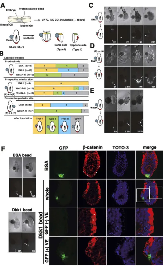
Dkk1 and Wnt Can Function as Attractive and Repulsive Guidance Cues, Respectively, Controlling Directional Migration of Visceral Endoderm Cells The aforementioned findings lend support to the regu-lation of visceral endoderm cell migration by Wnt/ β-catenin signaling. One possible mechanism by which Wnt/β-catenin signaling controls cell migration involves attractive mediation by Dkk1 and/or repulsive media-tion by Wnt, which funcmedia-tion as guidance cues. In order to address this issue, migratory behavior of DVE cells was analyzed with recombinant protein-soaked beads in whole-embryo culture. Results reveal that Dkk1 attracts and that Wnt3A repels migratory DVE cells ( Fig-ure 6).
To identify migratory DVE cells, Cerl-GFP transgenic embryos were employed; this strain permits labeling of migrating visceral endoderm cells from E5.5 to E6.5 via the fluorescent microscopy (Belo et al., 1997; Mesnard et al., 2004). E5.5–E5.75 transgenic embryos were em-bedded in a gel-matrix medium for three-dimensional culture (see Experimental Procedures); beads coated with a recombinant protein were located at the proximal side of the embryos (Figure 6A). During the whole-embryo culture for 5–48 hr, GFP-positive cell behavior was analyzed under the fluorescent microscope; sub-sequently, behavior was classified into four types ( Fig-ures 6A and 6B, Types I–IV). In the case in which a BSA-soaked bead was embedded in the proximal side of the embryo prior to migration of GFP-positive cells, DVE cells migrated randomly irrespective of the position of BSA beads (Figure 6B) (the same side of the bead, Type I, n = 4/10; the opposite side of the bead, Type II, n = 4/10). Notably, when a Dkk1-soaked bead was embed-ded in the proximal side of the embryo prior to cell mi-gration, DVE cells consistently migrated to the same side of the Dkk1 bead (Figures 6B and 6C) (Type I + III, n = 13/16); these cells migrated to the same side
exclusively (Type I, n = 7/16), or they migrated in two distinct directions, the same and opposite sides (Type III, n = 6/16). This finding suggests that Type III migra-tion occurs as a result of the attracmigra-tion of DVE cells by two Dkk1 sources: endogenous Dkk1 expression at the opposite side and embedded Dkk1.
Next, a Dkk1-soaked bead was embedded in the prospective posterior side of the embryo after initiation of anterior migration of GFP-positive cells (Figure 6B). As a result, migrating visceral endoderm cells altered direction and migrated to the same side of the Dkk1 bead (Type I + III, n = 9/10); the entire population of these cells migrated to the same side, the prospective posterior side (Type I, n = 3/10), or they migrated in two distinct directions, the prospective anterior and poste-rior sides (Figures 6B and 6D; Type III, n = 6/10). In contrast, when a Dkk1 bead was embedded in the prospective anterior side after initiation of anterior mi-gration of cells (Figure 6B), the Dkk1 bead did not alter the direction of approaching DVE cells (Type I, n = 9/9). These observations clearly demonstrate that Dkk1 pro-tein can attract DVE cells prior to and during movement. On the other hand, in the case in which a bead soaked with Wnt3A, a canonical Wnt ligand, was em-bedded in the proximal side prior to cell migration, GFP-positive cells did not migrate to the same side of the bead (Type I, n = 0/10); rather, these cells migrated to the opposite side (Type II, n = 5/10), or they failed to migrate (Type IV, n = 5/10;Figures 6B and 6E). When the 40 ng/l Wnt3A-soaked bead was embedded in the prospective anterior side, the direction of migrating vis-ceral endoderm cells was unaffected (Figure 6B) (Type I, n = 8/11). However, a bead soaked at a 10-fold lower concentration (4 ng/l) efficiently repelled the migrating DVE cells (Type II + III + IV, n = 12/19). Additionally, when the Wnt3A bead was embedded in the prospective pos-terior side after initiation of anpos-terior migration of cells (Figure 6B), the Wnt3A bead did not alter the direction of receding DVE cells (Type III, n = 6/7). These findings suggest that Wnt3A can repel DVE cells prior to and during movement.
In order to determine whether embedded protein-soaked beads mediate cell migration via alteration of activity of Wnt/β-catenin signaling, β-catenin expres-sion in explants containing the beads was examined by confocal microscopy (Figure 6F). After a 5 hr incubation, the BSA bead failed to alter symmetricalβ-catenin ex-pression in the visceral endoderm layer (Figure 6F). However, after a 5 hr incubation with the Dkk bead, the visceral endoderm layer displayed asymmetricalβ-catenin expression along the prospective A-P axis; the Dkk1 side exhibited dramatic reduction ofβ-catenin expres-sion, whereas the opposite side demonstrated upregu-lation ofβ-catenin (Figure 6F). These findings strongly suggest that asymmetrical Wnt/β-catenin signaling in the prospective A-P axis of the pregastrula embryo pro-vides the morphogenetic force that drives the direc-tional displacement of the DVE cells.
Discussion
The present investigation demonstrates that Otx2 specifies A-P polarity via attenuation of Wnt/β-catenin signaling in the prospective anterior side of the mouse
Figure 6. Dkk1 and Wnt3A Act as Guidance Cues Controlling the Migratory Direction of DVE Cells
(A and B) Experimental strategy and summary of bead explants. Cerl-GFP transgenic embryos were embedded in the medium gel with protein-soaked beads. After culture, the positions of GFP-positive DVE cells were classified into four types as illustrated (Types I–IV): Type I (orange), same side as the explanted bead; Type II (green), opposite side of the explanted bead; Type III (light blue), both the same and the opposite sides of the explanted bead; Type IV (gray), distal side of the embryo. Two different concentrations of Wnt3A beads were used: 40 ng/l (Wnt3A–H) and 4 ng/l (Wnt3A–L).
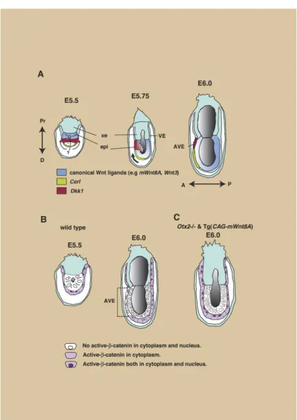
Figure 7. Schematic Model of the Mecha-nism Governing A-P Axis Development via Wnt/β-Catenin Signaling in the Mouse Embryo (A) Schematic representation of wild-type mouse embryos between E5.5 and E6.0. At E5.5, DVE cells are thought to possess the ability to migrate in the proximal direction marked by the Dkk1-positive domain (dotted arrows). At E5.75, Wnt/β-catenin signaling is attenuated by anterior restriction of Dkk1 ex-pression. Direction of DVE cell migration is indicated by an arrow. By E6.0, Dkk1 expres-sion is detected in the most anterior portion of AVE; furthermore, axis conversion is com-pleted in the A-P direction.
(B and C) Schematic diagrams regarding ex-pression of active-β-catenin in the wild-type at E5.5 and E6.0 and in the Otx2−/− and
Tg(CAG-mWnt8A) embryos at E6.0. (B) At
E5.5, cytoplasmic and nuclearβ-catenin is localized in the visceral endoderm layer, but not in the epiblast layer; at subsequent E6.0, cytoplasmic and nuclearβ-catenin expres-sion is reduced anteriorly and upregulated posteriorly in the wild-type. (C) In the mi-gration-defective embryos, cytoplasmic and nuclear β-catenin expression is symmetri-cally distributed in the entire visceral endo-derm even at E6.0. A, anterior; AVE, anterior visceral endoderm; D, distal; epi, epiblast; P, posterior; Pr, proximal; VE, visceral endo-derm; xe, extraembryonic ectoderm.
embryo. The Wnt antagonist Dkk1, which is a crucial downstream target of Otx2, acts as an attractive guid-ance cue controlling the directional migration of vis-ceral endoderm cells. Moreover, to our knowledge, this genetic evidence provides novel insights into evolution-arily conserved mechanisms governing primary body axis formation in metazoans.
Wnt/-Catenin Signaling Regulates A-P Axis Development
Our findings led to the proposal of a model describing the role of Wnt/β-catenin signaling in A-P axis develop-ment of the mouse embryo (Figure 7). Normally, prior to
(D) Example of the Dkk1 protein-soaked bead embedded in the prospective posterior side of the embryo (Type III).
(E) Example of the Wnt3A-H protein-soaked bead embedded in the proximal side of the embryo (Type II). GFP-positive cells are indicated by white arrows in (C)–(E).
(F) The BSA or Dkk1 protein-soaked beads embedded in the proximal side of the embryo forβ-catenin expression analysis. After a 5 hr incubation, Cerl-positive cells do not migrate to one side in the BSA-embedded explant, while these cells start to migrate to one side in the Dkk1-embedded explant. Cerl-GFP (green), anti-active-β-catenin (red), TOTO-3 (nuclei, blue), and the merged image. The areas shown in the magnified views are marked in the upper merged images (white squares).
AVE migration, around E5.5, a canonical Wnt antago-nist, Dkk1, is expressed in the proximal portion of DVE cells in a circular, symmetrical pattern (Figures 7A). On the other hand, canonical Wnt ligands, e.g., mWnt8A and Wnt3, are expressed in the proximal epiblast ( Fig-ure 7A). At this stage, any proximal direction may be competent with respect to migration of DVE cells, as suggested by symmetrical Dkk1 expression. However, β-catenin expression does not appear to be downregu-lated in these Dkk1-positive domains. This delayed at-tenuation ofβ-catenin expression is probably attribut-able to the time lag associated with protein degradation ofβ-catenin or to insufficient levels of Dkk1 expression.
By E5.75, attenuation of Wnt/β-catenin signaling in the AVE is closely linked with the axis conversion process from the P-D to the A-P orientation. Axis rotation may occur primarily as a result of asymmetrical expression of Wnt antagonists, including Dkk1. Concurrently, Dkk1 expression is downregulated in the prospective poste-rior side and upregulated in the anteposte-rior side, whereas β-catenin expression displays the opposite profile ( Fig-ures 7A and 7B).
Wnt signaling has been classified into two pathways: the canonical and noncanonical pathways, or b-catenin-dependent and -inb-catenin-dependent pathways, respectively (Logan and Nusse, 2004). The current study demon-strates that the canonical Wnt antagonist, Dkk1, and its nuclear target,β-catenin, participate in DVE migration. Additionally, misexpression of a canonical Wnt ligand, mWnt8A, prevented correct A-P axis conversion (Figure 4). Coincidentally, asymmetrical distribution ofβ-catenin expression was not observed in the AVE of Otx2-defi-cient and Tg(CAG-mWnt8) embryos (Figures 5 and 7C). These results, in concert, strongly support the crucial role of asymmetricalβ-catenin localization in A-P axis conversion. However, Dkk1 homozygous mutant em-bryos appeared to undergo normal A-P axis develop-ment (Mukhopadhyay et al., 2001). To account for this disparity, other inhibitory molecules of Wnt/β-catenin signaling, which complement the Dkk1 function in vis-ceral endoderm at different levels of the signaling path-way, may be present, albeit at reduced levels in Otx2-deficient embryos, e.g., Cerl, Axin, TCF3, Wise, and ICAT (Logan and Nusse, 2004).
Wnt/-Catenin Signaling Guides Directional Migration of Visceral Endoderm Cells
This investigation provides for the evidence that the ca-nonical Wnt ligand and its antagonist function as guid-ance cues in the DVE cell migration. Migrating visceral endoderm cells possess unique morphology distinct from that of adjacent endoderm cells, as well as active migratory character; these cells continuously change shape and project filopodial processes (Kimura et al., 2000; Srinivas et al., 2004). Comparative expression analysis of Dkk1 with Cerl suggests that Dkk1-positive cells are located at the forefront of migratory DVE cells (Figure 1). These findings suggest that Dkk1 may deter-mine the migratory direction of DVE cells. Moreover, ap-parent normal expression of Nodal and its antagonists in Otx2-deficient embryos (Figure 1) might afford evi-dence in support of the previously described observa-tion that Otx2−/−DVE cells display no significant change with respect to cell proliferation; rather, they exhibit a defect associated with directional migration ( Perea-Gomez et al., 2001). Further BrdU incorporation experi-ments with Wnt8A-misexpressing embryos and Dkk1/ Wnt3A-embedded explants revealed that Wnt ligands and Dkk1 failed to mediate asymmetrical cell prolifera-tion along with the A-P axis (C.K.-Y., D.O., Y.M., and I.M., unpublished data). In a manner consistent with these notions, Dkk1 can attract Cerl-positive DVE cells and a canonical Wnt ligand, Wnt3A, can repel them as a migratory guidance cue (Figure 6). Furthermore, at-tenuation ofβ-catenin expression in the cytoplasm and the nucleus during DVE cell migration appears to be
linked to asymmetrical expression of Dkk1 and the em-bedded Dkk1 bead (Figures 5 and 6). In conjunction, these findings directly support mediation of the direc-tional migration of DVE cells by the combination of Wnt-mediated repulsion and its antagonist-mediated attraction.
Otx2 May Regulate Asymmetrical Distribution of b-Catenin Activity in Conjunction
with the Primary Body Axis
This study indicates that localization of the dephos-phorylated form ofβ-catenin is dynamically regulated during A-P axis specification (Figures 5, 7B, and 7C). In the wild-type visceral endoderm layer, cytoplasmic and nuclear β-catenin expression are specifically reduced in the prospective anterior side. Notably, in both Otx2-deficient and Tg(CAG-mWnt8A) embryos, which dis-play failure of axis rotation, the expression is not down-regulated; rather, it is upregulated throughout the entire visceral endoderm layer (Figures 5 and 7). Although fur-ther molecular analysis is necessary in order to eluci-date the precise molecular mechanism by which Dkk1 expression is initially induced in the most proximal por-tion of DVE and subsequently downregulated in the prospective posterior side, Otx2 expression is crucial for Dkk1 expression in the visceral endoderm (Figures S2 and S3). In addition, Dkk1 alone can rescue axis ro-tation failure attributable to Otx2 deficiency (Figure 2). These findings suggest that Otx2 specifies A-P axis development primarily via regulation of Wnt/β-catenin signaling pathways, including Dkk1, in the visceral en-doderm.
Surprisingly, mWnt8A transcripts driven by the CAG promoter are upregulated primarily in the epiblast, but not in the visceral endoderm (Figure 4A#), whereas ex-pression of the dephosphorylated form ofβ-catenin is not elevated in the epiblast layer of Tg(CAG-mWnt8A) embryos. This finding suggests the involvement of un-expected molecular mechanisms via which Wnt signal-ing can be transmitted toβ-catenin activity mainly in the visceral endoderm, but not in the epiblast layer.
To our knowledge, this genetic evidence affords novel insights into evolutionarily conserved mechanisms gov-erning primary body axis formation across the metazo-ans. The asymmetrical distribution ofβ-catenin activity along with the A-P axis plays a pivotal role in the speci-fication of A-P polarity throughout metazoan embryos. In amphibians, fish, ascidians, sea urchins, and cnid-arians,β-catenin is localized to cell nuclei preferentially at one pole of the cleavage-stage embryo (Imai et al., 2000; Logan et al., 1999; Schneider et al., 1996; Wikra-manayake et al., 2003). In these various organisms, nuclear activity of b-catenin is required for early axis specification and the subsequent establishment of crit-ical signaling centers, “organizers”, in the early embryo. The present investigation suggests that asymmetrical distribution ofβ-catenin expression serves as a primary mediator of axis specification in the mammalian embryo. Experimental Procedures
Construction of Targeting Vector for Dkk1 Knockin Mice In the targeting construct, Dkk1 cDNA with polyadenylation signals and the PGKneo cassette were inserted into the SmaI and the
EcoRI sites, located 220 bp upstream from the translation initiation site and in the first intron, respectively. Homologous recombinant TT2 ES cells and chimeric mice were obtained as described ( Mat-suo et al., 1995).
Generation of CAG-mWnt8A Transgenic Mice
Mouse Wnt8A cDNAs were isolated from a mouse cDNA library. In brief, mWnt8A cDNA fused with a CAG promoter (Niwa et al., 1991) was ligated to the lacZ gene flanked by two loxP sites. Subse-quently, a CAG-lacZ-mWnt8A transgene was constructed via liga-tion of the aforemenliga-tioned resultant vectors with the neo gene driven by the PGK1 promoter with a polyadenylation signal. TT2 ES cells were cultured, electroporated with linearized transgenic vectors, and selected in G418 (Figure S4).
Genotyping
Transgenic and knockin founders and their progenitors were rou-tinely determined by PCR. Primers and lengths of the products in the PCR analyses were as follows: in the transgenic mice,
CAG-lacZ-mWnt8A was identified with primers CAG-pro (5#-TAGAGC
CTCTGCTAACCATGTTCATGCCTT-3#) and CAG-lacZ (5#-AGTGTC CCAGCCTGTTTATCTACGGCTTAA-3#), yielding 270 bp. The
mWnt8 allele excised by Cre was determined with primers CAG-pro and CAG-mWnt8A (5#-GATGGCAGCAGAGCGGATGGCATGAAT
GAA-3#), yielding 500 bp. Dkk1 knockin mice were identified with the following primers: wild-type Otx2 allele with primers Forward1 (5#-GTATTTTCCTTGCTACCAAACTGCCGAGTG-3#) and Reverse1 (5#-CTGGAGGGAAGCCACACCTCTAAGGATTAA-3#), yielding 400 bp, and Dkk1 knockin allele with primers Reverse1 and SVDK-2 (5#-ACAGCAGAAACATACAAGCTGTCAGCTTTG-3#), yielding 200 bp. b-catenin mutant mice were obtained from the Jackson Laboratory and genotyped as described (Brault et al., 2001).
Expression Analysis and Semi- and Ultrathin Sections
In situ hybridization was performed as described (Wilkinson, 1998). For morphological analysis, embryos were fixed in 2% paraformal-dehyde plus 2.5% glutaralparaformal-dehyde in 0.1 M sodium cacodylate buffer (pH 7.3); subsequently, tissues were postfixed for 2 hr in 1% OsO4 and were embedded in Poly Bed 812. Semithin sections (0.55 m thickness) were produced with a glass knife and stained with 0.5% toluidine blue. Ultrathin sections (70 nm thickness) were ob-tained with a diamond knife and examined after staining with uranyl acetate and lead citrate.
Whole-Embryo Culture with Recombinant Protein Beads Female ICR mice were sacrificed between 10:00 and 17:00 hr on the fifth day of pregnancy. Embryos were dissected from decidual tissue in M2 medium lacking Phenol red. The GFP-positive em-bryos, which were collected under a fluorescent microscope (Leica), were cultured in the Mebiol Gel matrix (Mebiol, Inc.) con-taining 50% DMEM and 50% rat serum in 5% CO2at 37°C for 5–48 hr for three-dimensional culture. Images of migratory behavior of visceral endoderm cells were captured and recorded with a Ha-mamatsu chilled CCD camera (C5985). Beads (Affigel Bluo Gel, BioRad) were rinsed in PBS several times and soaked in 200 ng/l mouse Dkk1 recombinant protein (R&D system), 40 and 4 ng/l mouse Wnt3A recombinant protein (R&D System), or 50 mg/ml BSA-PBS solution overnight at 4°C or for 1 hr at 37°C. Prepared beads were transplanted to the proximal side of the embedded transgenic embryo.
Immunohistochemistry
Wild-type, Otx2−/−, Tg(CAG-mWnt8A), and Cerl-GFP embryos were
fixed as described (Ciruna and Rossant, 2001). Primary antibodies were applied at the following concentrations: 10 mg/ml for rat anti-E-cadherin (ECCD-2) (Takara Shuzo) and at a 1:300 dilution for anti-active-β-catenin (anti-ABC) (8E7) (Upstate). Appropriate spe-cies-specific, fluorophore-labeled secondary antibodies (Molecular Probes) were applied at 1:200 dilution. Nuclei were stained with TOTO-3 iodide (Molecular Probes) at a 1:500 dilution in the pres-ence of 100g/ml RNase A. Staining was examined with a confocal microscope (Leica).
Supplemental Data
Supplemental Data including five figures are available athttp:// www.developmentalcell.com/cgi/content/full/9/5/639/DC1/. Acknowledgments
We are grateful to Drs. E. De Robertis, R. Grosschedl, B.G. Herr-mann, B. Hogan, G. Martin, R. Nusse, M.M. Shen, and K. Yamamura for in situ probes. We also wish to thank Dr. C. Niehrs for Dkk1 cDNA plasmid; the Animal Resources and Genetic Engineering Team, RIKEN Center for Developmental Biology for housing the mice; and Ms. Naoko Inoue, Mr. Hiroshi Kiyonari, Ms. Rika Naka-yama, Ms. Ayako Nagao, and Ms. Kuniko Kitajima for their assis-tance. This work was supported in part by grants-in-aid for Scien-tific Research on Priority Areas and Young Scientists (B) from the Ministry of Education, Culture, Sports Science and Technology, Japan. Received: September 24, 2004 Revised: April 28, 2005 Accepted: September 22, 2005 Published: October 31, 2005 References
Batten, B.E., and Haar, J.L. (1979). Fine structural differentiation of germ layers in the mouse at the time of mesoderm formation. Anat. Rec. 194, 125–141.
Beddington, R.S., and Robertson, E.J. (1999). Axis development and early asymmetry in mammals. Cell 96, 195–209.
Belo, J.A., Bouwmeester, T., Leyns, L., Kertesz, N., Gallo, M., Follet-tie, M., and De Robertis, E.M. (1997). Cerberus-like is a secreted factor with neutralizing activity expressed in the anterior primitive endoderm of the mouse gastrula. Mech. Dev. 68, 45–57.
Brault, V., Moore, R., Kutsch, S., Ishibashi, M., Rowitch, D.H., McMahon, A.P., Sommer, L., Boussadia, O., and Kemler, R. (2001). Inactivation of theβ-catenin gene by Wnt1-Cre-mediated deletion results in dramatic brain malformation and failure of craniofacial development. Development 128, 1253–1264.
Brennan, J., Lu, C.C., Norris, D.P., Rodriguez, T.A., Beddington, R.S., and Robertson, E.J. (2001). Nodal signalling in the epiblast patterns the early mouse embryo. Nature 411, 965–969.
Ciruna, B., and Rossant, J. (2001). FGF signaling regulates meso-derm cell fate specification and morphogenetic movement at the primitive streak. Dev. Cell 1, 37–49.
Glinka, A., Wu, W., Delius, H., Monaghan, A.P., Blumenstock, C., and Niehrs, C. (1998). Dickkopf-1 is a member of a new family of secreted proteins and functions in head induction. Nature 391, 357–362.
Imai, K., Takada, N., Satoh, N., and Satou, Y. (2000). (β)-catenin mediates the specification of endoderm cells in ascidian embryos. Development 127, 3009–3020.
Kimura, C., Yoshinaga, K., Tian, E., Suzuki, M., Aizawa, S., and Mat-suo, I. (2000). Visceral endoderm mediates forebrain development by suppressing posteriorizing signals. Dev. Biol. 225, 304–321. Kimura, C., Shen, M.M., Takeda, N., Aizawa, S., and Matsuo, I. (2001). Complementary functions of Otx2 and Cripto in initial pat-terning of mouse epiblast. Dev. Biol. 235, 12–32.
Lewandoski, M., Meyers, E.N., and Martin, G.R. (1997). Analysis of Fgf8 gene function in vertebrate development. Cold Spring Harb. Symp. Quant. Biol. 62, 159–168.
Logan, C.Y., and Nusse, R. (2004). The Wnt signaling pathway in development and disease. Annu. Rev. Cell Dev. Biol. 20, 781–810. Logan, C.Y., Miller, J.R., Ferkowicz, M.J., and McClay, D.R. (1999). Nuclearβ-catenin is required to specify vegetal cell fates in the sea urchin embryo. Development 126, 345–357.
Mao, B., Wu, W., Davidson, G., Marhold, J., Li, M., Mechler, B.M., Delius, H., Hoppe, D., Stannek, P., Walter, C., et al. (2002). Kremen proteins are Dickkopf receptors that regulate Wnt/β-catenin signal-ling. Nature 417, 664–667.
Matsuo, I., Kuratani, S., Kimura, C., Takeda, N., and Aizawa, S. (1995). Mouse Otx2 functions in the formation and patterning of rostral head. Genes Dev. 9, 2646–2658.
Mesnard, D., Filipe, M., Belo, J.A., and Zernicka-Goetz, M. (2004). The anterior-posterior axis emerges respecting the morphology of the mouse embryo that changes and aligns with the uterus before gastrulation. Curr. Biol. 14, 184–196.
Mukhopadhyay, M., Shtrom, S., Rodriguez-Esteban, C., Chen, L., Tsukui, T., Gomer, L., Dorward, D.W., Glinka, A., Grinberg, A., Hu-ang, S.P., et al. (2001). Dickkopf1 is required for embryonic head induction and limb morphogenesis in the mouse. Dev. Cell 1, 423– 434.
Niwa, H., Yamamura, K., and Miyazaki, J. (1991). Efficient selection for high-expression transfectants with a novel eukaryotic vector. Gene 108, 193–199.
Perea-Gomez, A., Lawson, K.A., Rhinn, M., Zakin, L., Brulet, P., Ma-zan, S., and Ang, S.L. (2001). Otx2 is required for visceral endoderm movement and for the restriction of posterior signals in the epiblast of the mouse embryo. Development 128, 753–765.
Popperl, H., Schmidt, C., Wilson, V., Hume, C.R., Dodd, J., Krum-lauf, R., and Beddington, R.S. (1997). Misexpression of Cwnt8C in the mouse induces an ectopic embryonic axis and causes a trunca-tion of the anterior neuroectoderm. Development 124, 2997–3005. Schneider, S., Steinbeisser, H., Warga, R.M., and Hausen, P. (1996). β-catenin translocation into nuclei demarcates the dorsalizing cen-ters in frog and fish embryos. Mech. Dev. 57, 191–198.
Srinivas, S., Rodriguez, T., Clements, M., Smith, J.C., and Bedding-ton, R.S. (2004). Active cell migration drives the unilateral move-ments of the anterior visceral endoderm. Development 131, 1157– 1164.
Staal, F.J.T., van Noort, M., Strous, G.S., and Clevers, H.C. (2002). Wnt signals are transmitted through N-terminally dephosphorylated β-catenin. EMBO Rep. 3, 63–68.
Stern, C.D. (2001). Initial patterning of the central nervous system: how many organizers? Nat. Rev. Neurosci. 2, 92–98.
Thomas, P.Q., Brown, A., and Beddington, R.S. (1998). Hex: a ho-meobox gene revealing peri-implantation asymmetry in the mouse embryo and an early transient marker of endothelial cell precursors. Development 125, 85–94.
Wikramanayake, A.H., Hong, M., Lee, P.N., Pang, K., Byrum, C.A., Bince, J.M., Xu, R., and Martindale, M.Q. (2003). An ancient role for nuclear beta-catenin in the evolution of axial polarity and germ layer segregation. Nature 426, 446–450.
Wilkinson, D.G. (1998). In Situ Hybridization: A Practical Approach, Second Edition (Oxford/New York: Oxford University Press). Yamamoto, M., Saijoh, Y., Perea-Gomez, A., Shawlot, W., Behringer, R.R., Ang, S.L., Hamada, H., and Meno, C. (2004). Nodal antago-nists regulate formation of the anteroposterior axis of the mouse embryo. Nature 428, 387–392.
Zakin, L., Reversade, B., Virlon, B., Rusniok, C., Glaser, P., Elalouf, J.M., and Brulet, P. (2000). Gene expression profiles in normal and Otx2−/−early gastrulating mouse embryos. Proc. Natl. Acad. Sci. USA 97, 14388–14393.