Universidade de Lisboa
Faculdade de Ciências
Departamento de Biologia Vegetal
Aeromonas spp.:
evaluation of genomic diversity and biofilm
forming ability
Sara Sofia Pereira Craveiro
Dissertação
Mestrado em Microbiologia Aplicada
2013
Universidade de Lisboa
Faculdade de Ciências
Departamento de Biologia Vegetal
Aeromonas spp.:
evaluation of genomic diversity and
biofilm forming ability
Sara Sofia Pereira Craveiro
Dissertação
Mestrado em Microbiologia Aplicada
Dissertação orientada pela Doutora Teresa Semedo-Lemsaddek
e pela Professora Doutora Ana Maria Gonçalves Reis
Aeromonas spp.:
evaluation of genomic diversity and
biofilm forming ability
Sara Sofia Pereira Craveiro
This thesis was performed at the Department of Animal Production and Food Safety (DPASA), Faculty of Veterinary Medicine of the University of Lisbon, under the direct supervision of Teresa Maria Leitão Semedo Lemsaddek in Applied Microbiology of the Faculty of Sciences of the University of Lisbon.
This thesis was performed under the supervision of Ana Maria Gonçalves Reis (Faculty of Sciences of the University of Lisbon) and the co-supervision of Maria Teresa Barreto Crespo (Institute of Experimental and Technologic Biology (IBET) and Institute of Biological and Chemical Technology (ITQB).
“Aquele que nunca viu a tristeza, nunca reconhecerá a alegria.”
Agradecimentos
Em primeiro lugar, agradeço às “minhas orientadoras externas”. À Doutora Teresa Semedo Lemsaddek, que me orientou ao longo de todo trabalho, pela enorme partilha de conhecimentos, paciência e apoio; e à Doutora Teresa Crespo, na qualidade de co-orientadora, que perante uma abordagem (quase) desesperada tornou possível a minha integração neste projecto.
À minha orientadora interna, a Professora Doutora Ana Maria Reis, agradeço a paciência, a compreensão, apoio e disponibilidade constante. É sempre bom ouvir palavras tão reconfortantes.
Agradeço aos professores e colegas da Tecnologia: Professor Doutor António Salvador Barreto, Professora Doutora Maria João Fraqueza, Engenheira Maria José Fernandes, Maria Helena Fernandes, Irani, Seliza, Ana Rita, Marta e Ana Martins.
Às colegas de laboratório, Cynthia, Verónica e Mafalda, fica um agradecimento especial por terem acolhido tão bem a “enjeitadinha”! (obrigada Rainha, por aquela grande ajudinha)
À Mafalda, Vero, Patrícia e Ani, agradeço especialmente a AMIZADE, o apoio, o carinho e as BOAS ENERGIAS.
À Sónia e Marta, quero pedir desculpa pela constante recusa de convites e pela (quase) total ausência de notícias. À D. Cidália, agradeço a amizade e o “back up” na clínica.
Aos meus pais, manos e sobrinho, quero agradecer a compreensão pela minha constante indisponibilidade, cansaço e mau-humor. Todos, de diferentes formas, contribuíram para que eu pudesse finalizar esta etapa. Obrigada por isso também.
Ao mano, quero agradecer especialmente pela contribuição com o meio de transporte, sem o qual seria muito difícil a realização deste trabalho. À mana e ao sobrinho, obrigada sobretudo pelas visitas!
Aos meus pais agradeço tudo o que fizeram e fazem por mim, a todos os níveis. Ao meu pai agradeço especialmente todos os esforços e sacrifícios que fez para me poder educar da melhor forma e que ainda faz hoje, para me poder ajudar. À minha mãe agradeço todo o amor e palavras de conforto, mesmo quando não percebia muito bem o que se passava.
À minha companheira, menina dos meus olhos (também conhecida por Íris, Íris gatinis, Ír is saquetas, princesa, gordinha ou ceguinha) e a “quem” os artigos serviam de cama para se poder deitar ao meu lado. Obrigada pelos artigos literalmente mastigados, pelas tampas e canetas constantemente atiradas ao chão e pelos milhões de pêlos deixados no computador! Definitivamente, fui (e sou) muito mais feliz assim!
Ao meu príncipe e Sol da minha vida, obrigada por tudo. Mas especialmente por me fazeres sentir tão amada e protegida.
Abstract
Aeromonas spp. are ubiquitous bacteria widely distributed among aquatic environments. Their persistence in water distribution systems is related to their ability to form biofilms, even in the presence of residual chlorine. Once in distribution water systems, aeromonads can contaminate drinking water, food processing facilities and food products. Moreover, members of this genus are known to be responsible for a variety of intra and extra-intestinal diseases in humans, their dissemination occurring essentially through the consumption of contaminated raw food or drinki ng water.
Since the creation of this genus until the present days, Aeromonas taxonomy experienced several changes and numerous approaches have been applied attempting to resolve issues regarding aeromonads species allocation, but this important issue remains under debate.
The present investigation had two main objectives: (i) determine the genomic diversity of aeromonads isolated from food, food processing surfaces, water and clinical samples using a multilocus sequence scheme, previously described and (ii) assess for the biofilm forming ability of a restricted number of isolates representing the microbial collection, and evaluating the effects of three commercial disinfectants on biofilm removal and prevention of biofilm formation.
For multilocus sequence analysis PCR amplification of six housekeeping genes (gyrB, gltA, groL, metG, ppsA e recA) was performed for 118 Aeromonas spp., followed by nucleotide sequencing of the correspondent amplicons. Data analysis demonstrated the high genomic diversity of the isolates under study and further analysis, based on a dendrogram built with the concatenated sequences of five housekeeping genes, allowed aeromonads separation into five well-defined clusters, attributed to the species A. hydrophila, A. salmonicida, A. caviae and A. media.
Regarding biofilm forming ability, the selected isolates were able to form biofilm on stainless steel coupons, at both refrigeration (4 oC) and room (20 oC) temperature. The disinfectants under study demonstrated to be efficient in removing biofilms at both temperatures, but were unsuccessful in preventing biofilm formation.
Overall, the data obtained clearly demonstrated the high genomic diversity of the aeromonads under analysis and also showed promising results regarding species allocation. Furthermore, aeromonads ability to produce biofilm on stainless steel was proved, highlighting the importance of adequate cleaning and disinfection procedures, with emphasis on food processing settings.
Keywords:
Resumo
Os membros do género Aeromonas são ubíquos na natureza, podendo ser isolados de uma grande diversidade de ambientes aquáticos e estando associados a uma grande variedade de infecções intestinais e extra-intestinais em humanos e animais.
Desde a sua criação até à atualidade, o género Aeromonas tem sido alvo de grande controvérsia, especialmente no que diz respeito à taxonomia. A análise concomitante de características fenotípicas e moleculares é, normalmente, incoerente. A utilização de inúmeras técnicas moleculares tais como hibridação DNA-DNA, sequenciação do rRNA 16S, Amplified Lenght Polymorphisms -RFLPs-, Random Amplified Polymorphic DNA -RAPD- e Pulsed Field Gel Electrophoresis -PFGE-, tem sido levada a cabo com o principal objetivo de ultrapassar esta barreira e, indubitavelmente definir a identificação dos membros deste género a nível de espécie, mas sem grande sucesso.
A presença de aeromonas em ambientes aquáticos está intimamente relacionada com a sua resistência à cloração da água e à sua capacidade para produzir biofilmes. Uma vez sob a forma de biofilme, estes microorganimos tornam-se uma possível fonte de contaminação de águas para consumo e de alimentos, por contacto direto ou por contaminação dos loc ais de processamento dos mesmos; sendo que a disseminação para o Homem ocorre essencialmente por ingestão de água ou consumo de alimentos crus contaminados.
Assim, o presente estudo teve dois objetivos principais: (i) avaliar a diversidade genómica de Aeromonas spp. isoladas de alimentos, ambientes de processamento alimentar, diferentes fontes de água e amostras clínicas, através de Multilocus Sequence Analysis e, adicionalmente, avaliar o potencial identificativo desta metodologia; (ii) avaliar a capacidade de formação de biofilme por membros representativos da coleção acima descrita e avaliar a eficácia de três desinfetantes comerciais na remoção de biofilme e na prevenção da formação do mesmo.
De forma a cumprir o primeiro objetivo do trabalho, 118 estirpes de Aeromonas foram submetidas à amplificação de seis genes housekeeping, isto é, genes conservados no genoma de Aeromonas spp., que codificam para enzimas responsáveis por funções vitais na célula bacteriana. Esta técnica foi recentemente aplicada a membros do género Aeromonas por vários autores, embora a escolha dos genes a analisar não tenha sido consensual. No presente trabalho a escolha recaiu sobre o esquema delineado por Martino et al. (2011), que analisa os genes gyrB, gltA, groL, metG, ppsA e recA, uma vez que foi esta a metodologia que permitiu a criação da “Aeromonas MLST database”, disponível online através da plataforma NCBI- National Center for Biotechnology Information. Após amplificação dos genes selecionados por PCR, os respectivos amplicões foram enviados para sequenciação (Macrogen, The Netherlands) e os cromatogramas das sequências recebidas editados de forma a corrigir eventuais erros (SeqTrace, versão 0.81). De seguida, a sequência forward foi alinhada com a sequência reverse de forma a obter a sequência consensus.
tamanho superior ao fragmento genómico de interesse, foi necessário proceder à seleção desse fragmento. Para tal, foi realizado o alinhamento de todas as sequências obtidas com algumas das sequências constantes da base de dados Aeromonas MLST database de forma a selecionar o fragmento de interesse. De seguida as sequências obtidas foram comparadas com as já existentes na base de dados, de forma a determinar o perfil alélico e o Sequence Type-ST. Entre as aeromonas em estudo verificou-se que um elevado número possui novos perfis alélicos e apresenta STs ainda não descritos.
Adicionalmente procedeu-se à elaboração da sequência concatenada utilizando 5 genes, com a exceção de ppsA devido a problemas na amplificação/sequenciação. Com base no concatâmero e utilizando o software BioNumerics 6.6 (Applied Maths, Bélgica) foi possível obter um dendrograma em que algumas das estirpes de referência de diferentes espécies -A. hydrophila, A. salmonicida, A. media e A. caviae- agrupam com isolados da colecção em estudo formando clusters bem definidos; o que sugere que as aeromonas agrupadas nesses clusters pertençam às espécies acima mencionadas.
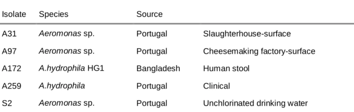
Para a concretização do segundo objetivo principal deste estudo, foram selecionados cinco isolados que haviam sido obtidos de origens distintas, nomeadamente locais de processamento de alimentos, água e amostras clínicas, como representantes da coleção anteriormente analisada. Adicionalmente foram introduzidas A. hydrophila subsp. hydrophila DSMZ 30187 e Aeromonas aeruginosa PAO1 como estirpes controlo dos ensaios referentes aos biofilmes.
De forma a avaliar a capacidade de formação de biofilme em discos de aço inoxidável foram estudadas duas temperaturas de incubação, refrigeração (4 oC) e temperatura ambiente (20 oC), e períodos de crescimento de 48 h. A quantificação do biofilme formado foi realizada através do cálculo de unidades formadoras de colónias (UFC’s).
Os resultados obtidos demonstraram que todas as estirpes em estudo possuem a capacidade de formar biofilme, tanto a 4 como a 20 oC, não tendo sido detectadas diferenças significativas na formação de biofilme entre estirpes ou temperaturas de incubação.
Subsequentemente foi avaliada a eficácia de desinfectantes comerciais à base de (A) tensioativos anfotéricos, (B) compostos clorados e (C) peróxido de hidrogénio, na remoção de biofilme, bem como a prevenção da sua formação. Os desinfetantes demonstraram ser eficazes na remoção do biofilme, à exceção do desinfetante (A), que foi ineficaz na remoção do biofilme formado pelas aeromonas A31 e S2 (isoladas de um matadouro de suínos e de água de captação do Rio Tejo, respetivamente). Relativamente à inibição da formação de biofilme, avaliada perante a incubação das estirpes na presença dos respetivos desinfetantes, durante 48 h a 20 oC, nenhum dos desinfetantes demonstrou ser eficaz o que, em contexto real, reflete a possibilidade de concentrações residuais de desinfetante não inibirem a formação de biofilme em superfícies de processamento de alimentos de aço inoxidável, o que constitui um risco agravado para possíveis contaminações cruzadas.
Em conclusão, os resultados obtidos demonstraram a elevada diversidade genómica da população em estudo, o que está de acordo com estudos realizados anteriormente por outros autores.
Adicionalmente, sugere-se que a metodologia utilizada possa ser utilizada na identificação de Aeromonas a nível de espécie. No que diz respeito à formação de biofilme e ação de diferentes desinfetantes, os resultados obtidos evidenciam a capacidade de formação de biofilme em discos de aço inoxidável por aeromonas de diferentes origens e da importância deste facto em ambientes de processamento de alimentos, reinterando a importância de adequados procedimentos de limpeza e desinfeção desses locais.
Palavras-chave:
Index
Chapter 1 – General introduction ... 1
1.1. Aims of the study ... 1
1.2. General characteristics of the genus Aeromonas... 3
1.2.1 Classification and taxonomy... 3
1.2.2. Ecology, etiology and transmission ... 4
Chapter 2 – Evaluation of genomic diversity ... 6
2.1. Introduction ... 6
2.2. Material and methods ... 8
2.2.1. Bacterial strains... 8
2.2.2. DNA extraction and optimization of PCR amplifications ... 8
Reagents concentrations ... 9
PCR amplification conditions ... 9
2.2.3. Data analysis ... 10
A. allosaccharophila/A. veronii (CECT 4199T) ... 11
A. hydrophila (CECT 398) ... 11
2.3. Results and discussion ... 12
Type strains ... 14
2.4. Conclusions ... 20
Chapter 3 – Biofilm forming ability ... 21
3.1. Introduction ... 21
3.1.1. General characteristics of biofilms ... 21
3.1.1.1. Factors influencing attachment and biofilm development ... 22
3.1.1.2. Advantages of biofilm mode of growth ... 22
3.1.2. Biofilms in the food-industry ... 23
3.2. Material and methods ... 25
3.2.1. Bacterial strains... 25
Isolate ... 25
Species ... 25
Source ... 25
3.2.2. Preparation of the inoculums ... 25
3.2.4. Effects of disinfectants on biofilms ... 28
3.2.4.1. Biofilm eradication ... 28
3.2.4.2. Inhibition of biofilm formation ... 28
3.3. Results and discussion ... 29
3.4. Conclusions ... 32
Chapter 4 – Final considerations ... 33
References ... 35
Appendix A ... 39
Index of Figures
Figure 1 - Representative scheme using groL amplicon and target sequence sizes as example. ... 9
Figure 2 - Amplification products of recA. ... 12
Figure 3 - Dendrogram obtained by multiple alignment of the concatenated sequences of five housekeeping genes ... 18
Figure 4 - Biofilm development as a five-stage process ... 21
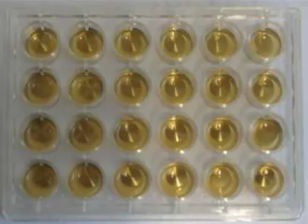
Figure 5 - Representative scheme of replicates organization in each plate. ... 26
Figure 6 - Representative scheme of SS coupons within the wells of a microtitre plate. ... 26
Figure 7 - SS coupons washing procedure ... 27
Figure 8 - Three replicates of a 10 μL drop of six tenfold serial dilutions, plated on BHI. ... 27
Figure 9 - Biofilm formation on SS coupons ... 29
Figure 10 - Inibition of biofilm formation on stainless steel coupons ... 31
Figure 11 - Dendrogram obtained by multiple alignment of the concatenated sequences of gltA ... 43
Figure 12 - Dendrogram obtained by multiple alignment of the concatenated sequences of groL ... 44
Figure 13 - Dendrogram obtained by multiple alignment of the concatenated sequences of gyrB ... 45
Figure 14 - Dendrogram obtained by multiple alignment of the concatenated sequences of metG ... 46
Figure 15 - Dendrogram obtained by multiple alignment of the concatenated sequences of ppsA .... 47
Figure 16 - Dendrogram obtained by multiple alignment of the concatenated sequences of recA ... 48
Index of Tables
Table 1- Primers used in PCR amplification reactions... 8Table 2 - PCR amplification conditions ... 9
Table 4 - Overview of the number of successful PCR amplifications and sequencing ... 14
Table 5 - Allele profile and sequence types of 24 aeromonads ... 15
Table 6 - Nucleotide diversity observed within the 24 aeromonads ... 16
Table 7 - Representative aeromonads chosen for biofilm assays ... 25
Table 8 - Specifications of the commercial disinfectants used in this study ... 28
Table 9 - Decimal log reduction (lg R) achieved by the commercial disinfectants ... 30
Table 10 - PCR amplifications and complete target sequences of the complete Aeromonas spp. collection ... 39
Chapter 1 – General introduction
1.1. Aims of the study
This study takes part of a major project named “AeroPath: Deciphering the pathogenicity potential of food-related Aeromonas”, carried out by Doutora Teresa Maria Leitão Semedo-Lemsaddek (Universidade de Lisboa, Faculdade de Medicina Veterinária) and Doutora Maria Teresa Barreto-Crespo (IBET – Instituto de Biologia Experimental e Tecnológica).
Briefly, a collection of 118 Aeromonas was previously gathered by Barroco (2013), comprising isolates from different sources:
70 isolates from Portugal, comprising 20 isolated at a slaughterhouse, 19 isolated from a supermarket, 8 obtained at a cheesemaking factory, 13 isolated from superficial waters from rio Tejo, 4 from food samples and 6 clinical isolates;
18 isolates from Belgium, comprising 7 from food samples, 6 from drinking water and 5 clinical isolates;
9 clinical isolates from Brazil;
6 clinical isolates from Denmark;
5 clinical isolates from Bangladesh;
3 isolates from Vietnam, comprising 1 from fish, 1 from water and 1 obtained from human stool;
1 isolate from Thailand obtained from human stool;
6 type strains from Deutsche Sammlung von. Mikroorganismen und Zellkulturen GmbH (DSMZ): A. bestiarum 13956T; A. enteropelogenes 6394T; A. veronii 7386T; A. caviae 7323T; A. hydrophila subsp. hydrophila 30187T and A. schubertii 4882T.
Aeromonads from the slaughterhouse, the cheesemaking factory and the supermarket were isolated and identified at genus level by Barroco (2013); aeromonads from superficial waters were isolated and identified a genus level by the author of the present study. Isolates identification was undertaken using both phenotypic (Gram, catalase and oxidase tests) and molecular [duplex-PCR described by Marques (2011)] methods. The remaining isolates were gently provided by collaborating laboratories (further details in Appendix A). Isolates from Denmark, Bangladesh, Vietnam, Thailand and Belgium were characterized through several biochemical tests and identified at Hybridization Group (HG) level through, Amplified Fragment Length Polymorphism (AFLP’s) and gas–liquid chromatographic analysis of cellular fatty acid methyl esters (FAMEs) (Rahman et al. 2007; Pablos et al. 2011).
In previous work, the diversity of the isolates was assessed by RAPDs with primers OPC19 and 1281 and further evaluated regarding the pathogenicity potential namely antibiotic resistance and virulence profile (Barroco 2013). Additional work regarding adhesion to caco-2 mammalian cells, as well as their invasion ability were also screened by another member of the team.
In the present study we aimed to evaluate the genomic diversity of the collection described above using a multilocus sequence approach previously described by Martino et al. (2011). Additionally, we attempted to determine if this approach had the potential to identify Aeromonas at species level. We also aimed to evaluate the biofilm forming ability of five representative aeromonads isolated from water, food-processing facilities and clinical samples, on stainless steel coupons. Additionally we have assessed the effects of three commercial disinfectants in preformed biofilm removal, as well as in preventing biofilm formation.
The present work is divided into four chapters: Chapter 1 – General introduction; Chapter 2 – Evaluation of genomic diversity among Aeromonas spp.; Chapter 3 – Biofilm forming ability of Aeromonas spp.; and Chapter 4 – Final considerations.
1.2. General characteristics of the genus Aeromonas
Members of the genus Aeromonas are essentially ubiquitous in the microbial biosphere and can be isolated from virtually every environmental niche where bacterial ecosystems exist (Janda & Abbott 2010). The genus Aeromonas comprises gram-negative straight rods with rounded ends but sometimes they appear as coccobacilli. Cells are 1.0 X 1.0-3.5 μm and, occasionally, can occur singly, in pairs or even as short chains. Most species are motile by means of a single polar flagella; lateral flagella occurs in some species. Even though their optimum growth temperature varies between 22 and 37oC, growth temperature ranges between 0and 45oC. With regards to pH and presence of NaCl, Aeromonas can growth with pH values between 4.5 and 9.0 and 0–4% NaCl. Aeromonas spp. are facultatively anaerobic and chemioorganotrophic, displaying oxidative and fermentative metabolism of D-glucose, with gas and / or acid production. They are catalase and oxidase positive, reduce nitrate to nitrite and are enzymatically very active, producing a variety of exoenzymes such as DNase, amylase, chitinase, elastase, esterases, peptidases, arylamidases and other hydrolytic enzymes (Martin-Carnaham & Joseph 2005).
1.2.1 Classification and taxonomy
The genus Aeromonas was first described in 1936 by Kluyver and van Niel, being Aeromonas liquefaciens originally proposed as the only and type species of this genus. Lately, three more species were recognized as belonging to the genus: Aeromonas hydrophila in 1943, Aeromonas salmonicida in 1953 and Aeromonas punctata in 1957 (Schubert 1967).
In the 7th edition of Bergey’s Manual of Determinative Bacteriology the genus Aeromonas was included in the family Pseudomonadaceae, however, in the 8th edition of the manual it was reclassified into the family Vibrionaceae. Subsequent analyses using 16S rRNA cataloging, 5S rRNA gene sequence comparisons and rRNA-DNA hybridization data revealed that aeromonads demonstrated an evolutionary divergence approximately equidistant from the Enterobacteriaceae and the Vibrionaceae, thereby justifying the reclassification of the genus Aeromonas in its own family Aeromonadaceae (Colwell et al. 1986; Martin-Carnaham & Joseph 2005). Further studies based 16S rRNA sequencing have also supported the proposal to recognize Aeromonadaceae as a separate family (Martinez-Murcia et al. 1992).
From the creation of the genus through the 1970’s, members of the genus Aeromonas could be divided in two groups: the mesophilic motile group, represented by A. hydrophila, and the psychrophilic nonmotile group, represented by A. salmonicida. In the early 1980’s became evident that the mesophilic group was heterogeneous in biochemical and structural properties resulting in the establishment of three phenotypic species, namely A. hydrophila, A. sobria and A. punctata/A. caviae. Attempting to redefine the genus, DNA-DNA hybridization studies have resulted in the recognition of different DNA HG’s. According to the 2nd edition of Bergey´s Manual of Systematic Bacteriology, the genus includes 14 phenospecies that correspond to 17 DNA HG’s. That means that certain HGs cannot be separated at the biochemical level (Carnaham & Joseph 2005; Janda & Abbott 2010).
The number of species attributed to the genus Aeromonas has increased during the last decade but in some cases their validity is not universally accepted. Several molecular methods have been applied to highlight aeromonads taxonomical relationships but however, controversies regarding species delineation still remain (Martin-Carnaham & Joseph 2005; Nhung et al. 2007).
Further phylogenetic studies leaded to the reclassification of the existing species and description of novel species. Currently there are twenty seven recognized Aeromonas species: A. punctata/A. caviae, A. salmonicida, A. hydrophila, A. sobria, A. media, A. eucrenophila, A. veronii, A. schubertii , A. enteropelogenes, A. ichthiosmia, A. allosachcharophila , A. jandaei, A. encheleia, A. bestiarum , A. popoffii, A. molluscorum, A. simiae, A. bivalvium, A. aquariorum, A. diversa, A. fluvialis, A. piscicola, A. sanarellii, A. taiwanensis, A. tecta, A. rivuli, A. australiensis (Euzéby, J.P., 2009. List of prokaryotic names with standing in nomenclature: genus Aeromonas. Available at: http://www.bacterio.cict.fr/a/aeromonas.html. Last access on October 2013).
1.2.2. Ecology, etiology and transmission
Aeromonas spp. can be found in all aqueous environments except thermal springs, hyper saline lakes, and extremely polluted waters (Janda & Abbott 1999 in United States Environmental Protection Agency 2006). Aeromonads have been isolated from rivers, lakes, ponds, seawater, groundwater, wastewater, chlorinated drinking water and bottled water and also from various food sources including raw milk, fish, shellfish, raw meats and fresh products (Martin-Carnaham & Joseph 2005; Janda & Abbott 2010). The incidence of Aeromonas in water exhibits a seasonal distribution, reaching a higher frequency of isolation in the summer months, due to a major proliferation of the mesophilic strains (Janda & Abbott 2010). Additionally, such distribution is also observed in clinical strains obtained from human stool samples (Sinha et al. 2004).
Members of the genus Aeromonas are regarded as the etiologic agents responsible for a wide range of infectious diseases in humans, especially in immunocompromised patients (Janda & Abbott 2010). Gastroenteritis is the disease most frequently associated with aeromonads, which in some cases evolves to cause peritonitis, colitis or colangitis. Additionally, an increasing number of extra-intestinal infections, such as wound, respiratory, genitourinary tract and ophthalmic infections have been documented (Janda & Abbott 1998; Janda & Abbott 2010; Parker & Shaw 2011). Nevertheless, aeromonads are not universally accepted as gastrointestinal pathogens since there are no evidences supporting the conclusion that aeromonads were responsible for a single clonally food-borne disease outbreak. Additionally, the inexistence of a animal model that can trustworthily reproduce Aeromonas-associated diarrheal disease inhibits Henle-Koch postulate number three to be fulfilled (Janda & Abbott 2010).
Colonization of the human gastrointestinal tract by aeromonads occurs most likely through the ingestion of contaminated drinking water and food. Additionally, aeromonads can also be acquired by exposure to aquatic environments, thus leading to infection through major or unapparent traumas on skin (Janda & Abbott 2010). Even though less frequently, there have been documented associations
between aeromonads infections and reptile, snake and bear bites (Angel 2002 & Kuminoto 2004 in Janda & Abbott, 2010).
Aeromonas spp. have the potential to resist when chlorine levels are low and form biofilms, even at refrigeration temperatures. The World Health Organization (WHO) listed Aeromonas in the third edition
of “Guidelines for drinking water quality” [Available at
http://www.who.int/water_sanitation_health/dwq/guidelines/en/index.html]. Their persistence in water distribution systems and food-processing environments will consequently act as a source of microbial contamination, increasing the food safety risk (Massa et al. 1999; Béchet & Blondeau 2003; Martin-Carnaham & Joseph 2005; United States Environmental Protection Agency 2006). Therefore justifying the need to assess the potential for biofilm formation of aeromonads isolated from different sources, namely from water, food environments and clinical samples.
Chapter 2 – Evaluation of genomic diversity
2.1. Introduction
Currently, species delineation is based upon the combined information of phenotypic and genomic characteristics of a group of individuals. The concept of species is defined as a group of organisms sharing a set of phenotypic characters, with 70% or higher homology in DNA-DNA hybridization and 16S rRNA gene sequence identity of 97% or more (Mende et al. 2013).
In the genus Aeromonas, however, lack of congruence between DNA-DNA hybridization and 16S rRNA gene sequencing have been found (Martinez-Murcia et al. 1992). The high conservation of the 16S rRNA gene sequence (97,8% - 100% of similarity) limits is usefulness for taxonomic analysis at the species level. Moreover, lateral gene transfer and recombination events seem to explain why rRNA operons aren’t effectively homogenized thus leading to intragenomic heterogeneity. Restriction Fragment Length Polymorphism (RFLP) of 16S rRNA has been reported as an alternative method but, besides discrepancies between biochemical identification and RFLP patterns, it cannot overcome the intraspecific heterogeneity found among 16S rRNA genes (Borrell et al. 1997; Martin-Carnaham & Joseph 2005; Morandi et al. 2005; Saavedra et al. 2006).
The limited usefulness of 16S rRNA gene sequences suggests that an appropriate identification approach for characterizing Aeromonas spp. shouldn’t rely on the analysis of a single gene. Thus, a number of other molecular chronometers have been recently used to evaluate phylogenetic relatedness among Aeromonas species, including gyrB (DNA gyrase, β-subunit), rpoD (RNA polymerase), rpoB (DNA dependent RNA polymerase, β-subunit) dnaJ (heat shock protein 40) and recA (recombinase A) housekeeping genes (Yanez 2003; Soler et al. 2004; Nhung et al. 2007; Silver et al. 2011). Sequence similarity values of 16S rRNA genes are less divergent (98,7%) than those within the housekeeping genes described above (89% to 92%), which means that their discriminatory power is higher than it is for 16S rRNA (Nhung et al. 2007). Although independent sequence analysis of these housekeeping genes has proved to be an excellent approach for characterizing Aeromonas spp., their simultaneous analysis improved the differentiation between closely related species (Soler et al. 2004).
Multilocus sequence analysis
The most widely used molecular typing methods rely on comparisons of DNA fragment patterns on agarose gels, either by (i) DNA restriction - Pulsed Field Gel Electrophoresis (PFGE) and RFLP or (ii) DNA amplification – AFLP and Random Amplified Polymorphic DNA (RAPD), among others.
Due to its high reproducibility and discriminatory power, PFGE is considered the gold standard method; however it is more laborious and expensive than the other techniques. These methods are very useful in short-term epidemiological studies, like for example, establishing clonally relationships among bacteria implicated in a outbreak, however they are not appropriate for long-term epidemiological studies (Cuenca et al. 2013; Pérez-Losada et al. 2013).
Multilocus Enzyme Electrophoresis (MLEE) has a great discriminatory power using genetic variation that accumulates relatively slowly, being appropriate for long-term epidemiological studies. MLEE
analyses the variances in electrophoretic mobilities of housekeeping enzymes and their combination defines the electrophoretic type. Multilocus Sequence Typing (MLST) was proposed in 1998 as an adaptation of MLEE with two outstanding advantages: increased discrimination power and accurate portable data (Maiden et al. 1998).
MLST is based on nucleotide polymorphisms within internal fragments of housekeeping genes. These genes are chosen regarding their high conservation, presence as a single copy and wide distribution across the bacterial chromosome. Fragments of approximately 450 bp to 500 bp are amplified and sequenced, usually for seven housekeeping genes. For each gene, different sequences are assigned as alleles and the alleles of the seven loci are combined into an allelic profile. Each isolate is then unambiguously defined by its allelic profile or Sequence Type (ST) (Maiden et al. 1998; Enright & Spratt 1999). Furthermore, bacterial strains can be clustered based upon concatenated sequences of the set of housekeeping genes. In this case, the term Multilocus Sequence Analysis (MLSA) is more appropriate (Martinez-Murcia et al. 2011).
Although the first MLST scheme was developed as a molecular typing method, its applications have increased. MLST has provide useful data that can be used to evaluate genetic diversity among bacterial pathogens, species delineation, population structure and dynamics, among others(Pérez-Losada et al. 2013).
Over the years, a number of molecular and phenotypic approaches have been applied to characterize aeromonads attempting to achieve a reliable species identification frame. However, despite all eff orts, identification of some species is still a serious problem because the conventional biochemical tests in automated or semi-automated commercial systems cannot accurately identify aeromonads to the species level and discrepancies remain between phenotypic and genetic groups (Janda & Abbott 2010).
Two years ago, the first multilocus scheme for studying Aeromonas spp. was proposed, attempting to analyze their phylogeny and resolve the taxonomic controversies within the genus (Martinez-Murcia et al. 2011). Meanwhile, two other schemes were developed and, presently, there are three different schemes that altogether comprise the following housekeeping genes: atpD, dnaJ, dnaX, gyrA, rpoD, groL, metG, ppsA, recA, gyrB, gltA, dnaK, radA, rpoB, tsf and zipA (Martinez-Murcia et al. 2011; Martino et al. 2011; Roger et al. 2012). Furthermore, Martino et al. developed and implemented a Web-based MLST sequence database (http://pubmlst.org/aeromonas) specific for the genus Aeromonas based on the analysis of six housekeeping genes: groL, metG, ppsA, recA, gyrB and gltA. In the present study we aimed to evaluate the genomic diversity of a collection of 118 Aeromonas isolated from water, food and clinical samples. For this purpose, we applied the previously described multilocus sequence typing scheme developed by Martino et al. (2011). Additionally, we also aimed to assess the potential of this approach for aeromonads species delineation. This scheme of housekeeping genes was chosen mainly due to the existence of a MLST database for comparison purposes. Moreover, the curator of this database (Barbara Cardazzo) is a collaborator of the undergoing investigation.
2.2. Material and methods
2.2.1. Bacterial strains
A collection of 118 aeromonads comprising 70 isolates from Portugal (51 obtained from food and food processing environments, 13 isolated from superficial waters and 6 clinical isolates), 42 isolates from different countries worldwide (8 obtained from food, 7 isolated from drinking water and 27 clinical isolates) and six type strains from DSMZ (A. bestiarum; A. enteropelogenes; A. veronii; A. caviae; A. hydrophila subsp. hydrophila and A. schubertii) were analyzed in this study (for further details consult Appendix A). All aeromonads were stored at -80oC in Brain Heart Infusion – BHI – broth (Scharlau, Barcelone, Spain) containing 20% (v/v) glycerol and routinely grown on BHI agar at 30
o
C.
2.2.2. DNA extraction and optimization of PCR amplifications
Genomic DNA for PCR reactions was extracted according to the guanidium thiocyanate method, described by Pitcher et al. (1989). Further quantification of DNA samples in NanoDrop 2000 (Thermo Scientific) allowed concentrations adjustment to 100 ng.
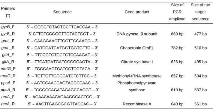
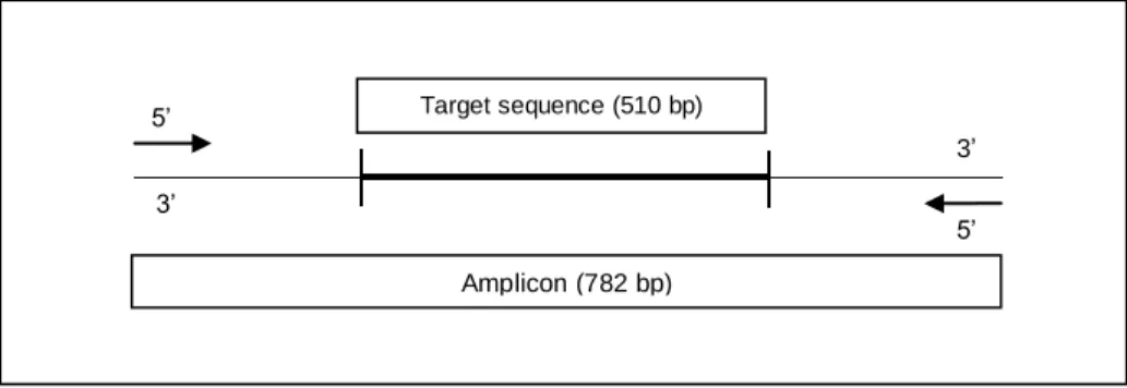
The PCR amplification protocol was optimized according to Martino et al. (2011) using the six type strains described above. Primers used for amplification were developed from the most conserved regions of the genes, in order to obtain amplicons with higher sizes than the sequence of interest. Primers are described in Table 1 and an explanatory scheme of the amplicon and corresponding target sequence sizes is presented in Figure 1.
Table 1- Primers used in PCR amplification reactions
Primers
(*) Sequence Gene product
Size of PCR amplicon Size of the target sequence gyrB_F 5’ – GGGGTCTACTGCTTCACCAA – 3’
DNA gyrase, β subunit 669 bp 477 bp
gyrB_R 5’ CTTGTCCGGGTTGTACTCGT – 3’ groL_F 5’ – CAAGGAAGTTGCTTCCAAGG – 3’ Chaperonin GroEL 782 bp 510 bp groL_R 5’ – CATCGATGATGGTGGTGTTC – 3’ gltA_F 5’ – TTCCGTCTGCTCTCCAAGAT – 3’ Citrate synthase I 626 bp 495 bp gltA_R 5’ – TTCATGATGATGCCGGAGTA – 3’ metG_F 5’ – TGGCAACTGATCCTCGTACA – 3’ Methionyl-tRNA synthetase 657 bp 504 bp metG_R 5’ – TCTTGTTGGCCATCTCTTCC – 3’
ppsA_F 5’ – AGTCCAACGAGTACGCCAAC – 3’ Phosphoenolpyruvate
synthase 619 bp 537 bp
ppsA_R 5’ – TCGGCCAGATAGAGCCAGGT – 3’
recA_F 5’ – AGAACAAACAGAAGGCACTGG – 3’
Recombinase A 640 bp 561 bp
recA_R 5’ – AACTTGAGCGCGTTACCAC – 3’
Figure 1 - Representative scheme using groL amplicon and target sequence sizes as example.
PCR amplifications were performed in independent reactions using 100 ng of each DNA sample in a final volume of 20 μL of PCR amplification mixture under the conditions described in Table 2.
Table 2 – PCR amplification conditions
Reagents concentrations
PCR amplification conditions (Doppio thermocycler, VWR, Radnor,
Pennsylvania, USA)
1 U of Taq DNA polymerase 94oC, 2 min
(Invitrogen, Life Technologies, Carlsbad, USA)
3 5 c y c le s 94 o C, 30 s 1X reaction buffer 56oC, 30 s 5 mM MgCl2 72oC, 1 min 0.2 mM of each dNTP 72oC, 5 min
150 mM of each forward and reverse primer 4oC, 10 min
For all the PCR products, 5 μL within 3 μL of a mixture (1:1) of bromophenol blue and GelRed (iNtRON Biotechnology, Korea) were resolved by agarose gel electrophoresis [1% (w/v)] in 0.5X TBE (0.1 M Tris, 0.1 M boric acid, 0.2 mM EDTA) at 110 V for 60 m. On each gel, a molecular weight marker (1 Kb Plus, Invitrogen, Life Technologies) was included at two positions. All gels were visualized and photographed in a UV transilluminator ImageMaster (PharmaciaBiotech, GE Healthcare, United Kingdom). All amplification products were sent for Macrogen Europe (Amsterdam, The Netherands) for sequencing purposes.
Target sequence (510 bp) Amplicon (782 bp) 3’ 5’ 3’ 5’
2.2.3. Data analysis
Data treatment was performed as follows:
(i) in order to rectify sequencing errors, all chromatograms were edited with SeqTrace software version 0.8.1.;
(ii) for each isolate, a consensus sequence was obtained by the alignment between forward and reverse sequences using the same software;
(iii) sequences of the reference strains used by Martino et al. (2011) were downloaded from the Aeromonas MLST database (reference strains are listed in Table 3);
(iv) for each gene, the consensus sequences obtained in “ii” were aligned with the sequences of the reference strains “iii”, using Mega 5.1 software;
(v) the target sequences were determined and exported in FASTA format;
(vi) the allelic profile/sequence type of each isolate was assessed using the Aeromonas MLST database;
(vii) diversity indices such as G+C content, number of polymorphic sites, Tajima’s D test, nucleotide diversity per site (π), average number of nucleotide differences per site (θ) and average number of synonymous and non-synonymous substitutions were calculated using Mega version 5.10;
(viii) for each isolate, the sequences of the housekeeping genes were manually concatenated according to the correct genomic order - gyrB, groL, gltA, metG, ppsA and recA;
(ix) single sequences and concatenated sequences were introduced in BioNumerics software (version 6.6, Applied Maths NV, Sint-Martens-Latem, Belgium);
(x) construction of dendrograms based on the concatenated sequences of the housekeeping genes and on single gene sequences were performed in BioNumerics. The similarity matrix was obtained by pairwise alignment of the sequences and cluster analysis was obtained by the unweighted pair group method with arithmetic mean algorithm (UPGMA);
(xi) the intra-specific diversity was calculated using Simpson‘s (D’) and Shanon (H’) diversity indexes, calculated using the following formulas:
S – total number of groups formed; N – total number of isolates analyzed; n – number of isolates in the group; The heterogeneity level is calculated by J’, a derivate of Shannon index: J’= H’/Hmax = H’/lnS.
Table 3 - Reference strains used by Martino et al. (2011)
A. allosaccharophila/A. veronii (CECT 4199T) A. hydrophila (CECT 398)
A. bestiarum/A. hydrophila (NCIMB 1134) A. media (DSMZ 4881T)
A. bestiarum (DSMZ 13956T) A. popoffii (DSMZ 19604T
)
A. caviae/A. punctata subsp. caviae (CECT 838T) A. salmonicida subsp. achromogenes (NCIMB 1109)
A. caviae (NCIMB 882) A. salmonicida subsp. masoucida (NCIMB 2020)
A. encheleia (DSMZ 11577T) A. salmonicida subsp. salmonicida (NCIMB 1102T)
A. enteropelogenes (CECT 4487T) A. schubertii (CECT 4240T)
A. enteropelogenes/A. trota (CECT 4255T) A. sobria (CECT 4245T)
A. eucrenophila (DSMZ 17534T) A. sobria (NCIMB 75)
A. jandaei (CECT 4228T) A. veronii bv. veronii (CECT 4257T)
A. hydrophila (ATCC 7966T) A. veronii bv. sobria (CECT 4246)
CECT: Colleción Española de Cultivos Tipo
NCIMB: National Collection of Industrial, Food and Marine Bacteria ATCC: American Type Culture Collection
In order to assess the reproducibility of the technique, 5% of replicates were performed. For that purpose we used the six type strains described above.
2.3. Results and discussion
In the present investigation we applied a multilocus sequence approach previously described by Martino et al. (2011) to 118 Aeromonas.
The isolates were submitted to PCR amplification of six housekeeping genes (Table 1) and the PCR amplification products were further visualized in agarose gels (Figure 2).
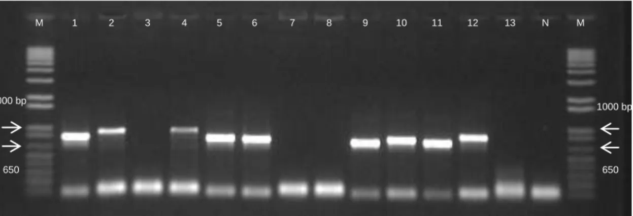
Figure 2 - Amplification products of recA. Lanes: 1. A97; 2. A143; 3. A147; 4. A150; 5. A161; 6. A163; 7. A174; 8. A179; 9.
A184; 10. A186; 11. A193; 12. A206; 13. A219; N. Negative Control; M. 1 Kb Plus molecular size standard ladder.
Visualization of PCR amplification products allowed the observation of different situations. Although amplicons with expected sizes were observed, most amplicons had higher sizes than expected. For example, amplicons of recA should be around 640 bp but, as we can observe in Figure 2, some presented higher sizes such as 850 or 1000 bp. This situation was observed in amplification products of all six housekeeping genes.
Additionally, lack of amplification was also observed in all genes. Attempting to resolve PCR amplification problems several rearrangements were undertaken, including: (i) changes in annealing temperature, ranging values between 56oC and 45oC, and time, from 30 s to 1 min; (ii) changes in MgCl2 concentration (6 mM and 8 mM); (iii) utilization of different enzymes – DYNAZyme II PCR
Master Mix (Thermo Fisher Scientific Inc, Langenselbold, Germany), Immolase DNA Polymerase (Bioline, United Kingdom) and NZYTaq 2x Green Master Mix (NZYTech Lda, Lisboa, Portugal), and (iv) use of distinct thermo cyclers – My CyclerTM
Thermal Cycler (BioRad, Berkeley, USA) and T3000 Thermo Cycler (Biometra, Goettingen, Germany) and (v) repetition of DNA extractions. Through all these changes, we were able to achieve a larger number of successful amplifications: gyrB – 97,76% (113/118); groL and gltA – 94,07% (111/118); metG and recA –93,22% (110/118) and ppsA – 90,68% (107/118). For further details see Appendix A.
650 bp 650 bp 1000 bp 1000 bp M 1 2 3 4 5 6 7 8 9 10 11 12 13 N M
Martino et al. (2011) also reported amplification problems with ppsA gene, which did not amplified in A. salmonicida subsp. masoucida, A. salmonicida subsp. salmonicida and A. veronii bv. sobria. Additionally, A. sharmana amplified only for gyrB gene, which can possibly be explained by the fact that, according to Saavedra et al. (2006), A. sharmana does not belong to the genus Aeromonas. In a personal communication, Martino et al. reported that they had to change annealing temperature for gyrB, groL and ppsA genes, which did not amplified in some species, namely A. jandaei and A. enteropelogenes.
Even though these are highly conserved genes, Martino et al. (2011) reported high nucleotide diversity among them and thus, the major explanation for amplification problems might be the existence of polymorphisms in the primer binding sites.
A total of 624 amplification products, corresponding to 104 isolates versus six genes (forward and reverse) were sent for sequencing purposes and 1248 chromatograms were obtained. Through the analysis of the obtained chromatograms, 105 were excluded due to lack of quality. Additionally, through the analysis of the remaining 1143 chromatograms, we confirmed that the sequences obtained had higher sizes than expected, as visualized in agarose gels (Figure 2).
Chromatograms were edited and subsequently forward and reverse sequences were aligned in order to obtain a consensus sequence. Through this alignment, we observed that consensus sequences had approximately the expected amplicon size, depending on the respective gene.
Then, consensus sequences obtained were aligned with Aeromonas MLST database sequences of the reference strains representing distinct species in order to obtain the desired target sequence (see Figure 1). Nevertheless, their alignment with the Aeromonas MLST database sequences, lead to two different situations: in most cases we were able to obtain the complete allele fragments; however, in other cases the fragments of interest were not complete.
One possible explanation for amplicons unexpected size is that it might be possible that there are insertions of mobile genetic elements between the primer binding sites and the target sequence. Additionally, we did not observe any gaps or insertions within this sequence. Thus, it seems to be more conserved than the upstream region making it a good selection for sequencing purposes.
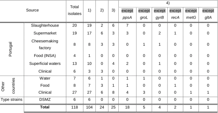
Thereafter, gene fragments were exported in FASTA format. For those genes which fragments were incomplete, we filled sequences endings with a nucleotide arbitrarily chosen – adenine (A) – so we can avoid a higher loss of sequence data. For example, target sequence of ppsA was incomplete in A24 and we filled it as follows: “AAAAAAAAAAAAAAAAACATCATGCGT...……TTCGGTAGCCCC”. Overall, 24 isolates had complete target sequences, 25 had complete target sequences by the addition of adenine and 31 had complete target sequences for five housekeeping genes (Table 4).
Table 4 - Overview of the number of successful PCR amplifications and sequencing Source Total isolates 1) 2) 3) 4) except ppsA except groL except gyrB except recA except metG except gltA P o rtu g a l Slaughterhouse 20 19 2 6 7 0 0 0 0 0 Supermarket 19 17 6 3 3 0 2 1 0 0 Cheesemaking factory 8 8 3 3 0 1 1 0 0 0 Food (INSA) 4 1 0 0 0 0 0 0 0 0 Superficial waters 13 10 0 4 2 0 1 0 0 0 Clinical 6 3 3 0 0 0 0 0 0 0 O th e r c o u n tr ie s Water 7 6 1 0 1 1 0 0 0 0 Food 8 7 3 1 1 0 0 1 0 0 Clinical 27 27 6 8 4 3 0 0 1 1 Type strains DSMZ 6 6 0 0 0 0 0 0 0 0 Total 118 104 24 25 18 5 4 2 1 1
1) Number of successful PCR amplifications;
2) Number of isolates with complete fragments for the 6 alleles;
3) Number of isolates with complete fragments for the 6 alleles through the addition of “ AAA”; 4) Number of isolates with complete fragments for 5 alleles.
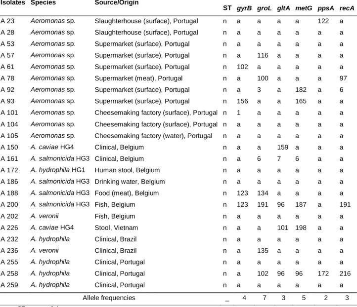
Sequence types and genetic diversity were assessed by the analysis of the 24 aeromonads with complete target sequences. Allelic profiles were assessed using the Aeromonas MLST database and are described in Table 5. In most cases we could not find any match with any allele and, in few cases, some alleles were the same. From the total of 24 aeromonads, 14 had a match with at least one allele representing all new sequence types and 10 didn’t have any match, representing sequence types different from those on the database. However, some of these 10 isolates could represent the same sequence type.
For isolates A200 and A258 we were able to allocate 5 alleles. The closest allelic profile encountered in the database was ST 217 for isolate A200, with 3 allelic sites in common (groL, metG and recA), and ST 189 for isolate A258, with 4 allelic sites in common (groL, gltA, metG and ppsA). Interestingly, ST 217 corresponds to an isolate from seafood and A200 was isolated from fish. The same correlation was found for ST 189 which corresponds to a clinical strain isolated from the gastrointestinal tract and A258 which is a clinical strain isolated in a Portuguese hospital. These findings suggest that allelic profiles might be identical in aeromonads isolated from the same sample type. However, Martino et al. 2013 observed that the same sequence type not always corresponded to strains isolated from identical samples.
Table 5 - Allele profile and sequence types of 24 aeromonads
Isolates Species Source/Origin Allele
ST gyrB groL gltA metG ppsA recA
A 23 Aeromonas sp. Slaughterhouse (surface), Portugal n a a a a 122 a A 28 Aeromonas sp. Slaughterhouse (surface), Portugal n a a a a a a
A 53 Aeromonas sp. Supermarket (surface), Portugal n a a a a a a
A 57 Aeromonas sp. Supermarket (surface), Portugal n a 116 a a a a A 61 Aeromonas sp. Supermarket (surface), Portugal n 102 a a a a a
A 78 Aeromonas sp. Supermarket (meat), Portugal n a 100 a a a 97
A 92 Aeromonas sp. Supermarket (surface), Portugal n a 3 a 182 a 6 A 93 Aeromonas sp. Supermarket (surface), Portugal n 156 a a 165 a a A 101 Aeromonas sp. Cheesemaking factory (surface), Portugal n 1 a a a a a A 104 Aeromonas sp. Cheesemaking factory (surface), Portugal n a a a a a a A 105 Aeromonas sp. Cheesemaking factory (water), Portugal n a a a a a a
A 150 A. caviae HG4 Clinical, Belgium n a a 159 a a a
A 161 A. salmonicida HG3 Clinical, Belgium n a 6 7 6 a a
A 172 A. hydrophila HG1 Human stool, Belgium n a a a a a a
A 186 A. salmonicida HG3 Drinking water, Belgium n a a a a a a
A 188 A. salmonicida HG3 Food (meat), Belgium n 123 134 a a a a
A 200 A. salmonicida HG3 Fish, Belgium n 123 191 96 187 a 191
A 202 A. veronii Fish, Belgium n a a a a a a
A 226 A. caviae HG4 Stool, Vietnam n a a 101 198 a a
A 232 A. hydrophila Clinical, Brazil n a a a a a a
A 236 A. veronii Clinical, Brazil n a 135 a a a a
A 255 A. hydrophila Clinical, Portugal n a a a a a a
A 258 A. hydrophila Clinical, Portugal n a 102 96 96 172 216
A 259 A. hydrophila Clinical, Portugal n a a a a a a
Allele frequencies _ 4 7 3 5 2 3
n: new ST; a: new allele
In our study, at least 14 out of 24 aeromonads represent all new different sequence types (58%). It also might be possible that the remaining 10 aeromonads represent different sequence types among them. Our results are in agreement with previous publications of different authors. Martino et al. 2011 identified 89 distinct ST’s among 96 Aeromonas spp. isolated from diseased fish, crustaceans and mollusks (93%), Martino et al. 2013 identified 250 distinct ST’s among 258 Aeromonas spp. isolated from (97%) and Roger et al. 2012, using another set of housekeeping genes, identified 175 distinct ST’s among 191 clinical and environmental strains of Aeromonas spp. Additionally, the database contains 252 different sequence types among 272 isolates (93%) [http://pubmlst.org/aeromonas; last access on October 2013]. All these results emphasize the high genetic variability among the genus Aeromonas.
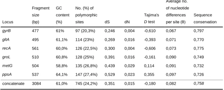
Table 6 –Nucleotide diversity observed within the 24 aeromonads
Average no.
Fragment GC No. (%) of of nucleotide
size content polymorphic Tajima's differences Sequence
Locus (bp) (%) sites dS dN D test per site (θ) conservation
gyrB 477 61% 97 (20,3%) 0,246 0,004 -0,610 0,067 0,797 gltA 495 61,1% 114 (23%) 0,269 0,016 -0,393 0,071 0,770 recA 561 60,0% 126 (22,5%) 0,300 0,004 -0,606 0,073 0,775 groL 510 60,8% 128 (25%) 0,391 0,016 -0,161 0,090 0,749 metG 504 58,8% 135 (26,8%) 0,439 0,029 0,114 0,091 0,732 ppsA 537 64,1% 147 (27,4%) 0,529 0,023 0,355 0,097 0,726 concatenate 3084 61,0% 745 (24,2%) 0,351 0,015 -0,180 0,082 0,758
dS: number of synonymous changes per site; dN: number of non-synonymous changes per site
The mean GC content of the genes varied from 58,8% for metG and 64,1% for ppsA. The number of polymorphic sites ranged values between 97 polymorphic sites for gyrB (20,3%) and 147 polymorphic sites for ppsA (27,4%); and the nucleotide diversity (the average number of nucleotide differences per site) ranged values between 0,067 for gyrB and 0,097 for ppsA. Tajima’s D test ranged values between -0,610 for gyrB and 0,355 for ppsA. Tajima’s D tests the hypothesis that all mutations are selectively neutral and is based on the differences between the number of polymorphic sites and the average number of nucleotide differences per site (Tajima 1989).
In what concerns the concatenated sequences of the 24 aeromonads, GC content was 61%, the similarity between isolates was 75,8% which corresponds to 745 polymorphic sites and the average number of nucleotide differences was 0,082.
Analysis of the results showed high genetic diversity was found in all genes. Additionally, genetic variance was found to be lower in gyrB locus and higher in ppsA locus.
Moreover, similar results supporting high genetic diversity were found by (i) Martino et al. 2011 and (ii) Martino et al. 2013: GC content varied from (i) 57,6% for metG and 63,7% for ppsA and (ii) 58% for metG and 64,1% for ppsA; the number of polymorphic sites ranged values from (i) 140 for gyrB and 233 for ppsA and (ii) 162 for gyrB and 263 for ppsA; nucleotide diversity ranged from (i) 0,057 for gyrB to 0,098 for ppsA and (ii) 0,058 gyrB to 0,106 for ppsA; Tajima’s D values ranged between (i) -1,109 and -0,336 and (ii) -1,170 and -0.302.
Attempting to clarify the relationships between the isolates of our collection we constructed a dendrogram with the concatenated sequences of 67 isolates five housekeeping genes in their genomic order: gyrB, groL, gltA, metG and recA. Attempting to include a greater number of aeromonads in the dendrogram construction, ppsA gene was excluded from the concatenation and
therefore, we were able to construct a dendrogram with 67 concatenated sequences. Additionally, 22 concatenated sequences of the reference strains mentioned on Table 3 were also included.
Considering the dendrogram’s global structure and reference aeromonads distribution, a cut off level of 95,8% similarity was chosen. Further analysis allowed the visualization of five well defined clusters attributed to A. salmonicida (A), A. hydrophila (B), A. caviae (C and D) and A. media (E) (Figure 4). A. salmonicida cluster included A. salmonicida subsp. salmonicida (NCIMB 1102T), A. salmonicida subsp. masoucida (NCIMB 2020), A. salmonicida subsp. achromogenes (NCIMB 1109), isolates A57, A61, A62, A184, A186, A188, A193, A195 and A200.
A. hydrophila cluster included ATCC 7966T, A85, A97, A98, A101, A105 and A163 A172, A232, A237 and A255. Accordingly, this last group of isolates was previously identified as A. hydrophila HG1. Two different clusters were formed for the species A. caviae. Cluster C included type strain CECT 838T, A78, A104, A258, S1, S3, S5, S6, A147, A150, A154, A157, A219, A222, A226, A230 and A253. Accordingly, isolates A147 to A230 were previously identified as A. caviae HG4. Clinical isolate A258 were previously identified as A. hydrophila, however, in clinical laboratories, aeromonads identification is based on their biochemical properties (Janda & Abbott 2010) rather than molecular methods. Once molecular methods are more reliable in aeromonads species allocation than biochemical methods, it is most likely that A258 is A. caviae rather than A. hydrophila. The other cluster (D) included NCIMB 882, A4, A5, A7, A8, A23, A24, A25, A27, A28, A31, A52 and A53. Interestingly, all these were isolated from the slaughterhouse, with the exception of A52 and A53 that were isolated from the supermarket. This is the only situation where isolates were clustered according to their source.
Finally, A. media cluster included type strain DSM 4881T and isolates A92, A95, A99, A161 and S10. A161 were previously assigned as A. salmonicida HG3, however, it clustered with A. media DSM 4881T with 99,6% similarity. This situation needs to be further evaluated using, for example, DNA-DNA hybridization studies.
In the absence of representative isolates of other species, some reference strains were found in the same clusters.
A major cluster constituted by A. veronii bv. sobria CECT4246T, A. veronii bv. veronii CECT4257T, A. sobria NCIMB75, A. hydrophila and A. allosacharophila/veronii CECT4199T was observed. In this cluster, isolate A259, previously identified in a clinical laboratory as A. hydrophila, clustered with A. sobria NCIMB75 with 96% similarity. This situation can be explained exactly as the situation found for isolate A258 and reinforces the idea that biochemical characterization is not suitable for aeromonads species allocation. Additionally, isolate A94 clustered with A. allosacharophila/veronii CECT4199T with 99,4% similarity. Isolate A202, previously identified as A. veronii, clustered with A. sobria CECT4245T. This result should be further confirmed.
Isolate S13 clustered with A. eucrenophila DSMZ17534T with 96,8% similarity, which means that this isolate should probably be allocated to A. eucrenophila species.
Isolate A231, previously identified as A. trota clustered with A. enteropelogenes CECT4487T and A. enteropelogenes/trota CECT4255T with 96,2% similarity. Since A. trota is an earlier heterotypic synonym of A. enteropelogenes, previous identification was confirmed by our study.
Figure 3 - Dendrogram obtained by multiple alignment of the concatenated sequences of five housekeeping genes grouped by the agglomerative clustering of unweighted pair group method with arithmetic mean (UPGMA).
Red rectangle (A): A. salmonicida cluster; Blue rectangle (C,D): A. caviae clusters; Green rectangle (E): A. media cluster; Yellow rectangle (B): A. hydrophila cluster.
A. bestiarum/hydrophila NCIMB1134 formed a cluster with A. bestiarum DSMZ13956T; single clusters were observed by A. jandaei CECT4228T, A. popoffi DSMZ19694T, A. encheleia DSMZ11577T, A. schubertii CECT4240T, A3, A6, A11, A13, A77, A143 and A252. Finally, A26 and A93, isolated from a supermarket and a slaughterhouse, clustered with 98,4% similarity, which means that they should probably be allocated to the same species; however, none of the represented in the present study. The inclusion of isolates representative of these species should allow the respective formation of well defined clusters. Additionally, it should also be included reference strains representing other Aeromonas species.
Additionally, to create a comparison between the six housekeeping genes and concatenated sequences, dendrograms based on single gene sequences were constructed (Appendix B). These dendrograms were constructed with sequences of the 22 reference strains (Table 3) and the 49 aeromonads with complete gene fragments. All six dendrograms formed the same five clusters as did the dendrogram of the concatenated sequences, even in the absence of ppsA gene. These findings suggest that all six genes would be able to distinguish members of these four species, even if they weren’t used combined. It also supports the species identification based on the five well defined clusters.
Single dendrograms based on the sequences of six type strains of the aeromonads collection (Appendix A) and the sequences of the 22 reference strains downloaded from the database (Table 3) were constructed. All six type strains used in the present study are from DSMZ; however, they all had homology with six of the 22 reference strains used by Martino et al. (2011). Dendrograms construction (data not shown) allowed the observation of clusters formed by homologous type strains sequences, with 100% similarity, thus conferring credibility to the technique.
Simpson’s diversity index (D) is the probabilities of two randomly selected individuals belonging to the same group. Nonetheless, D is inversely proportional to diversity and the complementary (D’) needs to be calculated to assess the probability of two randomly selected individuals belong to different groups. Samples diversity are considered acceptable if D˃0,90 and the maximum possible value is “1”, which corresponds to one individual per group (Hunter & Gaston 1988). Simpson’s diversity obtained was D’ = 0,906, which means that the sampling analyzed allow the isolation of distinct individuals.
Shannon’s diversity index (J’) measures population’s heterogeneity. It takes into account both abundance and evenness of species present in the community. Shannon’s diversity obtained was J’=0,83, which highlight population diversity.
2.4. Conclusions
In the present study we aimed to (i) evaluate the genomic diversity among a collection of 118 Aeromonas using a previously described multilocus sequence typing scheme and (ii) assess the potential of this approach in aeromonads species allocation.
Genetic diversity was found to be high in the population analyzed. In a total of 24 aeromonads, none had known STs and at least 10 new different ST’s are proposed. Additionally, several diversity indices confirmed the high level of sequence diversity detected.
The MLST scheme used allowed the separation of five well defined clusters attributed to A. salmonicida, A. hydrophila, A. caviae and A.media. This revealed to be a valuable tool in what regards to Aeromonas species allocation, however, the inclusion of representative isolates of other species would provide more reliable results.
Chapter 3 – Biofilm forming ability
3.1. Introduction
Aeromonas spp. have been isolated from chlorinated drinking water supplies in several countries (United States Environmental Protection Agency 2006) even during colder months, with temperatures below 14°C, and in food held at refrigerated temperatures (Massa et al. 1999; Manuel Pablos et al. 2009). Even though conventional water treatment processes are effective in removing or inactivating aeromonads, they may persist in distribution systems as biofilms when disinfectant levels are low (< 0.2 mg/L free chlorine residual). Aeromonads ability to survive and multiply at low temperatures and to persist in water distribution systems provides a reservoir for food contamination, being directly by water contact or through contaminated food processing surfaces.
3.1.1. General characteristics of biofilms
In their natural habitats, bacteria grow preferentially as biofilm complex communities comprising single or multiple species, adhered to wet surfaces and embedded in a self-produced slimy matrix composed by Extracellular Polymeric Substances (EPS), such as polysaccharides, lipids, proteins, nucleic acids and enzymes (Lasa 2006; Brooks & Flint 2008; Shi & Zhu 2009; Simões et al. 2010).
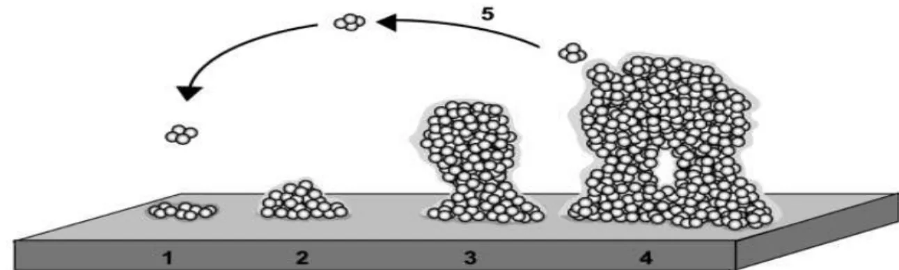
Biofilm development is described as a five-stage process of adaptation and changing genetic regulation (Stoodley et al. 2002; Lasa 2006; Pereira da Silva et al. 2012): (1) initial attachment of cells to the surface; (2) production of the EPS; (3) early development of the biofilm; (4) maturation of the biofilm; (5) dispersion of bacterial cells from the biofilm (Figure 5).
Figure 3 - Biofilm development as a five-stage process: (1) initial attachment of cells to the surface; (2) production of EPS; (3)
early development of the biofilm; (4) maturation of the biofilm; (5) dispersion of bacterial cells from the biofilm (Extracte d from Lasa, 2006)
(1) Initial attachment: Attachment is an initial weak interaction of bacteria with the substratum, involving Van der Waals and weak electrostatic forces and hydrophobic interactions (Chmielewski & Frank 2003; J. Brooks & Flint 2008; Pereira da Silva et al. 2012). Some components of the bacterial outer-membrane like flagella, fimbriae and lipopolysaccharides (LPS) confer hydrophilic properties to