Research Signpost
37/661 (2), Fort P.O., Trivandrum-695 023, Kerala, India
Vanadium Biochemistry
Chapter 7
DECAVANADATE INTERACTIONS WITH SARCOPLASMIC RETICULUM CALCIUM PUMP
Manuel Aureliano
Centre for Marine Sciences (CCMar), FCT, University of Algarve, 8005-139 Faro, Portugal
Abstract
Although not stable, once formed, decameric vanadate (V10) disintegration is in general slow enough to allow the study of its effects even in the micromolar range. Besides, it may become inaccessible to decomposition due to their specific interaction upon target proteins such as the Ca2+-ATPase from sarcoplasmic reticulum (SR). Characterization of the vanadate solutions and interactions with compounds containing phosphate as well as with the SR Ca2+-ATPase was analysed by
51
V NMR spectroscopy. Vanadate is a well known inhibitor of this E1E2 phosphohydrolases and it as been used as a probe in the study of the structure and in the function of the protein. Decameric vanadate species also interact with the SR Ca2+ -ATPase but clearly differs from monomeric vanadate by inhibiting sarcoplasmic reticulum calcium translocation at different calcium gradient conditions, coupled or not to ATP hydrolysis. Vanadium(IV) and (V) complexes, some known to have insulinomimetic properties, also inhibit the calcium ATPase activity, although at a lower extend than V10.
*Correspondence/Reprint request: Manuel Aureliano, FCT, University of Algarve, 8005-139 Faro, Portugal e-mail:[email protected]
1.INTRODUCTION
Thirty years ago vanadium was found in commercial ATP and rediscovered for biology as a muscle and Na+, K+-ATPase inhibitor [1-3]. Twenty years ago vanadium was
referred as specific inhibitor of E1-E2 ion transport ATPases [4] such as the sarcoplasmic reticulum (SR) Ca2+-pump. Currently, vanadium is used as a functional and structural tool
in many biological studies involving these enzymes since it is well known that vanadate mimics the transition states of phosphoryl transfer reactions [5]. However, the eventual role of vanadium in these energy transducing systems is not yet clearly understood, as discussed in the present review.
The majority of vanadate studies in muscle cells are related with insulin-mimetic properties and with the capacity to increase the muscle contraction (ionotropic effect) [6,7]. Other sound effects of vanadate includes stimulation of cell growth, induction of cell death and citotoxicity associated with reported vanadate effects on actin polymerization including sperm capacitation, detachment of pathogenic microorganisms and chemotherapy of solid tumors [8,9,10].
In skeletal muscle cells sarcoplasmic reticulum controls contraction and relaxation. Briefly, after cell membrane potential propagation within the transverse tubules (together with the two terminal cisternae constitutes the triads in sketeletal muscle fibers), changes in
membrane polarity induces conformational changes in specific receptor (the multimeric dihydropyridine receptor) produces changes in another receptor (ryanodine receptor) located at the terminal cisternae of the sarcoplasmic reticulum, that triggers a release of Ca2+ ions from the sarcoplasmic reticulum into the cytosol. The Ca2+ concentration in the myoplasm increases up to >1 µM and once calcium reaches to the myofibrillar apparatus (myosin and actin filaments forming the contractile system), the second messenger binds to troponin c and allows the interaction between myosin and actin filaments resulting in muscle contraction. Ca2+ is than pumped into the SR by the sarcoplasmic reticulum Ca2+ -ATPase which accumulates Ca2+ at expense of ATP splitting. Due to this energy dependent Ca2+-reuptake mechanism, the Ca2+ concentration in the myoplasm decreases < 0.1 µM inducing the dissociation of myosin-actin complex and leading to muscle relaxation.
Therefore, sarcoplasmic reticulum Ca2+-ATPase is responsible for initiating to the step of muscle relaxation. Besides sarcoplasmic reticulum Ca2+-ATPase, others systems are involved in calcium homeostasis such as a surface calcium pump and Na+/Ca2+ exchanger which is indirectly driven by the sarcolemmal Na+/K+-ATPase. Complex interactions between these systems with others also involved in calcium homeostasis such as calcium binding proteins and mitochondria, among others, provide the molecular basis which regulates muscle calcium homeostasis. It has been described that exposition of muscle cells to vanadate induces an increase of intracellular calcium leading to an increase of muscle contraction [11]. By affecting the systems involved in the regulation of calcium concentration such as the calcium ATPase from sarcoplasmic reticulum, mitochondria or by preventing the liberation of calcium from the calcium reservoirs, vanadate modulates muscle contraction/relaxation, besides others cellular processes. In the present review, it is focus the interactions of vanadate oligomers with the main regulator of muscle relaxation, the sarcoplasmic reticulum Ca2+-ATPase. Biochemical aspects related with vanadate and decavanadate effects on myosin structure and function are being reviewed in Chapter 5 of this book.
To the undefined role of vanadium in biochemistry contribute its complex chemistry, namely several oxidation states such as vanadium +4 (vanadyl) and +5 (vanadate), being the latter similar to phosphate and having the tendency to condense with itself forming vanadate oligomers and also with many other compounds of biological interested, such as for instance the ones containing phosphate. Among the vanadate oligomers, decavanadate (V10) as been referred of biochemical importance and is known to interact with many proteins besides affecting several biological processes [12,13,14,15].
In the last ten years, reported decavanadate studies includes modulation of voltage-dependent Ca2+-actived cation channel and mimicking noradrenaline action, besides the possibility to be used as a tool in the understanding of molecular mechanism of muscle contraction [16,17,18]. The decavanadate capacity to inhibit several ATPases, phosphohydrolases such as P-type ATPases (E1E2-ATPases), ABC-ATPases and ribonucleases suggests that the V10 interaction with the proteins maybe favored by the existence of an ATP binding site [15,18,19,20]. In the present review, it is described studies using vanadate and particularly decavanadate, as a tool in the study of calcium translocation by the ATPase from sarcoplasmic reticulum. Once vanadate was found in muscle and due to its similarities with phosphate, we might expect a physiological role of vanadium in the systems involved in the process of muscle contraction/relaxation, such as the Ca2+-ATPase from sarcoplasmic reticulum.
2.VANADATE COMPLEX CHEMISTRY
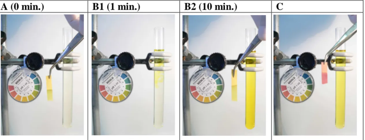
It is very important to be aware about the complex chemistry of vanadium in aqueous solution, before trying to understand the biological effects of vanadate. In fact, different vanadate species can occur simultaneously in vanadium(V) solutions, e.g. monomeric (V1), dimeric (V2), tetrameric (V4), pentameric (V5) and in some cases, with different states of protonation and structures, depending on several factors such as vanadate concentration, pH and ionic strength [21] Besides these vanadate species, decameric vanadate can also occur in solution, in particular at acidic pH values. However, to our knowledge, it is not possible to prevent the formation of decameric vanadate upon acidification of vanadate solutions, even if the global pH is maintained constant. In fact, many researchers working worldwide frequently observed the orange/yellow color in their vanadate solutions. This is due to the occurrence of decameric vanadate (V10), for instance in a cell culture, after an acidification procedure during protein purification, after the adjustment of the pH value of the reaction medium or even in methods for vanadium quantification. Thus, if after the preparation of a vanadate solution (10 mM) an acidification occurs, it is possible to observe at once the appearance of a yellow colour due to the occurrence of decameric vanadate species, even if the global pH value of the solution does not changes significantly (Figure 1).
A (0 min.) B1 (1 min.) B2 (10 min.) C
Figure 1. Upon acidification of a metavanadate solution 10 mM (A) it is possible to observe in minutes the
appearance of decameric species (B1 and B2) without changing dramatically the pH (B2). Further acidification, drops the pH to acidic values (C).
Thus, upon acidification, the vanadate solution slightly yellow (colourless in Fig. 1A), turns strong yellow due to decavanadate formation. The yellow colour diffuses to all the solution after 1 minute Fig 1B1) and 5 minutes (Fig 1B2), whether the global pH does not change significantly (Fig.1B2). Further acidification promotes decameric vanadate species formation (Fig. 1C). However, at this acidic pH the solution turns colourless in minutes, due to decavanadate decomposition (not shown). The formation of decameric species can be easily confirmed by 51V-NMR analysis. Even after a slight acidification, from 6.8 to 6.6, of a nominated metavanadate (or monovanadate, in some articles) solution containing V1, V2, V4 and V5 vanadate species, at 560 ppm, 574 ppm, 578 ppm and
-586 ppm, respectively (Figure 2A), as evaluate by 51V-NMR spectroscopy, it is possible to observe NMR signals ascribed to decavanadate species at -515 ppm, -500 ppm and -420 ppm (Figure 2B). Further acidification with HCl up to pH near 4, it is obtained the nominated decavanadate solution containing mainly decameric vanadate species (Figure 2C). At this pH value, decavanadate is very stable at 4 ºC. Those two vanadate solutions (Fig 2A and Fig 2C) where used in the studies described below concerning the sarcoplasmic reticulum ATPase inhibition by decavanadate. Therefore, even at physiological pH values, if an eventual local acidification occurs that will induce the formation of V10 species.
Figure 2. 105.2 MHz 51V NMR spectra, at room temperature, of 10 mM metavanadate solution, pH 6.8, before (A) and after addition of HCl up to pH 6.6 (B) and to pH 4.4 (C). Decavanadate solution corresponds to spectra C. V1, monomeric; V2, dimeric; V4, tetrameric; V5, tetrameric and V10, decameric vanadate [adapted from ref. 23]. Insert in the panels A and B are, respectively, structures of decameric and monomeric vanadate species.
Although used in an increasing number of biological studies, it is well known that decameric vanadate is instable. However, peculiarly very few studies referred to its stability at the different experimental conditions and when it does, they are often not quantitative.
C: Decavanadate, pH 4.4 B: Metavanadate, pH 6.6 A: Metavanadate, pH 6.8 V10C V10B V10A V1 V2 V4 V5
Only recently, it is has been clearly associated the biological effects with the kinetics of decavanadate decomposition, assessed by measurements of the absorption at 400 nm, as a first order kinetics; with a half-life time up to 3 hours (measured at 37 ºC with 100 µM decameric vanadate species) [14]. Additionally, it was described that V10 decomposition may be prevent due to specific interactions with target proteins such as the calcium pump from sarcoplasmic reticulum [14,22]. In fact, upon addition of sarcoplasmic reticulum vesicles, the half-life time of decameric vanadate as low as 10 µM decameric species (100
µM total vanadate), increases from 5 hours to 17 hours, at room temperature and pH 7.0 [22]. Therefore, once formed, decameric vanadate disintegration besides being in general slow enough to allow the study of its biological effects, even in the micromolar range and at physiological pH values, it may well be stabilized upon specific interactions with target proteins.
In addition the capacity to condense with itself to form oligomers, vanadate esterification with phosphate or hydroxyl groups of biological molecules are particularly important once they justified many biochemical effects mediated by vanadate [12,13,15]. Furthermore, during the course of experiments with the proteins, the presence of several compounds in the reaction medium may favour vanadate interactions, affecting the composition in vanadate oligomeric species or eventually the effective concentrations of a certain compound. For instance, it was described that vanadate interact with acetylphosphate (ACP), pyruvate, sucrose and phosphoenolpyruvate (PEP) besides ATP and ADP [24]. Particularly interesting is phosphoenolpyruvate, often used in studies when regeneration of ATP is required, for instance, in kinetic studies using an enzymatic couple assay, for instance with the SR calcium ATPase. 51V-NMR spectroscopy is a well known tool in studies involving vanadate. Specific vanadate-phosphate interactions can be detected in the presence of compounds containing phosphate (Figure 3). In fact, the monomeric
V A B C D V1 V4 Figure 3. 105.2 MHz 51V NMR spectra, at room temperature, of 1 mM metavanadate solution, pH 7.0, in the absence (A) and in the presence of 1 mM of glucose-6-phosphate (B), phosphoenolpyruvate (C) or acetylphosphate (D) (adapted from ref 24).
vanadate signal, at -556 ppm, shifts and suffers a large broadening upon interaction with compounds containing phosphate such as glucose-6-phosphate (Fig 3B), PEP (Fig. 3C) and acetylphosphate (Fig 3D), due to an interaction with phosphate [24].
However, while it interacts with PEP, vanadate induces its cleavage with liberation of phosphate in the medium, as observed by phosphate analysis [24]. The mechanism of phosphoenolpyruvate cleavage excludes the formation of pyruvate as a product since the
51V-NMR spectra of the vanadate solution, in the presence of pyruvate, displays a decrease
of the monomeric resonance signal without being broadened or sifted [25], while several products of vanadate and pyruvate can be formed. In fact, in the presence of pyruvate three main bound vanadate signals were observed with chemical shifts at -490, -518 and -546 ppm [25] assigned to octahedral and trigonal bipyramidal vanadate, as observed for oxalate, succinate and lactate [26,27]. Eventually, upon the cleavage of the C-O bond of phosphoenolpyruvate by vanadate, vanadoenolpyruvate complex and phosphate are formed (Figure 4). The mechanism would be similar to phosphorolysis, except that the cleavage would be promoted by vanadate, instead of phosphate [28].
These studies are particularly interesting, since the biochemical effects of vanadate in the E1E2 transport ATPases are reported to be due to the interaction with the protein at the conformation that is phosphorylated by phosphate, in the E2 conformation [29]. It was establish that E1 state is prevalent in the presence of Ca2+and the E2 state in the absence of Ca2+ [30]. An important functional difference is that E1 can be phosphorylated by ATP but not by inorganic phosphate, and E2 can be phosphorylated by inorganic phosphate but not by ATP (Figure 5). In fact, the catalytic site of sarcoplasmic reticulum Ca2+-ATPase contains an aspartyl residue that is phosphorylated by ATP during the catalytic cycle forming an acyl phosphate anhydride [30]. At the conformation E1 and after being phosphorylated the protein captures calcium from the cytoplasm and changes the conformation to E2P (Figure 5). When that happens, the protein loses the affinity to calcium, releasing it into the lumen. Subsequently, the enzyme phosphorylated in the conformation E2 suffers hydrolysis (Figure 5). Furthermore, the enzyme phosphorylated
Figure 4. Propose mechanism for
phosphoenolpyruvate cleavage by vanadate. Vanadate induces the phosphoenolpyruvate cleavage through a mechanism similar to phosphorolysis.
with inorganic phosphate can bind Ca2+ with high affinity. On contrary, vanadate is well-known to reduce the affinity of the ATPase for Ca2+, as discussed below. Because vanadate can act as an analogue for the conformation of the phosphate group at the transition state, we would expect that during the mechanism of calcium translocation by the calcium pump, vanadate prevent the E2P hydrolysis (Figure 5). Apparently, if that is true, the enzyme in the presence of vanadate will present a state similar to E2-P, that is, the ATPase will be “vanadyzed” at conformation E2 and the mechanism will be blocked. We cannot exclude, however, a mechanism similar to phosphorolysis. In that case, metavanadate (VO3-) will be
transfer to the enzyme, leading to the release of phosphate. However, other possibilities of interaction leading to enzyme inhibition may occur depending on the vanadate oligomers species, as it will be discussed in the present chapter. Notice that the enzyme in discussion is not truly an ATPase but rather a phosphohydrolase since what occurs it is not the hydrolysis of ATP but instead the hydrolysis of the phosphorylated enzyme.
3. VANADATE INTERACTIONS WITH THE SR CALCIUM PUMP
With the SR Ca2+-ATPase, vanadate interacts particularly with the E2 conformation of the enzyme [31,32], inducing crystallization of SR Ca2+-ATPase dimmers [33,34], mediating photocleavage [35,36] and inhibiting the enzyme activity [29,33,35,36]. Particularly relevant were the studies describing the effects of decavanadate in inducing SR
Ca2+ ATPase crystallization that allowed to understand the protein conformations during the process of calcium translocation. Also relevant were the pioneer 51V-NMR studies on the interaction between oligovanadates and proteins, once the first ones were performed with the SR Ca2+-ATPase by 1985 [32]. In fact, besides the monomeric vanadate species, it has been reported that a strong affinity for SR ATPase in the E2 conformation for several oligomeric species, like decameric and tetrameric [32]. Nevertheless, monomeric and other oligovanadates may also interact with other forms of the ATPase than the E2 conformation [36, 37]. It was described that the presence of ATP in the medium induces important changes on the 51V NMR spectra of vanadate, upon addition of SR calcium pump, in a
medium containing calcium thus favouring the E1 conformation [15]. These changes in the NMR spectra are possibly consequence of interactions of the different vanadate species
LUMEN CYTOPLASM
Figure 5. Monomeric vanadate
effects on the mechanism of calcium translocation by the SR calcium ATPase. Vanadate acts as a phosphate analogue during the hydrolysis of phosphorylated enzyme in the E2 conformation.
with the Ca2+-ATPase. The relative order of interaction of the vanadate oligomers with the SR ATPase was described to be V4>V10>V1=V2=1 and V4=V10=V1>V2=1 in the absence and in the presence of ATP, respectively, whereas no changes were observed for dimeric vanadate [38]. Therefore, the interaction of monomeric vanadate with the enzyme is only possible upon the presence of ATP, whereas the interaction with tetrameric vanadate is decreased. These studies also demonstrated that the affinity of tetrameric vanadate is stronger for the E2 than for the E1 conformation of the enzyme, and that the affinity of tetrameric vanadate for both forms of the ATPase decreases upon phosphorylation by orthophosphate or by ATP [38].
Therefore, as well E2, E1 conformation of the enzyme may be also favourable for interaction with vanadate [35,36]. As referred above, the interaction of monomeric vanadate with sarcoplasmic reticulum ATPase is favoured during enzyme phosphorylation by ATP, probably by forming an ATP intermediate adduct or by condensation with the aspartylphosphate group [15, 38]. In fact, it was initially reported that monomeric vanadate species may bind to the ATPase at the vicinity of the phosphate acceptor (351 aspartyl residue), thus blocking the active site by preventing formation of the phosphoenzyme intermediate [39]. However, two possible binding sites for monomeric vanadate on the ATPase was described, being one of then not the ATP binding and with high affinity (Kd 2 µM) [40]. On the other hand, decameric vanadate binding site to the enzyme has been referred as the ATP binding site or close to it, since that it was described that the covalent inhibitor that binds specifically to Lys-515 within the ATP binding site, prevents decavanadate binding to the ATPase [40]. Other studies, described an uncompetitive inhibition, suggesting that decavanadate interaction with the calcium pump is favour upon ATP binding [15].
Recently described structural studies, using decavanadate to induce crystallization of the calcium pump, demonstrated that decavanadate binding site in the E2 conformation, is located in the intersection of the three cytoplasmic protein domains that includes the nucleotide, the phosphorylation and the β-strand domains [41]. Notice, however, that the presence of any natural ligand of the Ca2+-ATPase will induces dramatically conformational changes in the protein, thus affecting decavanadate interactions. In fact, although far away from these domains (approx. 40 Å), the binding of Ca2+within the transmembrane domain, induces differences in the arrangement of the cytoplasmic domains such as the ATP binding domain and the phosphorylation domain. Besides the conformational changes induce by Ca2+, phosphorylation also induces strong conformational transitions. In addition binding to the cytoplasmic domains, it has been referred other binding sites for decameric vanadate, such as the specific intermolecular binding sites described to be essential to induce crystal formation [41]. Therefore, once it has been used so far to clearly define the structure of enzyme in the E2 state, as determined by electron microscopy [42], decavanadate seems to behave like a natural ligand of the enzyme, eventually, with a specific biological role, for instance, by inducing the formation of Ca2+-ATPase oligomers within the membrane.
4. VANADATE EFFECTS IN THE SR CALCIUM PUMP
Since 1979, studies have been carried out using vanadate for the characterization of SR ATPase, after the original works reporting inhibition of the enzyme [43,44]. However,
the inhibition of the SR Ca2+-ATPase was observed when the SR vesicles were incubated with vanadate in the presence of a Ca2+ ionophore [29]. In these conditions, ATP
hydrolysis is uncoupled with Ca2+ accumulation once the accumulated calcium is leaking.
Conversely, in intact SR native vesicles, 200 µM vanadate did not inhibit Ca2+ uptake or
Ca2+ dependent ATP hydrolysis, only a lag of Ca2+ uptake caused by pre-incubation with vanadate was observed [29]. However, even in the absence of an ionophore, decavanadate clearly inhibits calcium accumulation by sarcoplasmic reticulum when coupled with ATP hydrolysis, conversely to other oligovanadates (Figure 6). In native vesicles, and in the absence of an ionophore, the measurements of Ca2+ accumulation by the sarcoplasmic reticulum calcium pump reflect simultaneously the uptake of Ca2+ through the pump and the Ca2+ efflux (Figure 7A). At this condition, where a gradient of calcium modulated the calcium ATPase activity, only decameric vanadate (V10) inhibits the calcium pump whereas metavanadate concentrations up to 5 mM, concentration in which significant amounts of cyclic tetrameric vanadate, V4O12, can be found in solution, no effects were
observed (Figure 6).
Figure 6. Inhibition of calcium accumulation coupled with ATP hydrolysis by decavanadate and
metavanadate solutions (adapted from [25]. Decavanadate solutions contains mainly decameric vanadate species whereas metavanadate solutions contains monomeric, dimeric, tetrameric and pentameric vanadate species [15].
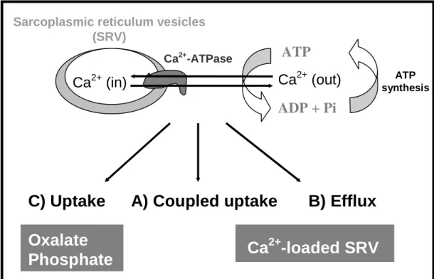
Using the SR vesicles it is possible to design experiments to observe only calcium uptake, using oxalate or phosphate in medium (Figure 7C), or only calcium efflux if we loaded the vesicles with calcium and put them in a medium without calcium (Figure 7B). At the latter experimental situation, the Ca2+ efflux could be passive when not associated with the pump activity or active when directly associated with intrinsic reactions to the SR Ca2+ pump mechanisms (Figure 7B). In a passive efflux of calcium the ATPase works as a Ca2+ channel (Figure 7B). For an active efflux of calcium, coupled to ATP synthesis (Figure 7B), it was demonstrated that in SR vesicles loaded with radioactive Ca2+, sediment by centrifugation and diluted into media containing EGTA, ADP and phosphate, Ca2+ active
Vanadate (mM) 0 1 2 3 4 5 C a 2 + a c c u m u la te d ( % o f c o n tr o l) 0 20 40 60 80 100 120 Decavanadate Metavanadate
efflux from the vesicles was strongly depressed (85%) by 40 µM decameric species while mM concentration of V1 has no effect [14].
Figure 7. Schematic representation of calcium translocation by sarcoplasmic reticulum at 3 different
experimental conditions: (A) coupled uptake (centre), (B) active efflux (right side) and (C) uncoupled accumulation (left side) (adapted from ref. 23).
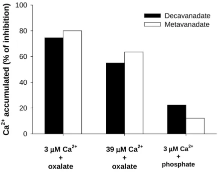
In another different experimental condition, it was observed that when the gradient of calcium is destroy, meaning using phosphate or oxalate to reduce the calcium concentration inside the vesicles to almost zero, and see only accumulation (Figure 7C, left side), the calcium ATPase is inhibited by both V10 and V1 solutions (1 mM total vanadium). However, it was observed that the inhibition of calcium accumulation by vanadate oligomers is dependent on the calcium gradient once it decreases 3 fold (from about 75 to 25%) by the increase of calcium inside the vesicles (presence of phosphate instead of oxalate) whereas the increase of calcium in the outside (comparison of the oxalate experiments) also prevent, although in a minor extend (from about 75 to 60%), the inhibitory effects induced by the vanadate solutions (Figure 8).
When the vesicles are damage, for example by the presence of a calcium ionophore that induces the leaking of calcium, the enzyme is working at full speed, and the ATP hydrolysis is not associated with calcium translocation. In this condition the ATPase activity is also inhibited by both decavanadate and metavanadate solutions with IC50 of 30
µM (meaning 3 µM decameric species) and 80 µM, respectively (not shown). Therefore, decavanadate exert noticeable effects, on comparison to metavanadate, on “physiological” calcium accumulation, e.g. coupled with ATP hydrolysis besides on the efflux of Ca2+ in particular when coupled with ATP synthesis. Being clearly different in inhibiting the SR calcium pump it is proposed a different mode of action for decavanadate in the mechanism
A) Coupled uptake
B) Efflux
C) Uptake
Oxalate
Phosphate
Ca
2+-loaded SRV
Ca
2+(in)
Sarcoplasmic reticulum vesicles (SRV)
ATP
ADP + Pi
Ca
2+(out)
Ca2+-ATPase ATP synthesisof calcium accumulation associated with ATP hydrolysis (Figure 5). Since decavanadate prevent both calcium accumulation and efflux, also both the processes 1 and 3 in the mechanism of calcium translocation by the SR calcium ATPase would be block (Figure 5).
Figure 8. Effect of SR vesicles calcium gradient on the inhibition of the calcium accumulation by
decavanadate and metavanadate solutions (1 mM total vanadium, meaning 100 µM decameric vanadate species). Specific experimental medium conditions: 5 mM oxalate, and 3 or 39 µM Ca2+; 10 mM phosphate, and 3 µM Ca2+ (adapted from ref. 25).
When the vesicles are damage, for example by the presence of a calcium ionophore that induces the leaking of calcium, the enzyme is working at full speed, and the ATP hydrolysis is not associated with calcium translocation. In this condition the ATPase activity is also inhibited by both decavanadate and metavanadate solutions with IC50 of 30
µM (meaning 3 µM decameric species) and 80 µM, respectively (not shown). Therefore, decavanadate exert noticeable effects, on comparison to metavanadate, on “physiological” calcium accumulation, e.g. coupled with ATP hydrolysis besides on the efflux of Ca2+ in particular when coupled with ATP synthesis. Being clearly different in inhibiting the SR calcium pump it is proposed a different mode of action for decavanadate in the mechanism of calcium accumulation associated with ATP hydrolysis (Figure 5). Because decavanadate prevent both calcium accumulation and efflux, also both the processes 1 and 3 in the mechanism of calcium translocation by the SR calcium ATPase would be block (Figure 5).
On the basis of these in vitro experiments, showing that V10 is able to inhibit SR calcium ATPase, it will be recognized that inhibition of Ca2+-ATPase and the following intracellular calcium release are responsible for decavanadate induced muscle contraction. Recently, characterization of decavanadate effect on cardiac miocytes described that decavanadate strongly affects calcium homeostasis [45]. Other authors described that concentration of decameric vanadate as low as 0.5 µM inhibits 50% of the SR calcium release induced by the second messenger inositol triphosphate (IP3) [46]. Moreover, the
C a 2 + a c c u m u la te d ( % o f in h ib it io n ) 0 20 40 60 80 100 Decavanadate Metavanadate 3 µµµµM Ca2+ + oxalate 39 µµµµM Ca2+ + oxalate 3 µµµµM Ca2+ + phosphate
described modulation of voltage-dependent Ca2+-actived cation channel point out to a sound physiological role for decameric vanadate [16]. In addition, besides affecting these systems involved in the regulation of calcium homeostasis, decavanadate also strongly inhibits cardiac mitochondria oxygen consumption with an IC50 of 1 µM (meaning 0.1 µM
decameric species) [47]. Once it was demonstrated that decameric vanadate induced rat liver mitochondrial depolarization at very low concentrations, half-depolarization with 39 nM decavanadate [47], it was suggested that this organelle is a potential cellular target for decavanadate. Biological aspects related with decavanadate in vivo effects are being reviewed in Chapter 9 of this book. We can not exclude that by affecting mitochondrial membrane potential, decavanadate may also indirectly modulate calcium homeostasis. Although mitochondria is clearly a potential subcellular target, the early event or the event that is first affect by V10 is yet to be clarify. Therefore, the role of decavanadate as an inhibitor of the systems involved in calcium homeostasis regulation, such as SR calcium pump and mitochondria, is very important because it links decavanadate with cell signalling. Moreover, its ability to act as an ATPases inhibitor can also indirectly affect diverse cellular processes (e.g. energy to copper transport).
5. EFFECTS OF VANADIUM COMPLEXES ON THE SR CALCIUM PUMP
SR calcium pump has proven to be an excellent model to study the effects of vanadium in Ca2+ homeostasis [15]. Besides vanadium (V), within the vanadate oligomeric species as described above, vanadium (IV), vanadyl is also known to affect contractile processes besides described as an antidiabetic and antyhipertensive agent, among other biological effects, although, in comparison to vanadate, it differs markedly in biochemical, pharmacological and toxicological properties [48, 49]. Concerning the calcium pump, it is considered to be of extreme importance the study with vanadium(IV) and vanadium (V) complexes for future mechanistic considerations once they provide insights into the mode of vanadium interaction with the calcium pump. Moreover, it is well-known that the use of organic ligands increased the vanadium effects [49]. In recent work, we consider the effects of three very different classes of vanadium compounds: i) two complexes with citric acid [50]; ii) two complexes of which are known to induce insulin-like effects [51] and iii) a compound that is a well-known analog of a vanadium-containing natural product [52].
It was observed that 1 mM of the two vanadium(V)-citrate complexes, namely vanadium(V)-citrate (VC) inhibits Ca2+ accumulation by 75 %, whereas the complex peroxovanadium(V)-citrate (PVC) inhibits the calcium pump activity by 33 % [53]. In contrast, a 1 mM metavanadate solution has no effect on Ca2+ accumulation. This latter observation was important because it was verified that both complexes partially decomposed into vanadate oligomers upon dilution in the medium. Moreover, it was described that the NMR signals from the VC complex (-548.0 ppm) and for PVC complex (-551.5 ppm) were broadened upon SR vesicle addition, being the relative order for the half width line broadening of the NMR signals, which reflect the interaction with the protein, V4>PVC>VC>V2=V1=1, with no effect observed for the V1 and V2 signals. On contrary, to the effects observed for the calcium accumulation, both complexes inhibited the ATPase activity with a similar IC50 of approx. 0.5 mM [50]. More potent inhibition of SR Ca2+
-ATPase activity were observed for the complexes pyridinedicarboxylate vanadium (V), oxovanadium (IV)-bis(maltolate) and amavadine with IC50 values of 25, 40 and 325 µM,
6. CONCLUDING REMARKS
Vanadium and skeletal muscle are strongly connected to each other, since almost thirty years ago vanadium was found in commercial ATP obtained from horse skeletal muscle. Vanadium is currently used as inhibitor of E1-E2 ion transport ATPases, e.g. the sarcoplasmic reticulum (SR) Ca2+-ATPase, besides being considered as a tool for the
comprehension of several biochemical processes. However, the complex chemistry of vanadium difficult the interpretation of the effects promoted in biological systems and concomitantly the understanding of the role of this element in life sciences. This is not surprising if we consider that among the chemical properties of vanadium, besides the multiple oxidation states, is the capacity of vanadium(V) species to condense with it self, forming vanadate oligomers, and with many compounds with biological interest.
In the present chapter, it is discussed the formation of decameric vanadate species, specific vanadate interactions with compounds containing phosphate and the effects of decavanadate on the calcium pump. It is clear that only decameric vanadate inhibits calcium accumulation promote by the SR calcium ATPase independently of the calcium gradient. It is proposed a different mode of interaction for decavanadate in the mechanism of calcium accumulation associated with ATP hydrolysis. A role for decavanadate might be recognized as an inhibitor of the systems involved in calcium homeostasis, considered to be very important because it links decavanadate to cell signalling. The use of vanadium complexes provides information on whether specific compounds inhibit SR calcium pump and are important for future considerations about vanadium interactions with sarcoplasmic reticulum Ca2+- ATPase, once it as been a useful tool in the understanding of the structure and function of the SR Ca2+-ATPase.
ACKNOWLEDGMENTS
This work has been supported by POCTI program financed through FEDER for the research project 38191/QUI/2001 (to M.A.). The author also thank to Joint Spanish-Portuguese CRUP project E-105/06.
REFERENCES
[1] Josephson, L., Cantley Jr., L.C. 1977, Biochemistry 16, 4572. [2] Beaugé, L.A., Glynn, I.M. 1978, Nature 272, 551.
[3] Cantley Jr, Josephson L.C., Warner L.R, Yanagisawa, M., Lechene, C., Guidotti, G., 1977, J. Biol. Chem. 252, 7421.
[4] Pedersen, P.L., Carafoli, E.1987, Trends Biochem. Sci.12, 146. [5] Davies, D.R., Hol, W.G.J., 2004, FEBS Lett. 577, 315.
[6] Shechter, Y., Diabetes 1990, 39, 1.
[7] Carmignani, M., Volpe, A. R., Masci, O., Boscolo, P., Di Giacomo, F., Grilli, A., Del Rosso, G. and Felaco, M.,1996, Biol. Trace Elem. Res. 51, 1-12
[8] E. Brener, S. Rubinstein, G. Cohen, K. Shternall, J. Rivlin, H. Breitbart, Biol. Reprod. 68 (2003) 837-845.
[9] Anaya-Ruiz, M., Perez-Santos, J. L., Talamas-Rohana, P., 2003, Int. J. Parasitol., 33, 663.
[10] Capella, L. S., Alcantara, J. S. M., Moura-Neto, V., Lopes, A.G., Capella, M. A. M., 2000, Tumor Biol. 21, 54.
[11] Sandirasegarane, L. and Gopalakrishnan, V., 1995, Life Sci. 56, 169.
[12] Chasteen, N.D. 1983, Struct. Bond., 53, 105. [13] Crans, D.C. 1994, Comments Inorg. Chem., 16, 35.
[14] Aureliano, M., and Gândara, R.M.C. 2005, J. Inorg. Biochem., 99, 979.
[15] Aureliano, M., and Madeira, V.M.C. 1998, Vanadium in the environment, J.O. Nriagu (Ed.), John Wiley & Sons, New York, 333.
[16] Nilius, B., Prenen J., Janssens, A., Voets, T., Droogmans, G., 2004, J. Physiol. 560, 753.
[17]Venkataraman, B.V., Ravishankar, H.N., Rao, A.V., Kalyani, P., Sharada, G., Namboodiri, K., Gabor, B., Ramasarma, T., 1997, Mol. Cell Biochem. 169, 27.
[18] Tiago, T., Aureliano, M., and Gutiérrez-Merino, C. 2004, Biochemistry, 43, 5551. [19] Messmore, J.M., Raines, R.T., 2000, Arch. Biochem. Biophys. 381, 25.
[20] Pezza, R.J., Villarreal, M.A., Montich, G.G., Argarana, C.E., 2002, Nucleic Acids Res. 30, 4700.
[21] Amado, A.M.; Aureliano, M.; Ribeiro-Claro, P.J.; Teixeira-Dias, J.J.C., J. Raman Spect. 1993, 24, 669-703.
[22] Ramos, S., Manuel, M., Tiago, T., Gândara, R.M.C., Duarte, R.O., Moura, J.J.G., Gutiérrez-Merino, C., and Aureliano, M. 2006, J. Inorg. Biochem., 100, 1734.
[23] Aureliano, M, Soares, S.S., Tiago, T., Ramos, S., Gutiérrez-Merino, C., 2007, ACS Symposium Series. 5th Vanadium (V) symposium (in press).
[24] Aureliano, M, Leta, J., Madeira, V.M.C., de Meis, L., 1994, Biochem. Biophys. Res. Comm., 201, 155.
[25] Aureliano, M., 1996, PhD thesis, University of Coimbra, Coimbra.
[26] Tracey, A.S., Gresser, M.J. and Parkinson, K.M., 1987, Inorg. Chem. 26, 629. [27] Tracey, A.S., Li, H. and Gresser, M.T., 1990, Inorg. Chem. 29, 2267.
[28] Cori, G.T., Colowick, S.P. and Cori, C.F.,1938, J. Biol. Chem. 123, 375. [29] Pick, U., 1982, J. Biol. Chem. 257, 6111.
[30] De Meis, L. and Vianna, A.L., 1979, Ann. Rev. Biochem. 48, 275. [31] Medda, P. and Hasselbach, W., 1983, Eur. J. Biochem., 137, 7.
[32] Csermely, P., Martonosi, A., Levy, G.C. and Ejchart, A.J., 1985, Biochem., J. 230, 807.
[33] Dux, L. and Martonosi, A., 1983, J. Biol. Chem., 258, 2599.
[34] Maurer, A. and Fleischer, S., 1984, J. Bioenerg. Biomembr., 16, 491.
[35] Vegh, M., Molnar, E. and Martonosi, A., 1990, Biochim. Biophys. Acta, 1023, 168. [36] Molnar, E., Varga, S. and Martonosi, A., 1991, Biochim. Biophys. Acta, 1068, 17. [37] Coan, C., Scales, D.J. e Murphy, A.J., 1986, J. Biol. Chem. 261, 10394.
[38] Aureliano, M., and Madeira, V.M.C. 1994, Biochim. Biophys. Acta, 1221, 259. [39] Varga, S., Csermely, P. and Martonosi, A., 1985, Eur. J. Biochem., 148, 119. [40] Csermely, P., Varga, S., Martonosi, A., 1985, Eur. J. Biochem., 150, 455. [41] Xu, C., Rice, W.J., He, W., Stokes, D.L. 2002, J. Mol. Biol. 316, 201-211
[42] Zhang, P., Toyoshima, C., Yonekura, K., Green, N.M., Stokes, D.L., 1998, Nature, 362, 835.
[43] Wang, T., Tsai, L-T, Solaro, R. J., De Gende, A.O.G. and Schwartz, A., 1979, Biochem. Biophys. Res. Commun., 91, 356.
[44] Bigelow, D.J. and Inesi, G.,1992, Biochim. Biophys. Acta.,113, 323.
[45] Soares, S.S., Henao, F., Aureliano, M., Gutiérrez-Merino, C. 2007, J. Biol. Chem., submitted.
[46] Fohr, K.J., Wahl, Y., Engling, R., Kemmer T.P., Gratzi, M., 1991, Cell Calcium, 12, 735
[47] Soares, S.S., Gutiérrez-Merino, C., Aureliano, M. 2007, J. Inorg. Biochem., in press. [48] Cadene, A, Grigorescu, F., Serrano, J-J., Cros, G., 1997, J. Pharmacol. Exp. Ther. 281,
491.
[49] McNeill, J.H., Yuen, V.G., Dai, S., Orvig, C., 2005, J. Mol. Cell. Biochem., 153, 175. [50] Kaliva, M, Raptopoulou, C.P.m Terzis, A., Saligoflou, A., 2003, J. Inorg. Biochem.
93, 161.
[51] Crans, D.C., Yang, L., Jakusch, T., Kiss, T, 2000, Inor. Chem. 39, 4409
[52] Garner, C.D. Armstrong, E.M., Berry, R.E., Beddoes, R.L, Collison, D., Cooney, J.J., Ertok, S.N., Helliwell, M., 2000, J. Inorg. Biochem., 80, 17.
[53] Aureliano, M, Tiago, T., Gândara, RMC, Sousa, A., Moderno, A., Kaliva, M., Salifoglou, A. Duarte, R.O., Moura, J.J.G., 2005, J. Inorg. Biochem, 99, 2355.
[54] Tiago T., Henao F., Gândara, R.M.C., Duarte R.O., Moura, J.J.G., Crans, D.C., Aureliano, M, abstract, 232nd ACS Meeting, 2006, S. Francisco, CA, USA.


![Figure 6. Inhibition of calcium accumulation coupled with ATP hydrolysis by decavanadate and metavanadate solutions (adapted from [25]](https://thumb-eu.123doks.com/thumbv2/123dok_br/18491871.901160/10.892.238.640.515.811/figure-inhibition-calcium-accumulation-hydrolysis-decavanadate-metavanadate-solutions.webp)