UNIVERSIDADE DO ALGARVE
Faculdade de Ciências e Tecnologia
Departamento de Química e Farmácia
Clinical Application of Extracellular
Vesicles in Vascular Calcification
Catarina Isabel Lousada Marreiros
Dissertação para obtenção do Grau de Mestre em
Ciências Farmacêuticas
Trabalho efetuado sob a orientação da Professora Doutora Dina Cristina Fernandes Rodrigues da Costa Simes e coorientação
da Professora Doutora Carla Alexandra São Bento Viegas
Clinical Application of Extracellular Vesicles in Vascular Calcification Page |ii Catarina Marreiros
“The scariest moment is always just before you start.” Stephen King
Acknowledgments
Clinical Application of Extracellular Vesicles in Vascular Calcification Page |iii Catarina Marreiros
Acknowledgments
Ainda que toda a dissertação se encontre escrita na língua inglesa, não poderia deixar de escrever as palavras de apreço que se seguem noutra língua que não na minha materna.
Em primeiro lugar, o meu maior agradecimento à Professora Doutora Dina Cristina Fernandes Rodrigues da Costa Simes e à Professora Doutora Carla Alexandra São Bento Viegas, que prontamente se disponibilizaram para me orientar e sem as quais não me imaginava a escrever esta dissertação.
À Universidade do Algarve e ao Mestrado Integrado em Ciências Farmacêuticas que me facultaram um ensino de excelência, baseado entre a proximidade do aluno com instituição e docentes. Destaco um especial agradecimento à Professora Doutora Isabel Ramalhinho por todo o esforço feito em prol do curso e por toda a dedicação para com os alunos do mesmo. A todos os restantes professores com quem tive o privilégio de me cruzar e trabalhar durante este período, um genuíno “obrigada”. Contribuíram para o meu enriquecimento pessoal e para que me tornasse na farmacêutica que sou hoje. O conhecimento de nada serve se não o soubermos transmitir aos que nos seguem. Espero saber fazê-lo daqui em diante tão bem quanto o fizeram comigo.
Às equipas de trabalho da farmácia hospitalar do Hospital Fernando Fonseca e da farmácia comunitária Holon-Pragal, agradeço por me terem disponibilizado a oportunidade de estágio para me preparar para o sector farmacêutico.
Não posso deixar de agradecer às pessoas que me acompanharam mais de perto neste percurso e que apesar da distância fizeram do Algarve a minha segunda casa. Aos meus colegas de turma, que tive o privilégio de conhecer e com os quais partilhei cinco anos repletos de momentos e aventuras que levo como recordações para a vida.
Ao Jorge, por estar sempre ao meu lado.
Por último, mas não menos importante, um enorme OBRIGADA a todos os meus familiares, em especial à minha irmã, pai, mãe e avó, por me amarem e apoiarem incondicionalmente na decisão de estudar longe de casa. Sem vocês nunca teria conseguido.
Abstract
Clinical Application of Extracellular Vesicles in Vascular Calcification Page |iv Catarina Marreiros
Abstract
Cellular communication and signaling are essential for the organization, preservation, and proper functioning of different cell types in multicellular organisms. Recently the study of the mechanisms for intercellular communication mediated by extracellular vesicles (EVs) has been the focus of research within the scientific community.
Extracellular vesicles exert diverse physiological and pathophysiological functions by serving as vehicles of horizontal transfer of protein, lipids, DNA, and RNA between cells. These cellular interaction processes can influence, among others, the developmental patterning of tissues, as the case of vascular smooth muscle cells (VSMCs) differentiation with the generation of EVs playing an important role in vessel calcification. These calcification-competent EVs are thought to create the first nidus for calcification through the clustering of calcium-phosphate (Ca/P) minerals within the extracellular matrix of blood vessels, resembling skeletal mineralization.
Extracellular vesicles are a promise and fresh therapeutic area for the delivery of different synthetic and biological molecules in cellular therapy. Indeed, EVs-based drug delivery is an experimental field that has evolved greatly in the past few years, with an especial attention for miRNA therapeutics. The potential advantages over the existing synthetic delivery systems are attracting much attention, adding a new pharmacological mean of intervention using EVs as an encouraging drug transportation system in the treatment of cardiovascular diseases.
However, in order to speed up the clinical application of EVs as the next generation of targeted gene delivery vehicles, much work still needs to be done focusing the features of EVs-mediated vascular calcification in order to highlight potential therapeutic targets within this pathology and achieve progress in diagnosis and treatment in the cardiovascular filed.
Keywords: Cellular communication; Extracellular vesicles; Vascular
Resumo
Clinical Application of Extracellular Vesicles in Vascular Calcification Page |v Catarina Marreiros
Resumo
A comunicação e sinalização celular são essenciais para a organização, preservação e bom funcionamento dos diferentes tipos de células dos organismos multicelulares. Recentemente, o estudo dos mecanismos de comunicação intercelular mediados por vesículas extracelulares têm sido foco de diversos estudos na comunidade científica.
As vesículas extracelulares exercem diversas funções fisiológicas e fisiopatológicas ao servir como veículos para a transferência horizontal de proteínas, lípidos, ADN e ARN entre as células. Estes processos de interação celular podem influenciar, entre outros, o padrão de desenvolvimento de tecidos, como é exemplo a diferenciação das células musculares vasculares lisas com a geração de vesículas extracelulares, que desempenham um papel crucial na mediação da calcificação vascular. Estas vesículas extracelulares, competentes para a calcificação, criam o primeiro sítio de calcificação através da aglomeração de minerais de cálcio-fosfato (Ca/P) na matriz extracelular dos vasos sanguíneos, similar à mineralização do osso.
As vesículas extracelulares representam uma promissora e nova área terapêutica para o transporte de diversas moléculas sintéticas e biológicas na terapia celular. O transporte de fármacos baseado nas vesículas extracelulares é um campo experimental que evoluiu muito nos últimos anos, com especial atenção para a terapêutica com miRNAs. As potenciais vantagens em relação aos existentes sistemas sintéticos de transporte estão a atrair muita atenção, revelando uma nova medida farmacológica de intervenção usando as vesículas extracelulares como um sistema encorajador de transporte de fármacos no tratamento de doenças cardiovasculares.
De modo a acelerar a aplicação clínica das vesículas extracelulares como a próxima geração de veículos de transporte direcionados, muito trabalho tem ainda que ser feito de modo a evidenciar as características da calcificação vascular mediada pelas vesículas com vista a destacar potenciais alvos terapêuticos dentro desta patologia e desta forma, progredir no diagnóstico e tratamento no campo cardiovascular.
Palavras-chave: Comunicação celular ;Vesículas extracelulares; Calcificação
Resumo Alargado
Clinical Application of Extracellular Vesicles in Vascular Calcification Page |vi Catarina Marreiros
Resumo Alargado
As células utilizam vários meios de comunicação de modo a assegurar a troca de materiais e a transferência de informações com vista o desenvolvimento, reparação e sobrevivência dos tecidos. Para alcançarem o sucesso na realização das suas funções celulares e manterem a homeostase dos seus tecidos, as células recetoras têm de possuir a capacidade de receber e interpretar os sinais celulares recebidos.
As vesículas extracelulares são uma família de partículas nano-esféricas, de tamanho variável, libertadas para o ambiente extracelular por meio de uma célula mãe. Retratam um novo tipo de sinalização intercelular que pode ser encontrada tanto em células procariotas como eucariotas. Estudos revelaram que as vesículas extracelulares são secretadas pela maioria, se não por todos os tipos de células humanas e podem, igualmente, ser encontradas em vários fluídos corporais, incluindo o sangue. De modo a proteger o conteúdo da carga interna da degradação enzimática existente nestes fluídos, a membrana exterior das vesículas é formada por uma bicamada lipídica protetora.
Apesar da sua descoberta remontar aos anos 60, apenas recentemente ficou claro que as vesículas extracelulares são veículos fundamentais para a sinalização celular, capazes de transmitir informações complexas a longas e curtas distâncias em relação à sua célula de origem.
Embora as vesículas extracelulares sejam amplamente classificadas em três classes principais, tendo em consideração o seu tamanho e mecanismos de biogénese (exossomas, microvesículas e corpos apoptóticos), existe alguma discrepância na literatura relativamente à sua nomenclatura. Isto é, podem ser encontradas algumas disparidades, onde frequentemente as vesículas extracelulares são utilizadas como sinónimo de matrix vesicles (MVs). Estas últimas são na verdade um subtipo de vesículas, que em condições fisiológicas normais são produzidas por células ósseas e têm a capacidade de reter cristais de Ca/P, promovendo a mineralização no tecido ósseo. Devido ao facto das vesículas extracelulares, envolvidas na deposição patológica de minerais nos vasos, se assemelharem às MVs encontradas nas células ósseas, elas são comummente designadas por MVs no contexto vascular.
No interior da camada média dos vasos sanguíneos residem as células musculares vasculares lisas que possuem a capacidade extraordinária de alterar o seu fenótipo entre
Resumo Alargado
Clinical Application of Extracellular Vesicles in Vascular Calcification Page |vii Catarina Marreiros
um estado celular contrátil e um sintético. Ao contrário de muitas outras células no corpo humano, tais como as cardíacas e ósseas, as células musculares vascular lisas não maturam totalmente, retendo esta capacidade de diferenciação fenotípica durante o seu tempo de vida. Este processo tem sido implicado como um dos principais mecanismos de libertação de vesículas extracelulares, com alto poder de calcificação, para o meio extracelular dos vasos sanguíneos.
As vesículas extracelulares secretadas para o espaço extracelular podem potencialmente transportar dentro do seu conteúdo celular proteínas, ácidos nucleicos e receptores de membrana, provenientes da sua célula mãe. Para além desta particularidade, o conteúdo celular transportado pelas vesículas varia ainda de acordo com o tipo de célula envolvida e com as condições fisiológicas ou patológicas existentes no momento da sua formação e secreção celular. Em condições normais, as vesículas extracelulares derivadas das células musculares vasculares lisas não apresentam um poder de calcificação porque contêm no seu interior inibidores da mineralização e microARN regulatório como forma de sinalização, que previne o processo de diferenciação destas células.
No entanto, sob um contexto patológico, a sinalização celular mediada pelas vesículas torna-se comprometida, assistindo-se a um desequilíbrio dos inibidores de calcificação e à promoção de um ambiente favorável à diferenciação das células musculares vasculares lisas. Na matriz extracelular dos vasos sanguíneos, estas células começam a libertar vesículas com alto poder de calcificação, isto é, vesículas capazes de reter cristais de Ca/P no seu interior, que formam um dos primeiros sítios de mineralização, com consequente propagação da calcificação vascular.
Previamente considerada como um processo passivo de significância fisiopatológica limitada e como uma consequência inevitável do envelhecimento, a calcificação vascular entende-se pela deposição inadequada e patológica de cristais de Ca/P nos tecidos vasculares, representando um grande contributo para a progressão da doença cardiovascular, uma das principais causas de morte nos países industrializados. A cada ano, as doenças cardiovasculares causam 3,9 milhões de mortes na Europa, representando 45% de todas as mortes. Dada a associação entre a calcificação e os outcomes cardiovasculares, é urgente uma melhor compreensão desta patologia, com
Resumo Alargado
Clinical Application of Extracellular Vesicles in Vascular Calcification Page |viii Catarina Marreiros
destaque principal sobre os mecanismos que levam à deposição de minerais e ao seu crescimento progressivo na parede dos vasos sanguíneos desde os primeiros estadios.
A calcificação vascular pode ainda ocorrer nos vasos sanguíneos, válvulas e tecidos cardíacos, manifestando-se sob a forma de calcificação aterosclerótica, calcificação da camada média, também conhecida por esclerose média de Monckeberg, calcifilaxia (um subtipo de calcificação da camada média) e calcificação das válvulas cardíacas.
Apesar dos vários estudos que focam os mecanismos básicos desta complexa patologia, ainda existem alguns processos que precisam de esclarecimento adicional. No entanto, a calcificação vascular é atualmente interpretada como uma resposta patológica multifacetada a diferentes estímulos que podem surgir por diversos mecanismos fisiopatológicos, tais como níveis séricos elevados de cálcio e fosfato, perda de inibidores de calcificação vascular, diferenciação e/ou morte das células musculares vascular lisas e a libertação de vesículas extracelulares com poder de mineralização/calcificação.
Uma vez que representam vetores naturais de informação biológica, capazes de alterar mecanismos fisiológicos através da transferência de mediadores benéficos ou prejudiciais à célula recetora, diversos são os estudos que evidenciam as vesículas no processo de calcificação vascular. De facto, pelo seu papel fundamental nos mecanismos de calcificação e pela capacidade de transferir o seu conteúdo celular através de mecanismos naturais de absorção endógena, a promissora aplicação clínica das vesículas como sistemas de transporte direcionado de fármacos tem sido uma área de investigação intensa. Adicionalmente, como o seu conteúdo celular reflete o estado fisiológico da célula mãe, estas vesículas podem igualmente ser aplicadas em diagnóstico clínico como biomarcadores patológicos.
Pesquisas futuras dentro desta linha de pensamento poderão levar a uma nova geração de terapias direcionadas para os múltiplos pontos de intervenção da formação da microcalcificação dos vasos sanguíneos, onde as vesículas serão uma escolha terapêutica capaz de atingir células específicas de modo a prevenir ou retardar os resultados cardiovasculares da calcificação vascular.
Resumo Alargado
Clinical Application of Extracellular Vesicles in Vascular Calcification Page |ix Catarina Marreiros
No entanto, é ainda necessária muita investigação com vista a completar a compreensão atual dos mecanismos de calcificação extracelular mediados pelas vesículas. Um passo adiante neste campo passará inegavelmente pelo desenvolvimento de protocolos padronizados para o isolamento das vesículas, a fim de assegurar que a caracterização documentada possa permanecer comparável entre diferentes laboratórios.
Contents
Clinical Application of Extracellular Vesicles in Vascular Calcification Page |x Catarina Marreiros
Contents
Acknowledgments ... iii
Resumo ... v
Figure Index ... xii
Abbreviations ... xiii
1 - Intercellular Communication ... 1
1.1 - Types of Intercellular Communication ... 1
1.1.1 - Extracellular Vesicles... 3
1.1.1.1 - Biological Properties and Relevance... 3
1.1.1.2 - EVs Research Field - Historical Notes ... 4
1.1.1.3 - EV’s characterization and current classification ... 5
1.1.1.4 - EVs Cellular Uptake Mechanisms ... 9
1.1.1.5 - Nomenclature controversy ... 10
2 - Vascular Anatomy and Physiology ... 12
2.1- General Characteristics of Blood Vessels ... 12
2.1.1 - Tunica Intima ... 13
2.1.2 - Tunica Adventitia ... 14
2.1.3 - Tunica Media ... 14
2.1.3.1 - Vascular Smooth Muscle Cells ... 14
3 - Calcification in Cardiovascular Disease ... 16
3.1 - Vascular Calcification ... 16
3.1.1 - Types of Vascular Calcification ... 17
3.1.1.1 - Intimal Calcification ... 17
3.1.1.2 - Medial Calcification ... 17
3.1.1.3 - Calcific uremic arteriolopathy ... 18
3.1.1.4 - Cardiac Valve Calcification ... 18
3.1.2 - Vascular Calcification Mechanisms ... 19
3.1.2.1- Osteochondrogenic Differentiation ... 19
3.1.2.2 - Loss of mineralization inhibitors ... 20
3.1.2.2.1 - Matrix- Gla Protein ... 20
3.1.2.2.2 - Fetuin-A ... 21
3.1.2.2.3 - Gla-Rich Protein ... 22
Contents
Clinical Application of Extracellular Vesicles in Vascular Calcification Page |xi Catarina Marreiros
3.1.2.4 - Role of Calcifying EVs ... 23
3.1.2.4.1 - The emerging role of RNA... 26
4 - Clinical Role of Extracellular Vesicles in Cardiovascular Disease ... 28
4.1 - EVs-mediated Delivery of Therapeutic Biomolecules... 28
4.1.1- Endogenous Drug-Loading Mechanisms ... 30
4.1.2- Exogenous Drug-Loading Mechanisms ... 30
4.1.3 - Therapeutic Delivery of Nucleic Acid-Based Drugs ... 32
4.2 - Extracellular vesicles as promising biomarkers ... 34
5 - Conclusion ... 36
Figure Index
Clinical Application of Extracellular Vesicles in Vascular Calcification Page |xii Catarina Marreiros
Figure Index
Figure 1.1- Cells produce different types of extracellular vesicles that vary in size. ... 7
Figure 2.1- Structure of blood vessels. ... 1212
Figure 2.2- VSMC’s phenotypic plasticity. ... 15
Figure 3.1- Different types of vascular calcification. ... 19
Figure 3.2- Mechanisms of vascular calcification. ... 23
Abbreviations
Clinical Application of Extracellular Vesicles in Vascular Calcification Page |xiii Catarina Marreiros
Abbreviations
ABs Apoptotic Bodies ANXs Annexins
ATP Adenosine Triphosphate BPM-2 Bone Morphogenic Protein 2 Ca Calcium
Ca/P Calcium-Phosphate
CaSR Calcium-Sensing Receptor CAD Coronary Artery Disease
CKD Chronic Kidney Disease
CPP Calciprotein Particles
CUA Calcific Uremic Arteriolopathy
CVD Cardiovascular Disease DM Diabetes Mellitus DNA Deoxyribonucleic Acid ECM Extracellular Matrix
ESRD End-Stage Renal Disease EU European Union
EV Extracellular Vesicle FMC Fetuin-Mineral Complex
GRP Gla-Rich Protein
HA Hydroxypatite
ISEV International Society of Extracellular Vesicles
miRNA Micro Ribonucleic acid mRNA Messenger Ribonucleic Acid MGP Matrix-Gla Protein
MMP Metalloproteinase MVs Matrix Vesicles MVB Multivesicular Body
Abbreviations
Clinical Application of Extracellular Vesicles in Vascular Calcification Page |xiv Catarina Marreiros
MVSMCs Mouse Vascular Smooth Muscle Cells Msx2 Msh Homeobox 2
NaPi Sodium-Phosphate Cotransporters
P Phosphate
PAD Peripheral Arterial Disease
PS Phosphatidylserine
RNA Ribonucleic acid RNAse Ribonuclease
Runx2 Runt-related transcription factor 2 SMCs Smooth Muscle Cells
VC Vascular Calcification
VKDP Vitamin-K Dependent Protein VSMCs Vascular Smooth Muscle Cells
Intercellular Communication
Clinical Application of Extracellular Vesicles in Vascular Calcification Page |1 Catarina Marreiros
1 - Intercellular Communication
During the course of evolution, both prokaryotes and eukaryotes developed elegant cell-to-cell communication strategies to adapt to stimuli created by the surrounding environment. The development of multicellular organisms most likely began when cells remained associated in small colonies after division, instead of separating into individual cells. A few prokaryotes and several unicellular eukaryotes, such as many fungi and slime molds, exhibit such rudimentary social behavior. The full flowering of multicellularity, however, occurred in eukaryotic organisms, whose cells became differentiated and organized into groups or tissues in which cells perform specialized functions (1).
Cellular communication is considered one of the most important regulatory mechanisms for cell growth, differentiation and tissue remodeling that allows multicellular organisms to maintain regular cellular functions. In fact, a key step in the evolution of multicellularity was indeed the ability of cells to contact tightly and communicate specifically with each other (1).
Cells use several means of communication for the exchange of materials and the transfer of information in order to maintain tissue homeostasis, development, repair and survival. To succeed their inner functions, an appropriate communication through signals must be present and capable of being interpreted by specific and complex machinery in the recipient cell (1). Regardless the nature of the signal, the target cell responds by means of a specific protein called receptor that specifically binds to the signaling molecule and then initiates a response in the target cell. In most cases the receptors are transmembrane proteins on the target cell surface, and when they bind to an extracellular signaling molecule, they become activated generating a cascade of intracellular signals that alter the behavior of that specific cell. In some cases, however, the receptors can be inside the target cell and the signaling ligand has to be incorporated by the cells for activation (1,2).
1.1 - Types of Intercellular Communication
Cells have evolved a variety of signaling mechanisms to accomplish the transmission of important biological information. Classically in cell biology, eukaryotic cells communicate directly, requiring cell to cell contact, or indirectly, via soluble
Intercellular Communication
Clinical Application of Extracellular Vesicles in Vascular Calcification Page |2 Catarina Marreiros
molecules secreted by one cell which are then carried away to target cells. Depending on the distance that the signaling molecule has to travel, intercellular communication can be classified in two types, the direct cell to cell communication and the distant cell communication (2).
The direct cell-to-cell signaling can be mediated by juxtacrine interactions between touching cells, in which signals can be transmitted by junctional complexes including tight junctions, desmosomes, adherens and gap junctions. On the other hand, distant cell communication is carried out by signaling molecules that can be carried far afield to act on distant targets (long-range) or by local mediators affecting the cells in the immediate environment of the signaling cell (short-range) (2). These soluble factors can act on the cell itself (autocrine signaling) or influence both neighboring (paracrine signaling) and distant cells (endocrine signaling) allowing cellular communication in the absence of physical contact (3).
Recently a distant intercellular communication mechanism mediated by extracellular vesicles has gained a growing interest among the scientific community. Extracellular vesicles (EVs) are a new type of intercellular signaling, representing an universal and highly conserved active cellular function that can be found in both prokaryotics and eukaryotics cells (3).
Despite their discovery decades ago, only now has become clear that these vesicles are important vehicles of cellular signaling, capable of carrying out complex information through autocrine, paracrine and even endocrine ways (3). EVs released by cells into the extracellular space can potentially reach distant tissues, transporting within their cargo proteins, lipids, nucleic acids and membrane receptors from their origin cell. This functional EV’s content differs not only according to its cell of origin cell function but also with the specific physiological and pathological conditions existing at the time of packaging and secretion (4). In the last decade, the number of studies recognizing EVs as a crucial and integral part of cellular microenvironment and communication has grown exponentially revealing a new scenario in terms of understanding signal and molecule transfer between cells, not only locally, but also over long distances (5,6).
Intercellular Communication
Clinical Application of Extracellular Vesicles in Vascular Calcification Page |3 Catarina Marreiros
1.1.1 - Extracellular Vesicles
1.1.1.1 - Biological Properties and Relevance
Extracellular vesicles are a family of heterogeneous and spherical nano sized particles released by cells into the extracellular environment, differing in terms of the contents, size and formation mechanisms. EVs are formed by a lipid bilayer membrane that encloses a small organelle free cytosol, containing proteins and nucleic acids present on the EV’s origin cell (7).
Due to their rich composition and capacity to interact with other cells, EVs play a functional role in many biological processes, having the remarkable capability to deliver combinatorial information to multiple cells all over the body. However, very little is known about the role of EVs in homeostasis maintenance in normal physiological conditions. EVs intrinsic cell functions and regulation only started to be highlighted recently (8).
Studies reveal that EVs are secreted by most, if not all, human cell types, like epithelial cells, fibroblast, hematopoietic cells, immune cells, tumor cells, and even stem cells. In addition, EVs can be found in several body fluids, including urine, saliva, nasal fluid, amniotic fluid, breast milk, seminal plasma, bronchoalveolar fluid, bile, cerebrospinal fluid, and in blood (3).
Depending on their biogenesis and size EVs are classically classified in exosomes, microvesicles, and apoptotic bodies (ABs). Their lipid bilayer membrane, containing various proteins and receptors, protects their bioactive internal cargo from the enzymatic degradation, present in the extracellular environment, conceding them the ability to deliver both physiological and pathological information (9).
It has been studied and documented that the transfer of RNA and miRNA between cells are involved in the change of target cell phenotype and microenvironment, reprogramming their functions by pleiotropic effects, potentially leading to several pathological conditions (10). Since EVs are capable of becoming enriched with molecules expressed by the cells of origin, current studies have focused on the EVs capacity to induce epigenetic changes in target cells, resembling the origin cell. In this context, several studies have been showing that EVs play a fundamental role in the transfer of genetic information between cells promoting cellular differentiation (11,12).
Intercellular Communication
Clinical Application of Extracellular Vesicles in Vascular Calcification Page |4 Catarina Marreiros
The understanding of why cells release vesicles, how vesicles play a role in intercellular communication, how vesicles may contribute to cellular homeostasis, and especially how vesicles can mediate pathophysiological conditions, reveals a very complex and sophisticated role of vesicles both in health and disease. A growing body of evidence suggests that EVs are truly involved in physiological as well as pathological processes, and the interest in their biological role and clinical application is increasing exponentially (13).
1.1.1.2 - EVs Research Field - Historical Notes
The discovery of cell derived extracellular vesicles followed the introduction of the transmission electron microscope, and their relevance occurred simultaneously in many physiological settings, without the realization that this form of cellular function and communication is an universally shared cell biological property (14).
In history of cell biology, the detection of these particles can be traced back to initial researches on blood coagulation. Originally reported in 1946 by Chargaff and West, EVs were observed as procoagulant platelet-derived particles in normal plasma (15). In the same area, Peter Wolf identified these particles as part of a disposal mechanism to discard unwanted materials from platelets, labeling them as “platelet dust”, in 1967 (12). In the same year, the releasing of extracellular vesicles during the physiological mineralization processes in bones was discovered and reported by and Bonucci (17), and in 1969 by Anderson (16), that originally termed them as matrix vesicles. Twenty years later, the research group led by Rose Johnstone introduced for the first time the term exosomes when studying reticulocytes undergoing maturation into red blood cells (11).
Later, Graça Raposo and colleagues demonstrated that these type of vesicles, isolated from Epstein-Barr virus transformed B lymphocytes, were antigen-presenting and able to induce T-cell responses. These studies provided the basis for the hypothesis that exosomes could play an active role in intercellular communication, particularly in the immune system, inciting the very first attempt of using EVs as a new type of anticancer therapy in humans (19).
The discovery of apoptotic bodies and microvesicles came later, in 1972 by John Kerr (20) and 1991 by Janet Stein (21), respectively. Since then apoptotic bodies have been largely related with programmed cellular death, and their biological role in cellular
Intercellular Communication
Clinical Application of Extracellular Vesicles in Vascular Calcification Page |5 Catarina Marreiros
communication is still unclear. By contrast, microvesicles have been widely investigated and found to be secreted by various cell types. Their role in cellular communication and differentiation is nowadays an extensive research field, alongside with exosomes.
Findings that EVs could in fact enclose RNA and microRNA led to an increasing interest of EVs as novel mediators of intercellular communication (22). In 2011, the International Society of Extracellular Vesicles (ISEV) was created as a need for standardization and clarification of several aspects concerning the EVs field, such as the nomenclature, purification and characterization methodologies (3).
Nowadays, intensive investigation has highlighted the role of EVs in many pathological conditions, such as lung injury (7), liver diseases (23), neurodegenerative diseases (24), cancer (25). The increased interest in EVs, allied to the development of improved isolation and detection methods, led to novel insights into possible clinical applications of these vesicles. In these last two decades, cardiovascular disease was one of the clinical areas most intensely studied in the extracellular vesicles field (26).
1.1.1.3 - EV’s Characterization and Current Classification
The small differences in physical properties and composition of EVs, the need of high sensitivity techniques to detect them, the lack of standard isolation methodologies and the fact that the same cell type may even secrete different types of vesicles makes EVs detection and isolation very challenging. Furthermore, the content and number of EVs secreted depends on the cells they originate, the stimulus of production and the mechanism of vesicle generation (27).
EVs can be categorized into three main classes based on their size and biogenesis pathways. Although in the literature diverse scales are used by different authors, it is commonly accepted that microvesicles diameter can range from 50 nm to 1000 nm, exosomes from 10 nm to 100 nm, and apoptotic bodies that greatly vary in size can have between 1000 nm and 5000 nm of diameter. Overall EVs comprise a wide variety of vesicles with different cargos, and with overlapping in size for different EVs types (13).
Apoptotic bodies also called apoptotic blebs, apoptotic bodies or apoptosomes are membrane limited vesicles that can be classified as a subclass of EVs. They are released through protrusions and fragmentation of the cell membrane of cells undergoing
Intercellular Communication
Clinical Application of Extracellular Vesicles in Vascular Calcification Page |6 Catarina Marreiros
apoptosis. These particles are the largest extracellular vesicles harboring cell organelles and nuclear fractions surrounded by a permeable membrane. Under specific conditions, ABs can be more abundant than exosomes or microvesicles. A feature present in these vesicles is the externalization of phosphatidylserine (PS), a phospholipid component of the cellular membrane, a phenomenon also seen in microvesicles (28).
Their rapid elimination promoted by phagocytic cells such as macrophages, as a respond to specific signaling molecules, makes this population less well characterized, and the information on the potential role of apoptotic bodies in cardiovascular diseases is very scarce (23).
Microvesicles also found in literature as membrane particles, ectosomes or shedding microvesicles, are cell surface derived EVs usually larger than exosomes. They are generated by direct budding and subsequent fusion of the plasma membrane into the extracellular environment, comprehending a cytoskeleton remodeling and externalization of PS, like ABs. When the plasma membrane is activated by extrinsic stimuli, membranous globular extensions are shed from the plasma membrane surface and subsequently released to the extracellular space, as illustrated in Figure 1.1. This releasing mechanism appears to be increased in inflammatory conditions (9).
Exosomes are the smallest extracellular vesicles although no clear cut-off value separates microvesicles from exosomes in terms of size. They are present in many, if not in all biological fluids, and are the extracellular vesicles that have received most attention over the past few years (28). Additionally, their physicochemical properties and biological function are well documented in contrast to other types of vesicles (30).
Exosomes are intraluminal nano vesicles generated from multivesicular bodies (MVBs), a late endosomal compartment from the cell trafficking machinery. The biogenesis of exosomes is typically thought to occur in a twostep process, firstly involving the formation of cytoplasmic MVBs that gather and package molecules into luminal membrane bound structures, and secondly by their subsequent fusion with the plasma membrane which releases these internal vesicles as exosomes enabling their diffusion into the extracellular environment (8), as represented in Figure 1.1.
Intercellular Communication
Clinical Application of Extracellular Vesicles in Vascular Calcification Page |7 Catarina Marreiros
In the majority of EVs studies, clinical interests have been focused on exosomes and microvesicles rather than ABs, given that vesicles and their compositions derived from living cells can potentially play more crucial functions in the development of pathological conditions (7).
In order to confirm the presence of EVs and identify the specific subtype in isolated preparations, researchers have struggled to identify what could be designated as EV’s marker proteins. To confirm the presence of EVs, there is a subset of biomarkers existing in either EV’s membrane or cytosol that helps to separate EVs from non-vesicular entities present in preparations. (31). However, studies have shown that these markers are not common to all EVs subtypes, which depend and correlate to the EV’s cell origin functions, revealing that EV’s marker proteins can be highly variable (31). Indeed, the main challenge of ISEV and research groups within this field confines in the identification of specific markers to distinguish each of EVs subtypes (32).
Although EVs components are different among cells, some proteins are thought to be essential EV’s constituents, and therefore, can be commonly found in all EVs
Figure 1.1 - Cells produce different types of extracellular vesicles that vary in size and mechanism of vesicle generation. Exosomes and microvesicles are produced by
normal and pathologic cells, while apoptosis triggers the release of apoptotic bodies. Exosomes have been identified to be released from multivesicular bodies during their fusion with the plasma membrane. Microvesicles and apoptotic bodies are shed from the plasma membrane through direct outward budding of the plasma membrane, which defines their diameter and molecular composition with the difference that ABs are generated from a cell undergoing apoptosis; adapted from (29).
Intercellular Communication
Clinical Application of Extracellular Vesicles in Vascular Calcification Page |8 Catarina Marreiros
regardless of their origin. These molecules include tumour susceptibility gene 101 (TGS101) (31), and testraspanins such as CD9 and CD63 (33).
Despite the field of EVs research has not developed enough in order to identify a list of EV-specific markers that clearly distinguish each EVs subtypes (34), some protein markers are often used in the literature as a mean to classify the EVs subtypes present in a mixed population.
ABs are distinctly different from exosomes and microvesicles because they abundantly contain histones associated with membranes that float at high sucrose densities and because they are very heterogeneous in size and morphology when observed by electron microscopy (35).
Microvesicles are enriched in phosphatidylserine, integrins, selectins, and CD40 ligand (36). However, distinguish between exosomes and microvesicles is very challenging. Although there are some differences in the size and composition of these EVs, in a mixed EVs population it remains impossible to completely separate exosomes and microvesicles with the currently available purification methods. These methodological issues represent one of the biggest problems in terms of EVs isolation and characterization (37,38).
Common protein markers used to identify the presence of exosomes in an EVs population are components of the endosomal sorting complex required for transport, like ALG-2-interacting protein X (39) and TGS101 (31), heat shock proteins like HSP70 and HSP90 and tetraspanins CD9, CD63, CD81 (40). Due to the fact that some of the previous proteins are markers for the detection of EVs in general, a combinatorial identification of these markers is preferred rather than a single biomarker for exosomes characterization (32).
Additionally, some authors defend that in order to achieve a rigorous EVs characterization, it is required a combination of the most common techniques used in EVs studies (39), such as flow cytometry, dynamic light scattering, nanoparticle tracking analysis, scanning and transmission electron microscopy, atomic force microscopy, and detection of several marker proteins, for proper assessment of EVs quantity, size and features.
Intercellular Communication
Clinical Application of Extracellular Vesicles in Vascular Calcification Page |9 Catarina Marreiros
As the method used for EVs isolation can affect numbers and composition of the obtained vesicles, it is important to choose a suitable isolation method that ensures reliable and comparable measurements of EVs. Although, currently there is no consensus on a “model” method to isolate and/or purify EVs, and therefore no optimal method is uniformly used by investigators (34), Several researchers support the use of differential ultracentrifugation for isolation purposes and size-exclusion chromatography coupled with membrane filtration for subtype EVs purification from plasma/serum (23).
To facilitate and improve the exchange of information between investigators, in 2012 the ISEV defined extracellular vesicles as a generic term that can be applied to all types of vesicles found in the extracellular space (41). However, the use of outdated isolation and detection techniques allied to in vitro studies and classification based on different criteria, conducted to inconsistencies found in older and some recent literature regarding EVs nomenclature and classification (42).
1.1.1.4 - EVs Cellular Uptake Mechanisms
Extensive evidence on all types of vesicles indicates that EVs are a key player in intercellular communication, capable of carrying out a range of signals that can have a significant impact on the phenotype of the recipient cells. However, for this phenotypic effect to occur, EVs need to fuse with target cell membranes and combine their content with the cytoplasmic compartment of target cells (8).
Both release and uptake mechanisms depend on the donor and recipient cell type, as well as their physiological state and the conditions of the existing microenvironment. The internalization mechanism is proved to be an energy dependent process that requires a fully functioning cell cytoskeleton. There are two distinct mechanisms that EVs can enter a cell. They can be internalized via the fusion of the EV membrane with the target cell membrane or they can enter the cell by endocytosis. The uptake through endocytosis can be categorized into the different types of endocytotic processes including clathrin-mediated endocytosis, caveolin mediated endocytosis, lipid raft-mediated endocytosis, macropinocytosis, and phagocytosis. Although endocytosis appears to be the principal uptake mechanism, there is little agreement as to which type of endocytic via is most important (43).
Intercellular Communication
Clinical Application of Extracellular Vesicles in Vascular Calcification Page |10 Catarina Marreiros
When EVs enter by endocytosis, in order to exert its cellular effects, their cargo must be released before being destroyed or discard by the recipient cell, since endosomes mature into lysosomes or are ejected out again through the MVB plasma membrane fusion pathway. However, this mechanism of transferring the EVs cargo out of the endosomal compartment is still unclear (8).
The second EVs entrance mechanism via direct fusion of the EV membrane with the cell plasma membrane requires the fusion of two distinct lipid bilayers – EV and recipient cell membrane - in an aqueous environment. The lipid bilayers are brought into close proximity and a fusion pore is created permitting the two hydrophobic cores to mix with the delivery of EVs cargo into the recipient cell (43).
Different cell types are able to take up EVs using various mechanisms resulting in either functional transfer or degradation of their cargo. In many cases functionality of the EVs content depends on entry into the cytoplasm and potentially even into the nucleus in order to stimulated the normal cellular course or to induce their differentiation. This, mean that the cellular interaction established between EV and the target cell can determine the fate of EV’s content. (8).
1.1.1.5 - Nomenclature Controversy
As already described, despite the efforts of ISEV to reach an accurate and clear classification of EVs, there is still a lot of discrepancy in the literature regarding EVs nomenclature. Before the developing of further chapters, where the mechanism of vascular calcification will be deepened, it is crucial to clarify some of these inconsistencies.
In this specific field of research is very common to found cell derived vesicles nomenclature according to their origin cell or tissue. For example, dexosomes are dendritic cell derived exosomes; oncosomes are tumor cells derived exosomes; prostasomes are prostrate-derived vesicles and matrix vesicles are vesicles originated from bone and cartilage (44).
Much of the knowledge regarding the role of EVs in cardiovascular calcification deeply relies on previously established evidence of MVs involved in physiological bone mineralization (14). Physiological mineralization is conducted in bone, dentin, and cartilage by vesicles released from specific regions of the outer membranes of bone
Intercellular Communication
Clinical Application of Extracellular Vesicles in Vascular Calcification Page |11 Catarina Marreiros
derived cells, such as chondrocytes, osteoblasts and odontoblasts. These cells mineralize the bone ECM through specialized spherical structures named matrix vesicles. Matrix vesicles have the ability to nucleate Ca/P crystals in the form of hydroxyapatite (HA) within bone ECM and are believed to be one of the sites of mineral nucleation that occur in the organic matrix of the skeletal tissues (45).
Matrix vesicles are a sub-population of EVs that are specific to the bone tissue ranging from 100 to 200 nm in diameter, and their biogenesis is thought to occur via budding process from its bone-parenting cell in a highly polarized manner (46).
Matrix vesicles that mediate normal mineralization within the bone tissue, have high similarities with VSMCs-derived EVs known to be involved in pathological mineralization of blood vessels (calcifying EVs) (45). Proteomic analysis through mass spectrometry, has identified some common features, such as similar surface receptors, calcium binding proteins (annexins), cytoskeletal proteins and ECM components (45). These resemblances justify the fact that in literature calcifying EVs found in vasculature are commonly referred as MVs, sometimes contributing to a muddy reading.
However, it is important to note that, while MVs exhibit these characteristics within a physiological state in skeletal tissue, calcifying EVs are features of a pathological environment such as the vascular mineralization process. Only in the presence of vascular assault, VSMCs-derived EVs are proved to become calcifying EVs acquiring functions that resemble MVs in bone tissue. In normal vascular settings, VSMCs secrete EVs that are not MVs-like, instead they promote the maintenance of cardiovascular homeostasis (45).
Interestingly these calcifying EVs, responsible for vascular mineralization, have been recently shown to comprehend a release mechanism with an exocytosis pathway through MVBs, suggesting that they are in fact exosomes (47).
As the nomenclature and methods used to isolate and purify membrane vesicles differ significantly between studies, in order to avoid future misreading, the term EVs as a collective term that encompasses all types of secreted vesicles, will be used throughout this work, in the same line of thought as ISEV and explicitly identify the subtype when necessary.
Vascular Anatomy and Physiology
Clinical Application of Extracellular Vesicles in Vascular Calcification Page |12 Catarina Marreiros
2 - Vascular Anatomy and Physiology
2.1- General Characteristics of Blood Vessels
In order to understand the pathophysiological mechanisms through which vascular calcification begins, an introduction of the vascular anatomy and physiology is crucial to better understand how EVs promote vascular calcification and induce VSMCs differentiation in blood vessels, which are known to be main triggers of vascular calcification. A deep and descriptive vascular anatomy and physiology is not intended, in this chapter, but rather a general approach to the main constituents of the vascular wall with emphasis on the main structures that will be useful to understand the mechanisms of vascular calcification.
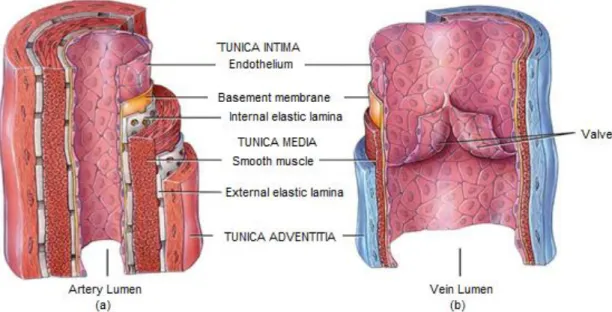
Blood vessels provide the main link between heart and tissues. They are the part of the circulatory system with the primarily function of transporting blood throughout the human body, playing a huge role in virtually every medical condition. The blood vessels are divided, depending on its function, location and size, into arteries, arterioles, capillaries, venules and veins. With the exception of capillaries and venules, the vascular wall is made up of three layers; the tunica intima (inner layer), the tunica media (middle layer) and the tunica adventitia (outer layer) (48).
Figure 2.1- Structure of Blood Vessels. (a) Arteries and (b) veins share the same general
features, but the walls of arteries are much thicker because of the higher pressure of the blood that flows through them. Adapted from (49).
Vascular Anatomy and Physiology
Clinical Application of Extracellular Vesicles in Vascular Calcification Page |13 Catarina Marreiros
2.1.1 - Tunica Intima
The tunica intima also designated by tunica interna, forms the inner lining of a blood vessel and is in direct contact with the blood as it flows through the lumen of the vessel. The endothelium, the innermost layer of this tunica, is a delicate sheet of flattened cells that lines the inner surface of the entire cardiovascular system (heart and blood vessels). Despite the fact that the endothelium is an extremely complex tissue from the metabolic point of view, its anatomical structure is extremely simple and linear, a single layer of mesenchymal cells. Until recently, endothelial cells were regarded as a passive barrier between the blood and the remainder of the vessel wall. It is now know that endothelial cells are active participants in a variety of vessel related activities, playing an important role in many physiological functions, including the control of vascular tone, blood cell trafficking, innate and adaptive immunity, among others (26,27).
The endothelium exerts its function in maintaining vascular homeostasis through the balanced release of a number of autocrine and paracrine substances in response to physical, biological, and chemical stimuli. These substances, known as vasoactive factors, can constrict or expand the smooth muscle cells (SMCs) within the vessels wall to increase or decrease the blood pressure, respectively. The endothelium forms an important part of the vasculature and is involved in promoting an atheroprotective environment via the balanced production of vasoactive factors. Disruption of vascular homeostasis can lead to the development of endothelial dysfunction which in turn contributes to hypertension and eventually cardiovascular disease (51).
The second component of the tunica intima is a basement membrane or basal lamina. The base membrane anchors the endothelium to the underlying connective tissue and also regulates molecular movement. It appears to play an important role in guiding cell movements during tissue repair of blood vessels walls. Finally, the outermost part of the tunica intima, which forms the boundary between the tunica intima and media, is the internal elastic lamina. The internal elastic lamina is a thin sheet of elastic fibers with a variable number of window-like openings that facilitate diffusion of materials through the tunica intima to the thicker tunica media (26,28), as illustrated in Figure 2.1
Vascular Anatomy and Physiology
Clinical Application of Extracellular Vesicles in Vascular Calcification Page |14 Catarina Marreiros
2.1.2 - Tunica Adventitia
The outer covering of the blood vessels, also known as tunica adventitia or externa, consists of elastic and collagen fibers. It contains numerous nerves and specifically in larger vessels, tiny blood vessels that supply the tissue of the vessel wall. The small vessels that supply the tissues of the vessel are called vasa vasorum, and they are easily seen on large vessels such as the aorta. In addition to the important role of supplying the vessel wall with nerves and self-vessels, the tunica adventitia is also a support structure, as it helps anchor the vessels to the surrounding tissues (48).
2.1.3 - Tunica Media
The tunica media, the thickest layer in arteries, is a muscular and connective tissue layer that displays the greatest variation among the different vessels types. It comprises mainly VSMCs and substantial amounts of elastic fibers. The primary role of the VSMCs, which extend circularly around the lumen like a ring, is the regulation of the vessel lumen diameter. These cells produce the elastic fibers that allow the vessels to stretch and recoil under the applied pressure of the blood. Contraction and relaxation of VSMCs decrease and increase the diameter of the vessel lumen, respectively. (48).
2.1.3.1 - Vascular Smooth Muscle Cells
Smooth muscle cells are found in many organs, comprising the blood vessels, trachea, stomach, small intestine, and uterus. Vascular smooth muscle provides the main support for the structure of the vessel wall and regulation of vascular tone in order to maintain intravascular pressure and tissue perfusion (52). However, in physiological conditions, VSMCs also perform other important functions during vessel remodeling such as in vascular injury.
Contrasting to other mature cell types of the human body, like skeletal and cardiac myocytes, VSMCs do not terminally differentiate, retaining a remarkable capability to modulate their phenotype during their live time. (53).
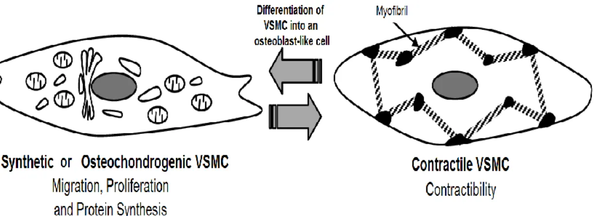
Vascular smooth muscle cells show different phenotypes according to external conditions, such as aging, developmental stage, angiogenesis state, and disease. Indeed, they have this unique ability to switch phenotype from a contractile to a synthetic, also designated as osteochondrogenic state, in response to environmental stimuli (33), as illustrated in Figure 2.2.
Vascular Anatomy and Physiology
Clinical Application of Extracellular Vesicles in Vascular Calcification Page |15 Catarina Marreiros
Figure 2.2 - VSMC’s Phenotypic Plasticity. VSMCs transform their phenotypes in response to
the surrounding environment. The contractile phenotype is a predominantly quiescent and anti-calcifying phenotype whereas synthetic phenotypes are associated with an increased propensity to promote vascular calcification. Adapted from (55).
Contractile VSMCs are characterized by low proliferation rates, high levels of cytoplasmic myofilaments, low rates of protein synthesis and a unique repertoire of contractile proteins including SM22α, SMα-actin, smoothelin, smooth muscle myosin heavy chain, among others. When VSMCs differentiate into the synthetic phenotype, normally found in embryonic and young developing blood vessels, they express relatively few contractile proteins, re-enter the cell cycle and become highly proliferative and migratory with high rates of protein synthesis and extracellular matrix secretion (56).
Interestingly, the synthetic phenotype confers a survival advantage since it allows VSMCs to proliferate, migrate and synthesize extracellular matrix components as a response required for vascular repair. However, an unfortunate consequence of this plasticity is that it predisposes VSMCs to environmental signals that can induce adverse phenotypic switching into an osteoblast-like cell type. This process promote the development and progression of vascular calcification, as it will be deepened in the following chapter (35,36).
Calcification in Cardiovascular Disease
Clinical Application of Extracellular Vesicles in Vascular Calcification Page |16 Catarina Marreiros
3 - Calcification in Cardiovascular Disease
3.1 - Vascular Calcification
Vascular calcification is nowadays a growing burden in Western countries, representing a major contributor to the progression and outcome of cardiovascular disease, one of the leading causes of death in industrialized countries. Each year cardiovascular disease (CVD) causes 3.9 million deaths in Europe accounting for 45% of all deaths. Overall CVD is estimated to cost the EU economy €210 billion a year. Of the total cost of CVD in the EU, around 53% (€111 billion) is due to health care costs, 26% (€54 billion) to productivity losses and 21% (€45 billion) to the informal care of people with CVD (59). Given the association between calcification and cardiovascular outcomes in both patients’ health and Europe’s health economic sustainability, there is an urgent need to better understand the mechanisms leading to the deposition and growth of calcium mineral deposits in blood vessels wall from its earliest stages.
Vascular calcification (VC) is defined as the inappropriate and pathological accumulation of mineral, most in the form of insoluble calcium-phosphate (Ca/P) salts in the medial and/or intimal layers of the vessel wall (60). Although calcification has been noted in the vasculature for many decades, it was first regarded as a passive process of limited pathophysiological significance, mostly viewed as a natural consequence of aging. The introduction of new non-invasively techniques to measure vascular calcification, such as electron beam computed tomography, has revolutionized our current thinking about the risks of VC. This pathology is directly linked with blood vessel wall stiffness, subsequent increased pulse wave velocity and altered arterial wall distensibility, ultimately leading to hypertension, left ventricular hypertrophy, compromised coronary perfusion and heart failure (61).
The challenges surrounding the ideal treatment of VC remain uncertain, and this is particularly pertinent as medicine continues to dedicate efforts in this fields to fully elucidate and discover novel treatment strategies to face this clinical problem (62).
Once established, vascular calcification is progressive, and its association with chronic comorbidities, including coronary artery disease (CAD), peripheral arterial disease (PAD), diabetes mellitus (DM), and chronic kidney disease (CKD) is well established. Ectopic calcification, meaning the inappropriate mineralization occurring in
Calcification in Cardiovascular Disease
Clinical Application of Extracellular Vesicles in Vascular Calcification Page |17 Catarina Marreiros
soft tissues, like arteries, vessels and heart-valves can indeed happen with normal aging, but it seems to be accelerated in these disease states, and related to an increased risk of morbidity and mortality (63). Nowadays, VC is no longer simply recognized as an inevitable consequence of aging, but as an active and highly complex process that cannot be ignored in patient’s cardiovascular clinical health.
3.1.1 - Types of Vascular Calcification
Vascular calcification can occur in the blood vessels, valves and cardiac tissues. Calcified deposits are found in distinct layers of the blood vessel and are related to underlying pathology. In general, vascular calcification can be categorized into four different types: intimal calcification, medial calcification, valvular calcification and calciphylaxis (62). VC is a pathologic response to environmental stimuli, which triggers a multifaceted process that may arise by different pathophysiological, non-mutually exclusive, mechanisms (64).
3.1.1.1 - Intimal Calcification
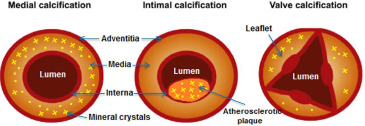
Intimal calcification, also known as atherosclerotic calcification, is the most common form of calcific vasculopathy. The pathologic mineral deposition is associated with the recruitment of inflammatory cells, such as macrophages and lipid deposits within atherosclerotic plaques (65).
Atherosclerotic microcalcifications are thought to derive from apoptotic VSMCs and from the accumulation of calcifying EVs within the internal elastic lamina (66). Moreover, macrophages associated with regions of calcified vascular structures have been shown to release EVs with high calcification and aggregation potential. (67).
Atherosclerotic calcification is linked with myocardial infarction derived from stenosis or acute thrombus, and with ischemia in both coronary and peripheral arteries (68).
3.1.1.2 - Medial Calcification
Vascular calcification may also occur in the medial layer of the vessels, known as Monckeberg’s medial sclerosis. The most extensive vascular calcification is a highly characteristic feature found in patients with type 2 DM and CKD patients. This type of calcification is connected with increased risk of sudden cardiac death and lower limb
Calcification in Cardiovascular Disease
Clinical Application of Extracellular Vesicles in Vascular Calcification Page |18 Catarina Marreiros
amputation due to vascular insufficiency, particular in type 2 DM and in end-stage renal disease (ESRD) (64).
Medial calcification is characterized by mineral deposition in the elastic lamina in the absence of classical atherosclerosis, this is, without lipid deposition and involvement of inflammatory cells (66).
The pathogenesis of vascular medial calcification is thought to also recapitulate skeletal bone formation, with the involvement of VSMCs differentiation and the release of calcifying EVs (69).
3.1.1.3 - Calcific uremic arteriolopathy
Calcific uremic arteriolopathy (CUA), commonly known as calciphylaxis, is a severely morbid and life-threatening form of vascular medial calcification with different clinical manifestation depending on the organ involved. It is a pathology that affects small arterioles (<0.6 mm diameter) leading to profound skin ulcerations due to ischemia being associated with an extremely high mortality rate in dialysis patients (70). CUA is a condition with high morbidity and mortality, especially in ESRD individuals. Skin nodules and painful ulcers rapidly progress to black eschar and demarcating cutaneous necrosis within these patients. (64)
3.1.1.4 - Cardiac Valve Calcification
Calcification of cardiac valves involves the pathological mineralization of the cardiac valve leaflets causing life-threatening stenosis. Worldwide, population-based studies have revealed that aortic valve disease is the most frequently observed valve pathology in patients diagnosed with valvular heart disease, and thus is the most studied heart valve (71).
Cardiac valve mineralization is similar to the vascular calcification process, including increased ECM degradation, differentiation of VSMCS and valvular intersticial cells, resembling physiologic mineralization in the bone tissue (69).
Calcification in Cardiovascular Disease
Clinical Application of Extracellular Vesicles in Vascular Calcification Page |19 Catarina Marreiros
Figure 3.1 - Different types of vascular calcification. Vascular mineralization processes can
occur in adventitia, media and interna layers within that vascular wall. It can also be observed in the leaflet of cardiac valves. Adapted from (72).
3.1.2 - Vascular Calcification Mechanisms
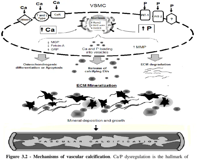
Vascular calcification has been widely described as a pathologic response to several stimuli (61). Although many aspects concerning the pathogenesis of VC are still uncertain, it is known that the base of this pathology comprehends multiple non-exclusively mechanisms, such as proliferation and differentiation of resident VSMCs, loss of mineralization inhibitors, release of calcification competent EVs, VSMCs apoptosis, endothelial dysfunction, oxidative stress, increased extracellular matrix (ECM) remodeling, and chronic inflammation (73). The synergistic effect of vascular calcification mechanisms is illustrated in Figure 3.2.
3.1.2.1- Osteochondrogenic Differentiation
Occasionally, structures that resemble bone tissue can be found in atherosclerotic lesions, suggesting that VC is an actively regulated process in which the vascular cells acquire osteoblast-like cell functions, ending up secreting osteoid-like matrix. Such finding, demonstrates that VC has a pattern that very much resembles some processes of bone formation (74). As explained in the previous chapter, VSMCs exhibit a remarkable phenotypic plasticity, which allows them to switch from a contractile into an osteoblast-like state.
In the cellular membrane vascular smooth muscle cells, have sodium-phosphate (NaPi) cotransporters known as PiT-1 and PiT-2, and calcium-sensing receptor (CaSR) as well voltage-activated channels (L and T type) that control phosphorus and calcium minerals entrance respectively, as illustrated in Figure 3.2. Recent studies have shown
Calcification in Cardiovascular Disease
Clinical Application of Extracellular Vesicles in Vascular Calcification Page |20 Catarina Marreiros
that the initiation of calcification requires an increased uptake of Ca/P by VSMCs, which leads to a pattern of cellular adaptations and damage that ultimately promote calcification (75). Indeed, vascular calcification’s most devastating manifestation transpires in CKD due to dysregulated mineral metabolism, the main pathologic characteristic within this patients, that conducts to long-term elevation of serum Ca/P levels (76). Vascular smooth muscle cells exposed to elevated Ca/P minerals, present loss of their contractibility, and upregulation of the expression of bone-related protein (osteochondrogenic expression) such as runt-related transcription factor 2 (Runx2), osteopontin, osteocalcin, alkaline phosphatase (ALP) ending up secreting calcifying EVs into the vessels ECM that promote mineralization sites and consequent calcification (47, 60).
Despite several studies aiming at understanding the complex mechanisms of calcification, there are still several processes that need further clarification. Whether differentiation of VSMCs or the release of calcifying EVs occurs first is a debatable issue that needs additional elucidation (10).
3.1.2.2 - Loss of mineralization inhibitors
In a normal physiological environment, VC is controlled because VSMCs synthesize or uptake from circulation natural mineralization inhibitors, counterbalancing mineralization promoters and therefore preventing ectopic calcification. This balance, however, seems to alter in certain pathophysiological environments, such as the increased levels of Ca/P serum levels, resulting in downregulation of the expression of typical vascular calcification inhibitors, as illustrated in figure 3.2. Decreased expression or activity of VC inhibitors, creates a setting that favors mineralization (77). In literature, several molecules have been identified as potential mineralization inhibitors. Within these inhibitors, matrix gla protein (MGP), fetuin-A and Gla-rich protein (GRP) have been reported to have a role in the mechanism of vascular calcification involving EVs, and therefore their relevance will be further described in the next chapters.
3.1.2.2.1 - Matrix- Gla Protein
Matrix-gla protein is considered one of the strongest mineralization inhibitors known to date. It is a vitamin K dependent protein (VKDP) containing 5 γ -carboxylated