INTRODUCTION
The twaite shad, Alosa fallax fallax, is an anadro-mous species that inhabits the North-East Atlantic
Ocean and adjacent waters, from the Christiana Fjord to the Iberian Peninsula and the coast of Morocco (Quignard and Douchement, 1991). Anadromous species are highly vulnerable and par-ticularly threatened by anthropogenic activities. Therefore, twaite shad is rare or even extinct in Europe, despite the existing legal protection for
SCI. MAR., 67 (3): 313-322
SCIENTIA
MARINA
2003Gross and histological observations of ovarian
development in twaite shad, Alosa fallax fallax, from
the Rivers Mira and Guadiana (Portugal)*
TERESA PINA, EDUARDO ESTEVES and JOSÉ PEDRO ANDRADE‡CCMar, Universidade do Algarve, Faculdade de Ciências do Mar e do Ambiente, Campus de Gambelas, 8000-810 Faro, Portugal. E-mail: jandrade@ualg.pt
SUMMARY: In order to describe the stages of oogenesis of twaite shad, Alosa fallax fallax, 265 females were collected between March and June 1997, February and June 1998 and January and April 1999 in the Rivers Mira and Guadiana. From the histological study of ovaries a total of eight developmental stages were delineated. Gross examination of paired ovary revealed that they could be placed into one of seven maturity stages according to their stage of development. Two stages of atresia, alfa and beta, were identified. Upon cessation of spawning, the ovaries still contained some oocytes at various stages of development but with a greater number of atretic oocytes. The simultaneous occurrence of oocytes at different stages of development in the ovary indicates asynchronous oocyte development. Oocyte size frequency distributions do not show a gap in size between cortical alveoli and vitellogenic oocytes during the spawning season. This may represent the ability of twaite shad to push oocytes through vitellogenesis from a previtellogenic condition during the spawning period. This has important implications for twaite shad fecundity, because fish with this type of oocyte development depend on estimates of batch fecundity and spawning frequency to determine potential annual fecundity.
Key words: oogenesis, oocyte dynamics, Alosa fallax fallax, Portugal.
RESUMEN: OBSERVACIONES DE VISU E HISTOLÓGICAS SOBRE EL DESARROLLO DEL OVARIO DE LA SABOGA, ALOSA FALLAX FALLAX, DE LOS RÍOSMIRA YGUADIANA(PORTUGAL). – Con objeto de describir las etapas de la oogénesis de la saboga, Alosa
fallax fallax, se recolectaron 265 hembras entre marzo y junio de 1997, febrero y junio de 1998 y enero y abril de 1999 en
los ríos Mira y Guadiana. A partir del estudio histológico de los ovarios se definieron ocho estadíos de desarrollo. La obser-vación de visu del ovario par reveló que, dependiendo de su estado de desarrollo, podría ubicarse en uno de los siete esta-díos de madurez . Se identificaron dos estaesta-díos de atresia: alfa y beta. Hasta que la freza finaliza, los ovarios tenían todavía algunos oocitos en diferentes estadíos de madurez, aunque presentado un gran número de oocitos atrésicos. La aparición simultánea de oocitos en diferentes estadíos de madurez en el ovario indica que su desarrollo es asincrónico. Las distribu-ciones de frecuencias de talla de los oocitos no mostró separación por tamaño entre los oocitos con alveolos corticales y los vitelogénicos durante la estación de puesta. Esto podría representar la capacidad de la saboga para forzar el paso de los ooci-tos en estado previtelogénico a vitelogénico durante la fase de puesta. Este hecho tiene implicaciones importantes para la fecundidad de la saboga, porque la determinación de la fecundidad potencial anual en un pez con este tipo de desarrollo oocitario dependerá de la estima de la fecundidad en cada tanda de puesta y de la frecuencia de las mismas.
Palabras claves: oogénesis, dinámica oocitaria, Alosa fallax fallax, Portugal.
‡ Corresponding author
migratory species (Lelek, 1980). This species still migrates into the River Mira and the River Guadi-ana, Portugal, to spawn. In these rivers, spawning migration is triggered by favourable environmental conditions, such as the increase in water tempera-ture, and starts between March and April, when adult twaite shad congregate in the sea near the mouth of the river. Subsequently, they enter with the rising tide, migrating to the spawning areas in the upper estuary to spawn. The spawning season can last until June (Pina, 2000).
Several studies have been undertaken on Alosa spp., mainly regarding age and growth (Walburg, 1960; Mennesson-Boisneau et al., 1986), migration patterns (Rameye et al., 1976; Loesch, 1987; Quinn And Adams, 1996), reproduction (Hoestlandt, 1958; Walburg, 1960; Nichols and Massmann, 1963;
Loesch and Lund, 1977; Leggett and Carscadden, 1978; Loesch, 1987; Manyukas, 1989; Jessop, 1993; Ross et al., 1993; Mylonas et al., 1995; Pina, 2000, Olney et al., 2001), and recruitment (Johnson and Loesch, 1983; Esteves, 1999). However, published information on the reproductive biology of A. fallax is restricted to the accounts of Manyukas (1989), Quignard and Douchement (1991) and Pina (2000), which provide no detailed description of the histo-logical development of the gonads. In Portugal, aspects of the feeding ecology and threats to survival (Assis, 1990; Assis et al., 1992), population genetics (Alexandrino et al., 1993, 1996), reproduction (Pina, 2000) and recruitment (Esteves, 1999) of twaite shad have been studied. This study was there-fore undertaken as part of a project studying the recruitment, reproduction, age and growth, and
genetics of shad populations in Portuguese rivers, and constitutes the first description of the oogenesis of twaite shad. The outcomes of this project will be used to develop conservation and management pro-grammes that endeavour to halt further declines and to improve twaite shad conservation status.
As a basis for further studies, the present investi-gation provides a detailed description of the differ-ent stages of oocyte developmdiffer-ent.
MATERIAL AND METHODS
Sampling was carried out between March and June 1997, February and June 1998 and January and April 1999 over the spawning areas of twaite shad in the upper estuaries of the Rivers Mira, South-west Portugal (Odemira) and Guadiana, South-east Portugal (Foz de Odeleite and Mértola) (Fig. 1). Fishing was carried out using trammel nets with an outer mesh size of 12.5 cm and an inner mesh size of 6.0 cm. A total of 265 females (106 in River Mira and 159 in River Guadiana) were mea-sured (total length, to the nearest 0.1 cm) and weighed (wet weight, to the nearest 0.1 g), and the ovaries were removed and weighed (to the nearest 0.1 g). The gonads were staged macroscopically according to a key developed in this study, as fol-lows: I - Immature/Resting; II - Early development; III - Maturing; IV - Ripe; V- Spawning; VI - Par-tially spent; VII - Spent (Table 1). Samples of gonads were fixed in San Felice solution (160 ml of 1% aqueous solution of chromic acid, 80 ml of 4% buffered formalin, 10 ml of acetic acid). After fixa-tion (36h), samples were dehydrated in ethanol, embedded in paraffin and sectioned at 6-8 µm. Ovaries were stained with Masson’s Trichrome (Martoja and Martoja-Pierson, 1967) and Toluidine blue (Bancroft and Stevens, 1990). After staining, the sections were covered with DePex mounting medium for microscopy (BDH Chemicals Ltd., Pools, England). Microscopic developmental stages of oocytes were categorised according to Janssen et
al. (1995). Histological classification of the atretic
oocytes and postovullatory follicles followed Hunter and Macewicz (1985) and Hunter et al. (1986). An oocyte frequency distribution (class interval: 50 µm) was produced for each maturity stage using counts of oocytes on 6 ovaries for each maturity stage. The maximum diameter of the oocytes was measured to the nearest 0.01 µm, using image analysis software (Image Pro Plus 3.0).
RESULTS
Developmental stages of the oocytes
Oogonia (Fig. 2A)
Cell diameter: 50 µm; nuclear diameter: 30 µm Oogonia were very small, round cells occurring singly or in groups with a relatively narrow zone of clear cytoplasm and a single, prominent nucleolus in the nucleus.
Chromatin nucleolus stage (Fig. 2A)
Cell diameter: 120 µm; nuclear diameter: 61 µm These newly formed oocytes appeared as small rounded cells and the cytoplasm was very thin and faintly basophilic
Early perinucleolus stage (Fig. 2A)
Cell diameter: 180 µm; nuclear diameter: 75 µm From this stage onwards, the oocytes were found as single cells, gradually migrating towards the centre of the lamellae. The cytoplasm was homogeneous and strongly basophilic. Numerous, relatively large, basophilic nucleoli appeared at the periphery of the nucleus, indicating increasing nuclear activity.
Late perinucleolus stage (Fig. 2A)
Cell diameter: 200 µm; nuclear diameter: 90 µm The chromatin material was dispersed through-out the nucleus, causing the nucleoplasm to appear granular. Numerous small round nucleoli were found in the periphery of the nucleus, quite close to the nuclear membrane. The cytoplasm was divided into two concentric zones: the inner dense and deeply basophilic and the outer less dense and only slightly basophilic. A flattened follicular layer sur-rounding the oocytes could be distinguished at the end of this stage, which is the last one of the prima-ry growth phase.
Cortical alveoli stage (Fig. 2B)
Cell diameter: 380 µm; nuclear diameter: 140 µm. During this growth phase, the cytoplasm had lost some of its basophilic property, and a narrow zone of small cortical alveoli (Ø 25 µm) was formed in the periphery of the cytoplasm. Several vacuoles
appeared in the basophilic layer of the cytoplasm around the nucleus. These vacuoles increased in number and volume and after a while formed a spongy-looking zone in the inner part of the cyto-plasm. The nucleus contained numerous nucleoli, which were arranged in a ring close to the nuclear membrane. The zona radiata and the theca had become visible.
Vitellogenesis (Fig. 2C)
Cell diameter: 550 µm; nuclear diameter: 170 µm.
Exogenous vitellogenesis started with the deposition of protein yolk granules at the periph-ery of the oocyte. These have multiplied increased in number and displaced most of cortical alveoli. The lipid droplets enlarged and occurred scattered between the yolk granules. The cytoplasm became less basophilic as vitellogenesis proceeded. Both the zona radiata, which assumed a fine, striated appearance, and the follicle epithelium were more prominent. Oocytes can remain in this stage until they either resume meiosis in response to
appro-FIG. 2. – Histological appearance of oocytes of the twaite shad. A, primary growth phase (O, oogonia; CN, chromatin nucleolus; EP, early perinucleolus; LP, late perinucleolus; N, nucleus; n, nucleoli) (Bar = 50 µm). B, cortical alveoli stage (CA, cortical alveoli; N, nucleus; C, cytoplasm; G, granulosa; ZR, zona radiata; L, lipids) (Bar = 110 µm). C, vitellogenesis (Bar = 60 µm). D, germinal vesicle migration (Bar =
75 µm). E, Germinal vesicle breakdown (Bar = 105 µm).
A
D
B
C
FIG. 3. – Histological appearance of A, alfa (arrows indicate wrinkled zona radiata) (Bar = 50 µm) and B, beta atresia (arrows) oocytes of the twaite shad. (Bar = 130 µm).
FIG. 4. – Photomicrograph of twaite shad ovaries at various magnifications. A, immature/resting; B, early development; C, maturing; D, ripe;
E, partially spent; F, spent (arrows indicate POF’s).
A
D
B
C
A
B
F
E
priate hormonal stimulus or are resorbed (oocyte atresia)
Germinal vesicle migration (GVM) stage (Fig. 2D)
Cell diameter: 850 µm; nuclear diameter: 180 µm The nucleus, or germinal vesicle (GV), migrat-ed towards the periphery of the oocyte and con-tained many nucleoli. The protein yolk granules and lipid droplets started to coalesce and the oocyte rapidly increased in volume due to hydra-tion. The cytoplasm was surrounded by a marked zona radiata.
Germinal vesicle breakdown (GVBD) stage (Fig. 2E)
Cell diameter: 950 µm
During this stage, a pronounced and rapid size increase occurred due to both hydration and limited protein uptake and the nuclear membrane disap-peared (germinal vesicle breakdown). Most of the oocytes in this stage collapsed during histological processing. Thus, photographs were possible only on an earlier phase of this stage (Fig. 2E).
Post-ovulatory follicles (POF’s)
The POF’s consisted of residual follicle layers, which remained in the ovaries after ovulation and degenerated during the following period (c.f. Fig. 4F).
Atretic follicles (alfa and beta)
From final spawning onwards, a number of post-vitellogenic oocytes failed to undergo maturation or ovulation and subsequently degenerated and were resorbed, i.e. became atretic. At the onset of atresia (alfa atresia), the zona radiata wrinkled and started to break up (Fig. 3A). The follicle granulosa cells proliferated and hypertrophied to form a compact, well-vascularised structure. These active granulosa cells invaded the oocyte through the broken down zona radiata and digested and resorbed the yolk con-tents by active phagocytosis. The phagocitic granu-losa cells in turn also degenerated, leaving behind a lightly staining fibrous mass surrounded by connec-tive tissue elements (beta atresia) (Fig. 3B).
The stages of gamma and delta atresia referred to by Hunter and Macewicz (1985) were not observed. The follicle was completely resorbed during the beta stage or a rapid evolution of atresia might have occurred after this stage, leaving no histological characteristics that could be identified.
Maturity stages of the ovary
Gross examination of ovaries revealed that they could be placed into one of seven maturity stages according to its stage of development (Table 1 and Fig. 4). This seven-stage classification is a modifi-cation of that given by Macer (1974).
TABLE1. – Macroscopic characteristics and histological description of the maturity stages of the ovary of Alosa fallax fallax.
Maturity stage Macroscopic description Histological description
I. Immature/resting Small thread-like ovary, pinkish in colour, Well-spaced ovigerous fold, orientated towards the no oocytes visible centre of the ovary, containing both oogonia and
chromatin nucleolus and early perinucleolus stage; oogonia generally occur in nests
II. Early development Ovaries one-third of ventral cavity, yellowish Oogonia still present, ovigerous folds fill cavity, late in colour, opaque oocytes visible through the tunica perinucleolus and cortical alveoli oocytes present III. Maturing Ovaries two-thirds of ventral cavity, yellowish Cortical alveoli and yolk granule oocytes
in colour, opaque and translucent oocytes visible, predominate; late perinuceolus and vitologenic increasing vascularization oocytes can also be observed
IV. Ripe Ovaries highly vascularized occupy most of Oocytes observed in previous stage still present; ventral cavity, opaque, translucent and predominance of GVM stage oocytes
dominant hyaline oocytes visible
V. Spawning Ovaries occupying most of ventral cavity, eggs Predominance of GVM oocytes and POFs released under slight pressure of the abdomen
VI. Partially spent Ovaries flaccid, deep red in colour, occupying Irregular convoluted ovigerous folds, conspicuous two-third of ventral cavity, with hyaline spaces in the septa and POFs in the lumen, early oocytes visible perinucleolus and atresia oocytes present VII. Spent Ovaries flaccid and fully empty, a few residual Ovigerous folds reorganizing, containing atretic
Oocyte size frequency distribution
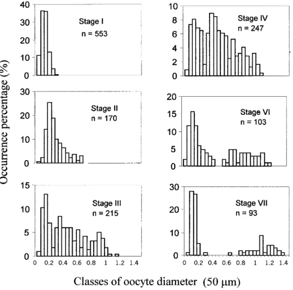
Figure 5 shows the oocyte size frequency distrib-ution in each maturity stage. In maturity stages I, II and III, only one modal class is identified. This dis-tribution is clearly polimodal from this stage onwards, when different cohorts of developing oocytes are identified. In ripe ovaries, oocyte diam-eter reaches 1.052 mm. Nevertheless, larger oocytes (1.415 mm) can be observed in partially spent and spent ovaries, which were not shed and will be resorbed.
DISCUSSION
The process of oocyte development in A. fallax
fallax follows the same basic progression as that
described for Alosa sapidissima (Mylonas et al.,
1995; Olney et al., 2001) and other teleost fish (Tyler and Sumpter, 1996).
In this work, oogenesis was divided into 8 stages according to size differences and the occurrence of new structures that are easily recognisable during the different phases of oocyte growth. Hence, oogo-nia give rise to immature oocytes with multiple nucleoli. The oocyte enlarges as vitellogenic yolk is deposited and its zona radiata thickens. The nucleus migrates to the animal pole prior to the breakdown of its membrane. Hydration precedes ovulation and the appearance of these hyaline oocytes indicates imminent spawning. The follicle collapses after the oocyte has been released, leading to the emergence of the postovulatory follicles.
The origin of the young oocytes that appear at the beginning of each reproductive cycle is a question of great biological interest, since those oocytes repre-sent the initial stage of oogenesis, and therefore the FIG. 5. – Oocyte size frequency distribution for each maturity stage of twaite shad; n, total number of oocytes measured.
reserve from which all oocytes will develop. There has been some controversy as to the origin of the new oocytes. Hann (1927), Matthews and Marshall (1956) and Ramos (1983) suggested that these oocytes originate from residual oogonia that remain in the ovary from year to year. Bara (1960) and Arruda (1988) concluded that oogonia were derived from germinal epithelial cells. According to Wheel-er (1930) and Yamamoto (1956), at least some oogo-nia are supplied each year by the transformation of certain cells remaining from the empty follicles of the previous year’s spawning. According to Howell (1983), the observation of mitotic activity in nia of mature fish suggests that a new stock of oogo-nia arises during each reproductive cycle. In this study, oocytes in the chromatin nucleolus stage were usually present in small groups but no mitotic divi-sions were apparent. Furthermore, at all stages, from late immature onwards and throughout the year, a “reserve fund” was present in the ovaries of the twaite shad. This “reserve fund” consisted of oocytes that had grown from the chromatin nucleo-lus stage up to a size of 0.37 mm, at which point they seem to suspend development for some time and resume growth towards maturation. This dynamics would agree with the conclusion of Foucher and Beamish (1977), indicating an apparent regeneration of the oocytes in the perinucleolus stage at the beginning of each reproductive cycle. On the other hand, the observation of oocytes in the chromatin nucleolus stage near the base of the septa supports an epithelial origin of these cells.
The presence of two differently stained zones in the cytoplasm of the oocytes in the cortical alveoli stage is related to the development of the Balbiani body, and has been described for a number of species (Bara, 1960; Arruda, 1988; Mayer et al., 1988; Coello and Grimm, 1990). According to Guraya (1986) and Coello (1990), it is composed of two parts, the yolk nucleus and the pallial substance. The pallial substance appeared inside the resting oocytes as the more deeply basophilic ring in the inner cytoplasm. The proportion of these oocytes showing Balbiani’s body increases with cell size. Similar results were described by Coello and Grimm (1990) in the oocytes of Scomber scombrus.
Migration of the germinal vesicle (GV) is an event associated with the onset of final oocyte mat-uration and was apparent in oocytes larger than 800 µm. Oocytes greater than 950 µm exhibited varying degrees of yolk coalescence, but the GV was not apparent after histological examination. According
to Mylonas et al. (1995), this suggests the dissolu-tion of the nuclear wall (GV breakdown).
Atresia followed one of the three patterns described by Hunter and Macewicz (1985), and the follicle was completely resorbed during the beta stage. Atresia affected only the oocytes that began the second growth phase and the younger oocytes were not affected in any way.
In post-spawning fish, the postovulatory folli-cles (POF’s) can be found embedded in the lamel-lae. The POFs degenerate rapidly, making it diffi-cult to access the percentage of oocytes spawned, unless samples are collected daily (Hunter and Macewicz, 1985). Also, the elastic nature of ovari-an tissue allows the ovary to contract as oocytes are spawned so that remaining oocytes are still closely grouped, making loss of oocytes less apparent. It is also difficult to determine whether the observed atresia represented premature resorption of oocytes that could have been released later or was part of the natural tissue resorption process after cessation of spawning.
According to Bye (1984), in wild populations the only factor that can cancel out or reverse the effect resulting from increased reproductive investment is temperature. Ovulation marks the commitment of considerable metabolic investment and can lead to successful reproduction only if critical external fac-tors (presence of a male and spawning substrate, low level of predation, etc.) are appropriate (Stacey, 1984). Thus, an overproduction of maturing oocytes can be part of a reproductive strategy of A. fallax
fal-lax and oocytes can be shed or not, depending on
both the abiotic and the biotic conditions. Further-more, fasting during anadromous migration can induce atresia, suggesting that some feedback mech-anism may exist to use resources, particularly yolk, efficiently so that there is a minimum requirement for resorption in migrating females. According to Bagenal (1978) and Guraya (1986), food shortening is one of the main factors determining increasing rates of atresia. Thus, the study of the direct effects of atresia in oocyte production is a major issue to be addressed in future studies, particularly in fecundity estimates.
Evidence that twaite shad are serial spawners releasing discrete batches of eggs over an extended spawning season includes macroscopic and histo-logical indications of recent spawning concurrent with mature vitellogenic oocytes. The development of oocytes in twaite shad is asynchronous because these fish are capable of bringing oocytes from an
immature condition through vitellogenesis during the spawning season. Eggs are recruited from a het-erogeneous population of developing oocytes and are subsequently ovulated in several batches during each spawning season. This is supported by the lack of a hiatus in the size frequency distribution of immature versus mature oocytes. Similar results were also reported for twaite shad by Le Clerc (1941) and Hass (1968) and for American shad, A.
sapidissima, by Mylonas et al. (1995) and Olney et al. (2001).
More information about the number of batches spawned can be obtained through the process of identifying and ageing the postovulatory follicles. Future studies will have to address this problem, since the delineation of the spawning frequency is essential for accurate fecundity estimates. Further research is also necessary to determine how the reproductive patterns affect the investment of energy and nutrients in the somatic and reproductive tissues of twaite shad.
This study indicates that there are still numerous gaps in our understanding of this complex process in twaite shad. Nevertheless, this study might serve as background information for future studies and may stimulate further research, leading to insights into the events and control mechanisms of egg produc-tion among vertebrates. Moreover, a greater under-standing of the relation between reproductive strate-gy and habitat characteristics is needed in order to evaluate exploitation techniques and permissible habitat changes.
ACKNOWLEDGEMENTS
This work was funded by the Fundação para a Ciência e a Tecnologia within the framework of the Praxis XXI research programme (Contract Nº 3/3.2/CA/1981/95) and grants to T. Pina (BIC/3852/96) and E. Esteves (BM/8427/96). Thanks are also due to Dr. Angel Guerra and Dr. Angel Gonzalez for the translation to Spanish. The manuscript benefited from the comments of two anonymous referees.
REFERENCES
Alexandrino, P.J., N. Ferrand and J. Rocha. – 1996. Genetic poly-morphism of a haemoglobin chain and adenosine deaminase in European shads: Evidence for the existence of two distinct genetic entities with natural hybridisation. J. Fish Biol., 48: 447-456.
Alexandrino, P.J., C. Sousa, A. Pereira and N. Ferrand. – 1993. Genetic polymorphism of adenosine deaminase (ADA; E.C. 3.5.4.4.) in allis Shad, Alosa Alosa and twaite shad, Alosa
fal-lax. J. Fish Biol., 43: 951-953.
Arruda, L.M. – 1988. Maturation cycle in the female gonad of the snipefish Macrorhamphosus gracilis (Lowe, 1839) (Gas-torosteiformes, Macro-rhamphosidae) off the western coast of Portugal. Invest. Pesq., 52(3): 355-374.
Assis, C.A. – 1990. Threats to the survival of anadromous fishes in the river Tagus, Portugal. J. Fish Biol., 37: 225-226. Assis, C.A., P.R. Almeida, F. Moreira, J.L. Costa, and M.J. Costa.
– 1992. Diet of the twaite shad Alosa fallax (Lacépède) (Clu-peidae) in the river Tagus estuary, Portugal. J. Fish Biol., 41: 1049-1050.
Bagenal, T.B. – 1978. Aspects of fish fecundity. In: S.D. Gerking (ed.), Ecology of Freshwater Fish Production, pp. 75-101. Blackwell Scientific Publications, Oxford.
Bancroft, J.D. and A. Stevens. – 1990. Theory and practice of
his-tological techniques. 3rd Edition. Churchill Livingstone
Pub-lishers, Avon, U.K.
Bara, G. – 1960. Histological and cytological changes in the ovaries of the mackerel Scomber scombrus, during the annual cycle.
Istamb. Univ. Fen Fak. Mecm., Ser. B 25: 49-91.
Bye, V.J. – 1984. The role of environmental factors in the timing of reproductive cycles. In: G. W. Potts and R. J. Woottoon (eds.),
Fish Reproduction: Strategies and Tactics, pp. 186-205.
Acad-emic Press, London.
Coello, S. and A.S. Grimm. – 1990. Development of Balbiani’s vitelline body in the oocytes of the Atlantic mackerel, Scomber
scombrus. J. Fish Biol., 36: 265-267.
Coello, S. – 1990. Reproductive biology of Atlantic mackerel. Ph. D. Thesis, University College of North Wales.
Esteves, E. – 1999. Factores que influenciam a abundância e
condição nutricional de larvas de peixe nos rios Mira e Guadi-ana. MSc Thesis, Faculdade de Ciências e Tecnologia da
Uni-versidade de Coimbra, Coimbra.
Foucher, R.P. and R.J. Beamish. – 1977. A review of oocyte devel-opment in fishes with special reference to Pacific hake
(Mer-luccius productus). Fish Mar. Serv. Techn. Rep., 755: 1-16.
Guraya, S.S. – 1986. The cell and molecular biology of fish
oogen-esis. Monographs in developmental biology 18. Basel, Karger.
Hann, H.W. – 1927. The history of the germ cells of Cottus bairdii Girard. J. Morph. Physiol., 43(2): 427-498.
Hass, H. – 1968. Untersuchungen uber die vertikale und horizontale verteilung der eier der finte, alosa fallax lac., in der elbe. Arch.
Fisch Wiss, 19: 46-55.
Hoestlandt, H. – 1958. Reproduction de l’alose atlantique (Alosa
alosa Linné) et transfert au bassin méditerranéen. Verh. Inter-nat. Ver. Limnol., 13: 736-742.
Howell, W.H. – 1983. Seasonal changes in the ovaries of adult yel-lowtail flounder, Limanda limanda. Fish. Bull., 81(2): 341-355. Hunter, J.R. and B.J. Macewicz. – 1985. Rates of atresia in the ovary of captive and wild Northern anchovy Engraulis mordax.
Fish. Bull., 83(2): 119-136.
Hunter, J.R., B.J. Macewicz and J.R. Sibert. – 1986. The spawning frequency of skipjack tuna, Katsuwonus pelamis, from the South Pacific. Fish. Bull., 84: 895-903.
Janssen, P.A.H., J.D.G. Lambert and H.J.Th. Goos. – 1995. The annu-al ovarian cycle and the influence of pollution on vitellogenesis in the flounder, Pleuronectes flesus. J. Fish Biol., 47: 509-523. Jessop, B.M. – 1993. Fecundity of anadromous alewives and
blue-back herring in New Brunswick and Nova Scotia. Trans. Am.
Fish. Soc., 122: 85-98.
Jonhson, J.R. and J.G. Loesch. – 1983. Morphology and develop-ment of hatchery-cultered American shad, Alosa sapidissima (Wilson). Fish Bull., 82: 323-339.
Le Clerc, M. – 1941. Note sur des essais de multiplication artifi-cialle de l’alose dans le bassin de le Loire. Bull. Fran.
Pisci-cult., 123: 27-37.
Legget, W.C. and J.E. Carscadden. – 1978. Latitudinal variation in reproductive characteristics of american shad (Alosa
sapidissi-ma): Evidence for population specific life history strategies in
fish. J. Fish. Res. Board Can., 35: 1469-1478.
Lelek, A. – 1980. Les poissons d’eau douce menacés en Europe. Conseil de l’europe. Sauvegarde de la Nature, 18.
Loesch, J.G. and W.A. Lund. – 1977. A contribution to the life his-tory of the blueback herring, Alosa aestivalis. Trans. Am. Fish.
Loesch, J.G. – 1987. Overview of life history aspects of anadro-mous alewife and blueback herring in freshwater habitats. Am. Fish. Soc. Symp., 1: 89-103.
Macer, C.T. – 1974. The reproductive biology of the horse macker-el Trachurus trachurus (L.) in the North Sea and English Chan-nel. J. Fish Biol., 6: 415-438.
Manyukas, Y.L. – 1989. Biology of the Atlantic shad, Alosa fallax fallax, in Kurshskiy Bay. J. Ichthyol., 29: 125-128.
Martoja, R. and M. Martoja-Pierson. – 1967. Initiation aux tech-niques de l’histologie animale. Masson et Cie Editeurs, Paris. Matthews, L.H. and F.H. Marshall. – 1956. Cyclical changes in the
reproductive organs of the lower vertebrates. In: A. S. Parker (ed.), Marshall’s physiology of reproduction, pp. 156-225. Longman, Green and Co, London.
Mayer, I., S.E. Shackley and J.S. Ryland. – 1988. Aspects of the reproductive biology of the bass Dicentrarchus labrax L. i. An Histochemical study of oocyte development. J. Fish Biol., 33: 609-622.
Mennesson-Boisneau, C., P. Boisneau and J.L. Baglinière. – 1986. Premières observations sur les caractéristiques biologiques des adultes de Grande alose (Alosa alosa l.) dans le cours moyen de la Loire. Acta Œcol., 7: 337-353.
Mylonas, C.C., Y. Zohar, B.M. Richardson and S. P. Minkkinnen .-1995. Induced spawning of wild American shad Alosa sapidissi-ma using sustained administration of gonadotropin-releasing hor-mone analog (GnRHa). J. World Aquac. Soc., 26(3): 240-252. Nichols, P.R. and W.H. Massmann. – 1963. Abundance, age, and
fecundity of shad, York River, VA., 1953-59. Fish. Bull., 63: 179-187.
Olney, J.E., S.C. Denny and J.M. Hoenig. – 2001. Criteria for deter-mining maturity stage in female America shad, Alosa sapidissi-ma, and a proposed reproductive cycle. Bull. Fr. Pêche Piscic., 362/363: 881-901.
Pina, T. – 2000. Aspectos da reprodução de savelha, Alosa fallax fallax (Lacépède, 1803), nos rios Mira e Guadiana. MSc
The-sis, Faculdade de Ciências e Tecnologia da Universidade de Coimbra, Coimbra.
Quignard, J.P. and C. Douchement. – 1991. Alosa fallax fallax, Alosa fallax lacustris, Alosa fallax nilotica, Alosa fallax rhoda-nensis. In: H. Hoestlandt (Ed.). The freshwater fishes of Europe. Aula-Verlag, Wiesbaden, 2: 225-295.
Quinn, T.P. and D.J. Adams. – 1996. Environmental changes affect-ing the migratory timaffect-ing of American shad and sockeye salmon. Ecology, 77: 1151-1162.
Rameye, L., A. Kiener, C.P. Spillmann. and J. Biousse. – 1976. Aspects de la biologie de l’alose du Rhone peche et difficultes croissantes de ses migrations. Bull. Franc. De Pisc., 263: 50-76. Ramos, J. – 1983. Contribución al estudio de la oogenesis en el lenguado Solea solea (Linneo, 1758) (Pisces, Soleidae). Invest. Pesq., 47(2): 241-251.
Ross, R.M., R.M. Bennett and T.W.H. Backman. – 1993. Habitat use by spawning adult, egg, and larval American shad in Delaware River. Rivers, 4: 227-238.
Stacey, N.E. – 1984. Control of the timing of ovulation by exoge-nous and endogeexoge-nous factors. In: G. W. Potts and R. J. Woot-toon (eds.), Fish Reproduction: Strategies and Tactics, pp. 207-222. Academic Press, London.
Tyler, C.R. and J.P. Sumpter. – 1996. Oocyte growth and develop-ment in teleosts. Rev. Fish Biol. Fish., 6: 287-318.
Walburg, C.H. – 1960. Abundance and life history of shad St. Johns River, Florida. Fish. Bull., 60: 487-501.
Wheeler, C.F. – 1930. The growth of the eggs in the dab (Pleu-ronectes limanda). Quart. J. Micr. Sc., 68(272): 641-660. Yamamoto, K. – 1956. Studies on the formation of fish eggs. i.
Annual cycle in the development of ovarian eggs in the floun-der Liopsetta obscura. J. Fac. Sc. Hokkaido Univers., Ser. Vi, 12(3): 362-374.