Tânia Raquel Rodrigues Grainha
Insights into interspecies interactions,
phenotypic profile and antimicrobial
susceptibility in an artificial
ventilator-associated pneumonia microbiome
Universidade do Minho
Escola de Engenharia
October 2016 Tânia R aq uel R odrigues Gr ainhaInsights into inter
species interactions, pheno
typic profile and antimicrobial
suscep
tibility in an ar
tificial ventilator-associated pneumonia microbiome
Minho | 20
1
6
Tânia Raquel Rodrigues Grainha
Insights into interspecies interactions,
phenotypic
profile
and
antimicrobial
susceptibility in an artificial
ventilator-associated pneumonia microbiome
Master Dissertation
Master in Bioengineering
Work Supervised by:
Professor Maria Olívia Pereira And Co-supervised by:
Doctor Susana Patrícia Lopes Doctor Maria Elisa Rodrigues
DECLARAÇÃO
Nome: Tânia Raquel Rodrigues Grainha
Endereço eletrónico: [email protected]
Título da tese: Insights into interspecies interactions, phenotypic profile and antimicrobial susceptibility in an artificial ventilator-associated pneumonia microbiome
Orientadora: Professora Maria Olívia Pereira Co-orientadoras: Doutora Susana Patrícia Lopes Doutora Maria Elisa Rodrigues
Ano de conclusão: 2016
Mestrado em Bioengenharia
DE ACORDO COM A LEGISLAÇÃO EM VIGOR, NÃO É PERMITIDA A REPRODUÇÃO DE QUALQUER PARTE DESTA TESE/TRABALHO.
iii
ACKNOWLEDGMENTS/AGRADECIMENTOS
Terminada esta importante jornada, não posso deixar de reconhecer que a realização desta dissertação teve de forma direta ou indireta, o contributo fundamental de muitas pessoas. Em primeiro lugar queria agradecer às minhas orientadoras, Professora Maria Olívia Pereira, Doutora Susana Lopes e Doutora Elisa Rodrigues pela orientação, pelos conhecimentos transmitidos e por todas as sugestões essenciais para a elaboração do trabalho.
Não posso deixar de agradecer também a todo o grupo MOP, em particular à Diana e à Paula, pela cooperação e disponibilidade demonstradas e pelos conhecimentos transmitidos ao longo do ano.
À Vanessa porque teve sempre presente, pelas nossas brincadeiras e pela amizade. Às colegas de laboratório (Vanessa, Carla, Sílvia), pela boa disposição e companheirismo.
Gostaria de agradecer à Ângela França pelas úteis sugestões e contributos na extração e análise do RNA e ao Luís Melo pelo seu imprescindível contributo na citometria de fluxo, pela disponibilidade e aprendizagem transmitida.
Não poderia deixar de fazer um agradecimento especial à Andreia que foi uma ajuda importante na realização deste trabalho, pela amizade, apoio e motivação nos momentos mais complicados. Por fim agradeço à minha família e ao meu namorado pelo apoio e carinho.
v This work was supported by ESCMID Research Grants 2014 and the Portuguese Foundation for Science and Technology (FCT), under the scope of the strategic funding of UID/BIO/04469/2013 unit and COMPETE 2020 (POCI-01-0145-FEDER-006684), by FCT and the European Community fund FEDER, through Program COMPETE, under the scope of project RECI/BBB-EBI/0179/2012 (FCOMP-01-0124-FEDER-027462).
vii
ABSTRACT
Ventilator associated pneumonia (VAP) is the second most common nosocomial infection in the intensive care units (ICU) and the most common in mechanically ventilated patients. VAP presents a serious problem in ICU due to high mortality and morbidity rates associated, because it is often biofilm-mediated and polymicrobial. Therefore, understanding the impact of microorganisms in VAP and their interaction is a major challenge posed. Additionally, the ineffective current treatment strategies have led to the emergence of new approaches to fight these polymicrobial consortia, with a great number intervening in the quorum-sensing (QS) intercellular communication.
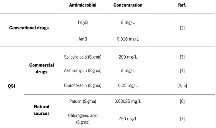
This work aimed to give insights into the behavior of bacterial-fungal communities involving Pseudomonas aeruginosa and Candida albicans associated to VAP, when exposed to different antimicrobial approaches. For this, single- and mixed-species biofilms were thoroughly characterized in terms of cultivable cells and biomass after 24 h treatment with conventional drugs (amphotericin B, AmB; polymyxin B, PolyB) and alternative agents, in particular QS inhibitors (QSI) from different sources (commercial drugs: salicylic acid, ciprofloxacin (CIP), azithromycin (AZT); natural sources: chlorogenic acid, farnesol, linalool, patulin) and enzymes (alginate lyase, desoxirribonuclease), tested alone or in combination.
Results showed that the combination AmB+PolyB did not affect the pre-established P. aeruginosa and C. albicans consortia. Interestingly, excepting for patulin, QSI agents were effective at reducing biofilm-encased cells, in particular single-species biofilms. CIP showed a great potential to inhibit both single-and mixed-species biofilms. Linalool was also effective in disturbing C. albicans in single and mixed biofilms. Contrariwise, enzymes had no effect against biofilms. Regarding double combinations, the addition of farnesol or linalool to CIP led to similar results from that obtained with CIP alone, with reductions observed in biofilm-encased cells. In general, the addition of a third agent - particularly in the case of chlorogenic acid - did not significantly improve the effect of AmB+PolyB or farnesol/linalool+CIP combinations.
Additionally, efforts were made to characterize the un- and treated dual-species biofilms by flow cytometry and RNA sequencing (RNA-seq), however no accurate results were obtained due to unexpected methodological hitches.
In conclusion, the use of new approaches seems to be a promise in treating bacterial-fungal consortia often involved in VAP. This work showed that combining different agents from distinct sources is a valuable option to control P. aeruginosa and C. albicans biofilms. Nevertheless, optimization on the antimicrobial doses and further clinical studies are urgently required to improve therapy effectiveness and avoid additional costs.
ix
RESUMO
A pneumonia associada à ventilação (PAV) é a segunda infeção nosocomial mais comum em unidades de cuidados intensivos (UCI) e a mais comum em pacientes sob ventilação mecânica. A PAV apresenta um problema grave na UCI devido às elevadas taxas de mortalidade e morbilidade associadas, porque muitas vezes é mediada por biofilmes e tem caráter polimicrobiano. Compreender o impacto dos microrganismos em PAV e suas interações é um grande desafio que se coloca. Além disso, a ineficiência das estratégias de tratamento atuais levaram ao surgimento de novas abordagens para combater esses consórcios polimicrobianos, com um grande número intervindo na comunicação intercelular quorum-sensing (QS).
Este trabalho teve como objetivo o conhecimento do comportamento das comunidades bacterianas-fúngicas envolvendo Pseudomonas aeruginosa e Candida albicans associadas a PAV, quando expostas a diferentes estratégias antimicrobianas. Para isso, os biofilmes simples e mistos foram caracterizados em termos de células cultiváveis e biomassa após 24 h de tratamento com medicamentos convencionais (anfotericina B, AmB; polimixina B, PolyB) e com agentes alternativos, em particular, inibidores de QS (QSI) obtidos de diferentes fontes (medicamentos comerciais: ácido salicílico, ciprofloxacina (CIP), azitromicina (AZT); fontes naturais: ácido clorogénico, farnesol, linalol, patulina); enzimas (alginato liase, desoxirribonuclease), testados isoladamente ou em combinação.
Os resultados mostraram que a combinação AmB+PolyB não afetou os consórcios pré-estabelecidos de P. aeruginosa e C. albicans. Curiosamente, com exceção de patulina, os agentes IQS foram eficazes na redução de células de biofilme, em particular nos biofilmes formados por uma única espécie. A CIP mostrou grande potencial para inibir biofilmes simples e mistos. O linalol também foi eficaz contra C. albicans em biofilmes simples e mistos. Pelo contrário, as enzimas não tiveram efeito contra os biofilmes. No que se refere às combinações duplas, a adição de farnesol ou linalol à CIP conduziu a resultados semelhantes obtidos apenas com CIP, com reduções observadas em células envolto-biofilme. Em geral, a adição de um terceiro agente - em particular no caso do ácido clorogénico - não melhorou significativamente o efeito das combinações AmB+PolyB ou farnesol/linalol+CIP.
Adicionalmente, foram feitos esforços para caraterizar os biofilmes de dupla espécie, não tratados e tratados, por citometria de fluxo e sequenciação de RNA (RNA-seq), contudo não foram obtidos resultados fiáveis devido a dificuldades metodológicas inesperadas.
Em conclusão, o uso de novas estratégias parece ser uma promessa no tratamento de consórcios bacterianos-fúngicos frequentemente envolvidas em PAV. Este trabalho mostrou que a combinação de diferentes agentes obtidos a partir de fontes distintas é uma opção valiosa para controlar biofilmes de P. aeruginosa e C. albicans. No entanto, é necessário a otimização das doses de antimicrobianos e mais estudos clínicos para melhorar a eficácia da terapia e evitar custos adicionais.
xi
AIMS AND OUTLINE OF THE THESIS
This study proposed to look into the behavior of inter-kingdom biofilms, as well as to find new approaches to treat these polymicrobial infections. In this regard, it was intended to characterize the communities involving a bacterial species, P. aeruginosa, and a fungal species, C. albicans, often found in VAP, as well as its susceptibility towards different treatment strategies involving combinations of antimicrobials with QS inhibitors (QSI). Further, a transcriptomic analysis of the un- and treated dual-species biofilms was attempted.
This thesis is organized into three chapters. Chapter 1 briefly reviews relevant clinical aspects of VAP, emphasizing the composition of microbial communities involved in VAP. The microbial biofilms, including their relevant particularities and their importance for VAP are also summarized in this chapter.
In Chapter 2, the microorganisms, culture conditions, materials and techniques used in the work presented herein are described.
Chapter 3 reports studies on mono- and dual-species biofilms of Pseudomonas aeruginosa and Candida albicans, often associated to VAP. These studies include microbial compositions of mono- and dual-species populations, before and after antimicrobial exposure.
Chapter 4 includes the main conclusions of the work displayed and proposes future research lines finalizing the thesis.
xiii
CONTENTS
ACKNOWLEDGMENTS/AGRADECIMENTOS ... iii
ABSTRACT ... vii
RESUMO ... ix
AIMS AND OUTLINE OF THE THESIS ... xi
ABBREVIATIONS AND ACRONYMS ... xix
SCIENTIFIC OUTPUT ... xxi
Chapter 1
... xxiiiGENERAL INTRODUCTION ... xxiii
1.1 Ventilator-associated pneumonia: a general overview of the disease ... 3
1.2 Microorganisms involved in VAP infection ... 4
1.3 Quorum-sensing: A Sophisticated Communication System ... 6
1.3.1 Pseudomonas aeruginosa quorum-sensing mechanisms ... 12
1.3.2 Candida albicans quorum-sensing mechanisms ... 13
1.3.3 Pseudomonas aeruginosa and Candida albicans interaction ... 14
1.4 Biofilms in VAP ... 16
1.5 VAP therapy ... 19
1.5.1 Conventional therapy ... 20
1.5.2 Alternative approaches in biofilm control: the QS inhibitors ... 22
REFERENCES ... 24
Chapter 2
... 32.1 Microorganisms and culture conditions ... 37
2.1.1 Biofilm pre-inoculum ... 37
2.1.2 Biofilm formation assay ... 37
2.2 Antimicrobial susceptibility ... 38
2.3 Methodologies ... 39
2.3.1 Biofilms analysis ... 39
2.3.1.1 Determination of cultivable cells ... 39
2.3.1.2 Biomass quantification ... 40
xiv
2.3.2. Extraction of biofilm matrix ... 41
2.3.3. RNA extraction ... 41
2.3.4. RNA yield and quality ... 42
2.3.5. RNA sequencing ... 42
2.4 Statistical analysis ... 42
REFERENCES ... 43
Chapter 3
... 75RESULTS AND DISCUSSION ... 75
3.1 Combined activity of AmB and PolyB against single- and mixed-species biofilms ... 47
3.2 Effect of QSI in single and mixed-species biofilms ... 49
3.3 Effect of double combination of QSI agents against single- and mixed-species biofilms ... 54
3.4 Effect of triple combination of QSI agents and conventional drugs against single- and mixed-species biofilms ... 56 3.5 Flow cytometry ... 62 3.6 RNA extraction ... 67 3.7 RNA sequencing ... 72 REFERENCES ... 73
Chapter 4
... 77xv
LIST OF FIGURES
Figure 1 Microorganisms involved in VAP infection and respective frequency... 5 Figure 2 Molecular mechanisms of the interactions between P. aeruginosa and C. albicans ... 15 Figure 3 Stages of biofilm formation. Initial attachment of microorganisms to the surface... 16 Figure 4 Photomicrographs captured by scanning electron microscopy of biofilms developed on the VAP ETT ... 19 Figure 5 Characterization of single- and mixed-species biofilms of P. aeruginosa and C. albicans in terms of (A) cultivable cells and (B) biomass, before and after treatment with the combination of AmB and PolyB at 0.016 mg/L and 8 mg/L respectively ... 47 Figure 6 Effect of different alternative compounds in single- and mixed-species biofilms of P. aeruginosa and C. albicans. Values of log10 CFU cm-2 were determined before and after 24 h treatment ... 51 Figure 7 Effect of different alternative compounds in single- and mixed-species biofilms of P. aeruginosa and C. albicans. Values of biomass were determined before and after 24 h treatment ... 52 Figure 8 Effect of double combinations of QSI agents in single- and mixed-species biofilms of P. aeruginosa and C. albicans ... 55 Figure 9 Effect of double combinations of QSI agents in single- and mixed-species biofilms of P. aeruginosa and C. albicans ... 56 Figure 10 Effect of triples combinations of QSI agents and conventional drugs in single- and mixed-species biofilms of P. aeruginosa and C. albicans. Values of log10 CFU cm-2 were determined before and after 24 h treatment ... 57 Figure 11 Effect of triples combinations of QSI agents and conventional drugs in single- and mixed-species biofilms of P. aeruginosa and C. albicans ... 59 Figure 12 Heat map showing biofilm-associated cells reduction after 24 h treatment using single and combinatorial approaches. ... 61 Figure 13 Schematic representation of dot plots obtained for P. aeruginosa by flow cytometry.. 62 Figure 14 Schematic representation of dot plots obtained for C. albicans by flow cytometry ... 63 Figure 15 Schematic representation of dot plots obtained for C. albicans 547096 by flow cytometry ... 65 Figure 16 Schematic representation of dot plots obtained for C. albicans 547096 by flow cytometry ... 66 Figure 17 Schematic representation of dot plots obtained for P.aeruginosa by flow cytometry ... 66
xvii
LIST OF TABLES
Table 1 Positive microbial interaction models between microorganisms frequently associated with pneumonia ... 7 Table 2 Negative microbial interaction models between microorganisms frequently associated with pneumonia ... 10 Table 3 Comparison of recommended initial empiric therapy for VAP according to time of onset ... 20 Table 4 Recommended therapy for suspected or confirmed multidrug resistant organisms and fungal VAP ... 21 Table 5 All antimicrobials tested and respective concentration ... 38 Table 6 RNA yield and purity obtained for the RNA extractions performed. RNA extractions were made by using the RNeasy Mini kit (Qiagen) ... 69
xix
ABBREVIATIONS AND ACRONYMS
°C: Celsius degrees %: Percent
μL: microliter
AI: Auto inducer
ANOVA: Analysis of variance AmB: Amphotericin B AZT: Azithromycin CIP: Ciprofloxacin
CFU: Colony-forming units CV: Crystal violet
DNA: Deoxyribonucleic acid cDNA: complementary DNA eDNA: extracelular DNA
e.g.: (exempli gratia) for example EPS: Extracellular polymeric substances et al.: (et alli) and others
ETT: Endotracheal tube g: Relative centrifugal force h: hour
ICU: Intensive Care Units kHz: kilo-Hertz
L: Liter
log10: logarithm with base 10
m/s: meter per secound
MBC: Minimum inhibitory concentration
MBEC: Minimum biofilm eradication concentration MBIC: Minimum biofilm inhibitory concentration MDR: Multidrug resistant
mg: milligram
MIC: Minimum Inhibitory Concentration min: minute
mL: milliliter M: Molar
MRSA: Methicillin resistant Staphylococcus aureus MSSA: Methicillin sensitive Staphylococcus aureus NaCl: Sodium chloride
OD: Optical density
OD570 nm: Optical density at 570 nm OD640 nm: Optical density at 640 nm P: probability
PBS: Phosphate-buffered saline PCL: Periciliary liquid layer pH: potential hydrogen PI: Propidium iodide
xx
PNA: Peptide nucleic acid PolyB: Polymyxin B
PQS: Pseudomonas quinolone signal PS: Polystyrene
Psl: Polysaccharide synthesis locus Q2: Rate of epithelial oxygen consumption QS: Quorum-sensing
QSI: Quorum-sensing inhibitors rpm: revolutions per minute RNA: Ribosomal Ribonucleic Acid
RNA-seq: Ribosomal Ribonucleic Acid sequencing RPMI: Roswell Park Memorial Institute
RQI: RNA quality indicator s: Second
SD: Standard deviation
SDA: Sabouraud dextrose agar SDB: Sabouraud dextrose broth TSA: Tryptic soy agar
TSB: Tryptic soy broth
TTSS: Type three secretion system UP: ultrapure
VAP: Ventilator-associated pneumonia VF: Virulence factors
V: volume
V/V: volume-to-volume W/V: weight-to-volume
xxi
SCIENTIFIC OUTPUT
Abstracts in conferences
Grainha, Tânia; Rodrigues, M. Elisa; Lopes, Susana P.; Lourenço, Anália; Henriques, Mariana; Pereira, Maria Olívia; Insights into Pseudomonas aeruginosa and Candida albicans consortia challenged by antimicrobials. Biofilms 7 - Microbial Works of Art. No. P3: 76, Porto, Portugal, June 26-28, 2016.
Chapter 1
3
1.1 Ventilator-associated pneumonia: a general overview of the disease
Mechanical ventilation can be understood as ventilatory assistance and can be seen as the maintenance of the oxygenation and/or ventilation of patients in an artificial manner [1]. In general, mechanical ventilation is crucial when clinical signs indicate that the patient cannot maintain an airway or adequate oxygenation or ventilation [2]. Clinical situations when assisted ventilation is crucial to include: exacerbation of chronic obstructive pulmonary disease (COPD), cardiogenic pulmonary edema, acute hypoxemic respiratory failure and in some immunosuppressed patients with pulmonary infiltrates and respiratory failure [3, 4].
Mechanical ventilation can be noninvasive, involving various types of face masks, or invasive, involving endotracheal intubation [2]. Whenever possible, the patient is only subjected to noninvasive ventilation; however, there are cases where it is necessary to reccur to invasive therapy. In the latter case, a plastic tube is usually inserted through the nose or mouth into the trache. If mechanical ventilation is needed for more than a few days, the tube may be inserted directly into the trachea through a small incision in the front of the neck, in a procedure named tracheostomy. A tracheostomy is safer and more comfortable for longer periods of ventilation [5].
Mechanical ventilation is a life-saving procedure, but it is also associated with some complications, presenting risks for the patients [6]. The presence of an endotracheal tube (ETT) in invasive ventilation causes risk of sinusitis, tracheal stenosis, vocal cord injury, and, very rarely, tracheal-esophageal, tracheal-vascular fistula or ventilator associated pneumonia (VAP) [2]. The presence of the ETT is the principal risk factor for the development of VAP [7], because the existence of this tube results in a change of natural defense mechanisms against microaspiration around the tube [8]. The tube modifies the anatomy of the larynx, causing damage in the mucociliary system, and acting as an entry point for microorganisms to the lower airways, through microbial adherence and multiplication on the tracheal surface [9]. Infectious bacteria obtain direct access to the lower respiratory tract via: microaspiration, which can occur during intubation itself; development of a biofilm (typically encompassing Gram-negative bacteria and fungal species) within the ETT; pooling and trickling of secretions around the cuff; and impairment of mucociliary clearance of secretions with gravity dependence of mucus flow within the airways [10, 11].The pathogens involved in this disease can be of endogenous or exogenous origin. The exogenous sources include aerosols of the contamineted air and medical devices (ventilatory circuit, catheter, bronchoscope and humidifier).
4
The endogenous sources are mostly from the oral, pharyngeal and gastric flora of the patient [12, 13].
Naturally, the defense mechanisms against lung infection include: the anatomy of airways as a first line of defense, mucus production, cought reflex, mucociliary clearance, lactoferrin, basement membrane and the immune system [12, 14].
By definition, VAP is a nosocomial infection that is developed as a consequence of intubation and mechanical ventilation, presenting serious problems in the intensive care units (ICU). It is the second most common nosocomial infection in the ICU and the most common in mechanically ventilated patients [15], presenting in some cases non-specific symptoms or clinical signs [16]. VAP contributes to approximately half of all cases of hospital-acquired pneumonia [4], being one common cause of mortality and morbidity within the ICU. It occurs after 48 hours or more of tracheal intubation, affecting 9 to 27% patients receiving mechanical ventilation [4]. Frequently, VAP may be classified as early onset or late onset, depending on the days when it occurs after the start of ventilation. Early onset pneumonia occurs within four days of intubation and mechanical ventilation and late onset pneumonia only develops after four days [8].
It is very difficult to clinically diagnose VAP infection. Nevertheless, it is suspected when certain conditions are identified in addition to the presence of a persistent pathogen on chest radiography. It is necessary to check, at least, two of the following conditions: temperature higher than 38 °C, leukocytosis (>12×109 white blood cells/L) or leukopenia (<4×10⁹ white blood cells/L) and purulent tracheal secretions [17].
1.2 Microorganisms involved in VAP infection
A diverse range of microorganisms are often involved in VAP infection, with bacteria predominating in the VAP microbiome. Microorganisms responsible for the infection differ according to the population of patients in the ICU, the times of hospital and ICU stays, and the specific diagnostic method(s) used [18].
According to the time of infection, microorganisms colonizing the ETT can be divided into two groups. Typically, early onset VAP is caused by pathogens that are sensitive to antibiotics (e.g.: Streptococcus spp., Haemophilus spp., methicillin sensitive Staphylococcus aureus (MSSA), antibiotic-sensitive enteric Gram-negative bacilli, Escherichia coli, Klebsiella pneumonia,
5 Enterobacter spp., Proteus spp. and Serratia marcescens). Conversely, late onset VAP is caused by multidrug resistant (MDR) pathogens (e.g.: methicillin resistant Staphylococcus aureus (MRSA), Acinetobacter spp., P. aeruginosa), which are extremely difficult to treat upon colonization [6, 8, 11]. Whilst there is no clinical information about the cause of the infectious agent, this information is relevant to initiate an adequate antibiotic therapy, normally using broad-spectrum antibiotics [16].
Such as in many diseases, not all microorganisms cause infection with the same frequency (Figure 1).
Figure 1Microorganisms involved in VAP infection and respective frequency (based in 24 studies). †Distribution when specified: methicillin-resistant S. aureus, 55.7 %; methicillin-sensitive S. aureus, 44.3 %. *Distribution when specified: Escherichia coli, 24.1%; Proteus spp., 22.3 %; Enterobacter spp., 18.8 %; Klebsiella spp., 15.6 %; Serratia spp., 12.1 %; Citrobacter spp., 5.0 %; Hafnia alvei, 2.1 %. S. maltophilia: Stenotrophomonas maltophilia. Adapted from [18].
P. aeruginosa and S. aureus are both important bacterial opportunistic pathogens and are frequently isolated from catheters, including the VAP ETT, as well as skin, eyes, and respiratory tract infections [19]. One of the most prevalent pathogen causing VAP is P. aeruginosa [4, 18], which contributes to high morbidity and mortality rates in ICUs and makes the treatment very difficult due to its intrinsic antibiotic resistance mechanisms (e.g.: multidrug efflux pumps, the presence of an outer membrane, enzymatic drug changes and target changes). The existence of
24.4 20.4 14.1 9.8 8 7.9 4.1 3.8 2.6 1.7 0.9 0.9 0 5 10 15 20 25 F req ue nc y (%)
6
the outer membrane is an important resistance factor of this pathogen due to existing porin channels that mediate the uptake of the antibiotic [20].
In addiction, numerous reports have also indicted the coexistence of bacterial and fungal species, namely P. aeruginosa and C. albicans, in a variety of different opportunistic infections, namely in VAP infections [21].
P. aeruginosa is a Gram-negative bacteria found in environments such as soil and water but being able to live even in hostile environments, its occurrence is common in other environments [22, 23]. This opportunistic pathogen is frequently isolated from healthy humans as part of the human microbiota and can coexist in mixed infections with C. albicans [24]. C. albicans, another opportunistic human pathogen is a polymorphic fungus that can grow either as yeasts (ovoid-shaped), as pseudohyphae and hyphae morphologies [25]. This microorganism is frequently found as part of the normal microbiota of the skin, gastrointestinal tract and female genital tract [26] and is a major cause of opportunistic infections that range from superficial infections to life-threatening systemic infections [27].
1.3 Quorum-sensing: A Sophisticated Communication System
When a microorganism interferes with other microorganism, it is called inter-cellular communication. This communication may result in the worsening of the infection (e.g.: increase of antibiotic resistance) or even bring benefits to the host (e.g.: microbial competition at the site of infection).
The situation of negative association can be said that is the 'ideal situation' as it is the most advantageous condition for the host. This means that one of the microorganisms in the surrounding environment will act to the detriment of the other, leading to a lower escalation of the disease and the patient own clinical state.
Different interactions can occur in a polymicrobial infection, including those between pathogens and between the pathogens and the host. If the severity of polymicrobial infection is equivalent to the sum of infection with each alone then it present an additive interaction. If the gravity of polymicrobial infection is greater than the sum of infection with each pathogen alone, then can be termed ‘synergism’ or if on the contrary the severity of polymicrobial infection is less than in case of infection with each alone that there is an antagonistic interaction [28].
7 Bacteria and fungi influence each other directly or indirectly in different ways (e.g.: use of metabolic by-products, physical interactions, chemical exchanges or changes in the environment) [28]. Several studies aim to evaluate the interaction of P. aeruginosa and C. albicans. The interaction of C. albicans and P. aeruginosa is often mediated by quorum sensing (QS) molecules produced by both microorganisms [29].
Biofilm formation by many pathogens is related to a form of inter-bacterial communication known as QS [30]. Microorganisms generally use QS to develop the biofilm by sensing the bacterial load and constructing a well-organized community with intimate relationships of competition or cooperation [31]. QS is a mechanism of cell-to-cell communication in which small diffusible signaling molecules, called autoinducers (AI), globally regulate gene expression. The bacteria sense their population density through these molecules and when a particular threshold concentration of AI is reached these signal molecules can bind and activate receptors inside bacterial cells. These receptors can alter gene expression to activate behaviors that are beneficial under the particular condition [32, 33]. Signaling by AI in the QS system forms the basis for alterations in various gene expressions including virulence factors VF, secretion system, motility, sporulation, and biofilm formation [34]. The production of VF by QS is achieved by high cell densities within a biofilm [35]. Using QS, bacterial populations can switch from acting as individual cells to operating in a concerted to a multi-cellular fashion [30].
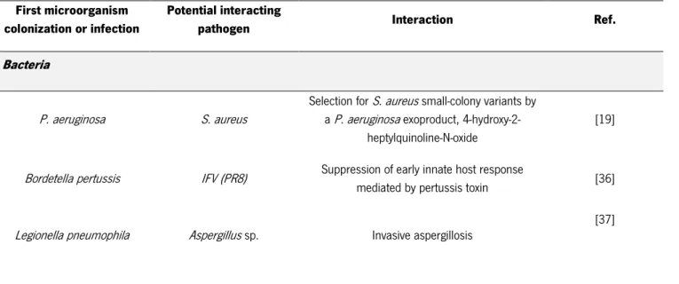
Some examples of microbial interaction models between microorganisms frequently associated with pneumonia are summarized in Tables 1 (positive interactions) and 2 (negative interactions).
Table 1 Positive microbial interaction models between microorganisms frequently associated with pneumonia First microorganism
colonization or infection
Potential interacting
pathogen Interaction Ref.
Bacteria
P. aeruginosa S. aureus
Selection for S. aureus small-colony variants by a P. aeruginosa exoproduct, 4-hydroxy-2-
heptylquinoline-N-oxide
[19]
Bordetella pertussis IFV (PR8) Suppression of early innate host response
mediated by pertussis toxin [36]
Legionella pneumophila Aspergillus sp. Invasive aspergillosis
8
Table 1 (Continued) Positive microbial interaction models between microorganisms frequently associated with pneumonia
Fungi
Candida spp. P. aeruginosa
Candida impedes alveolar macrophage reactive oxygen species production and is correlated
with an increase of P. aeruginosa
pneumonia
[38]
C. albicans P. aeruginosa C. albicans ethanol stimulates P. aeruginosa
strain PAO1 biofilm formation on airway cells [39]
Viruses
Adenovirus (types 1,2,3 and
5) S. pneumoniae
The virus particles increase bacterial adherence
to respiratory tract epithelial cells [40]
Coronavirus NL63 S. pneumoniae
Increased adherence of S. pneumoniae to infected cells correlated with an increased expression level of the platelet-activating factor
receptor
[41]
CMV A. actinomycetemcomitans Increased susceptibility for bacterial adherence
to cells [42]
IFV type A H. influenzae
Increase in bacterial adherence to epithelial cells due to increased cell surface eukaryotic
receptors
[43]
IFV type A N. meningitidis Enhancement in meningococcal adhesion to
epithelial cells due to viral neuraminidase [44]
IFV type A S. pneumoniae Inhibition of pneumococcal clearance due to
platelet-activating factor receptor [19]
IFV type A S. pyogenes
Increase in bacterial adherence to epithelial cells by the presence of GAS (group A Streptococcus) capsule and secondary bacterial
superinfection
[45]
IFV type A Ia group B streptococci Secondary bacterial infection by influenza [46]
IFV type A (H1N1) B. parapertussis Impairment of bacterial clearance mediated by
chemokine MIP-2 [47]
IFV type A (H1N1) S. aureus Viral hemagglutinin increase the efficiency of
9
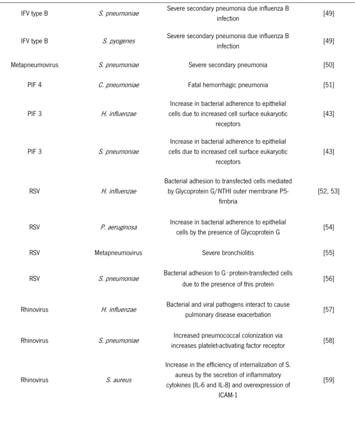
Table 1 (Continued) Positive microbial interaction models between microorganisms frequently associated with pneumonia IFV type B S. pneumoniae Severe secondary pneumonia due influenza B
infection [49]
IFV type B S. pyogenes Severe secondary pneumonia due influenza B
infection [49]
Metapneumovirus S. pneumoniae Severe secondary pneumonia [50] PIF 4 C. pneumoniae Fatal hemorrhagic pneumonia [51]
PIF 3 H. influenzae
Increase in bacterial adherence to epithelial cells due to increased cell surface eukaryotic
receptors
[43]
PIF 3 S. pneumoniae
Increase in bacterial adherence to epithelial cells due to increased cell surface eukaryotic
receptors
[43]
RSV H. influenzae
Bacterial adhesion to transfected cells mediated by Glycoprotein G/NTHI outer membrane
P5-fimbria
[52, 53]
RSV P. aeruginosa Increase in bacterial adherence to epithelial
cells by the presence of Glycoprotein G [54]
RSV Metapneumovirus Severe bronchiolitis [55]
RSV S. pneumoniae Bacterial adhesion to G‑protein-transfected cells
due to the presence of this protein [56]
Rhinovirus H. influenzae Bacterial and viral pathogens interact to cause
pulmonary disease exacerbation [57]
Rhinovirus S. pneumoniae Increased pneumococcal colonization via
increases platelet-activating factor receptor [58]
Rhinovirus S. aureus
Increase in the efficiency of internalization of S. aureus by the secretion of inflammatory cytokines (IL-6 and IL-8) and overexpression of
ICAM-1
10
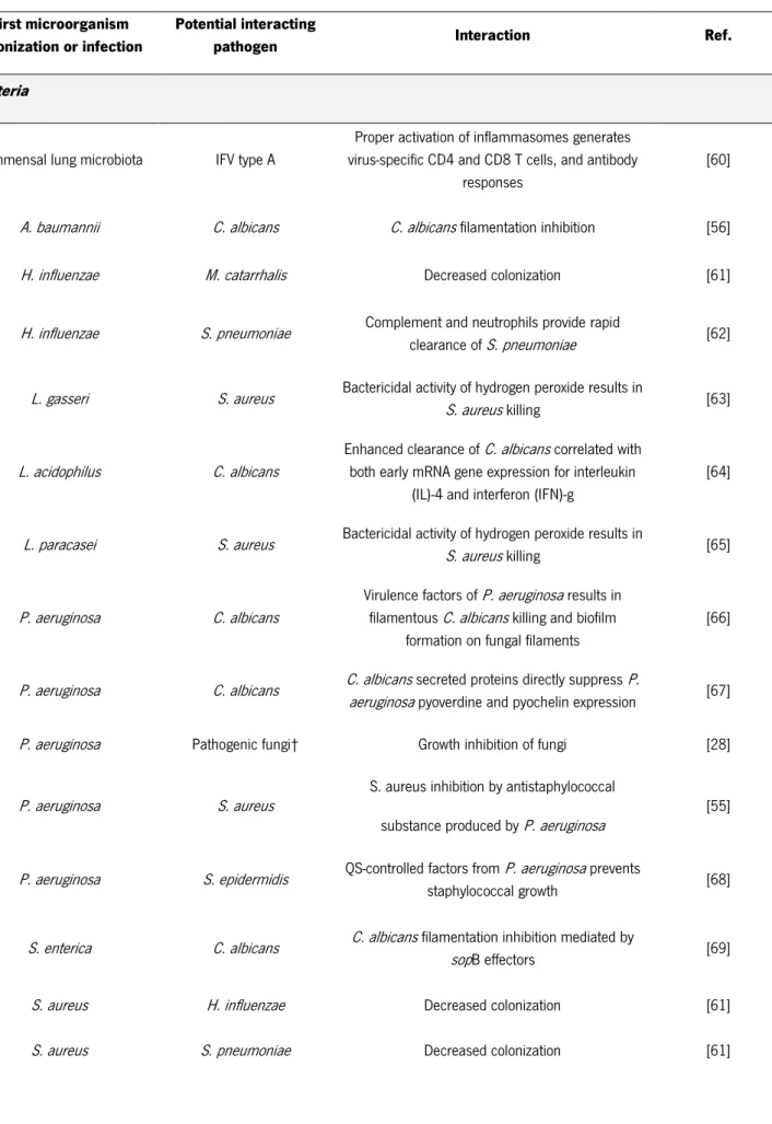
Table 2 Negative microbial interaction models between microorganisms frequently associated with pneumonia First microorganism
colonization or infection
Potential interacting
pathogen Interaction Ref.
Bacteria
Commensal lung microbiota IFV type A
Proper activation of inflammasomes generates virus-specific CD4 and CD8 T cells, and antibody
responses
[60]
A. baumannii C. albicans C. albicans filamentation inhibition [56]
H. influenzae M. catarrhalis Decreased colonization [61]
H. influenzae S. pneumoniae Complement and neutrophils providerapid
clearance of S. pneumoniae [62]
L. gasseri S. aureus Bactericidal activity of hydrogen peroxide results in
S. aureus killing [63]
L. acidophilus C. albicans
Enhanced clearance of C. albicans correlated with both early mRNA gene expression for interleukin
(IL)-4 and interferon (IFN)-g
[64]
L. paracasei S. aureus Bactericidal activity of hydrogen peroxide results in
S. aureus killing [65]
P. aeruginosa C. albicans
Virulence factors of P. aeruginosa results in filamentous C. albicans killing and biofilm
formation on fungal filaments
[66]
P. aeruginosa C. albicans C. albicans secreted proteins directly suppress P.
aeruginosa pyoverdine and pyochelin expression [67]
P. aeruginosa Pathogenic fungi† Growth inhibition of fungi [28]
P. aeruginosa S. aureus
S. aureus inhibition by antistaphylococcal substance produced by P. aeruginosa
[55]
P. aeruginosa S. epidermidis QS-controlled factors from P. aeruginosa prevents
staphylococcal growth [68]
S. enterica C. albicans C. albicans filamentation inhibition mediated by
sopB effectors [69]
S. aureus H. influenzae Decreased colonization [61]
11
Table 2 (Continued) Negative microbial interaction models between microorganisms frequently associated with pneumonia
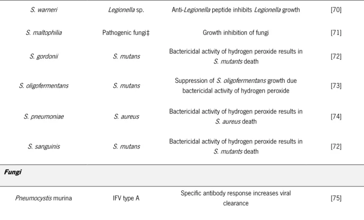
S. warneri Legionella sp. Anti-Legionella peptide inhibits Legionella growth [70]
S. maltophilia Pathogenic fungi‡ Growth inhibition of fungi [71]
S. gordonii S. mutans Bactericidal activity of hydrogen peroxide results in
S. mutants death [72]
S. oligofermentans S. mutans Suppression of S. oligofermentans growth due
bactericidal activity of hydrogen peroxide [73]
S. pneumoniae S. aureus Bactericidal activity of hydrogen peroxide results in
S. aureus death [74]
S. sanguinis S. mutans Bactericidal activity of hydrogen peroxide results in
S. mutants death [72]
Fungi
Pneumocystis murina IFV type A Specific antibody response increases viral
clearance [75]
In the literature, a wide set of interaction with different organisms have been studied. Tables 1 and 2 presented previously show that positive interactions mainly occur with by viruses the first microorganism colonization or infection. On the other hand, when bacteria are the first microorganism colonization it generally leads to negative interaction with potential interacting pathogen. This study is very important in clinical setting since it allows developing therapies considering these consortia in infections.
In polymicrobial synergism infection, the combined effect of two or more microbes on the disease progression can be more dramatic that any of the individuals alone and it display enhanced pathogen persistence in the infection site, increased disease severity, and increased antimicrobial resistance [76, 77]. Synergetic interactions lead to biofilm development with increase of antibiotic tolerance, defense against competitors, adaptation to changing environments, increased tissue damage and declined pulmonary infection [76, 78].
However, in some cases the antagonistic interactions between organisms within a community are unavoidable due to competition for limited resources, with effects on the growth or viability of competitors [79].
12
In VAP infections these both situations can occur as have seen in the tables and in case of inter-kingdom consortia of P. aeruginosa and C. albicans these situations were also observed [29, 66, 80–82].
Understanding the mechanisms of increased pathogenicity in polymicrobial infections is the first step in the development of an effective therapy. Inter-cellular communication is a challenge which has been working in an attempt to minimize the injury from infection to the host.
1.3.1 Pseudomonas aeruginosa quorum-sensing mechanisms
There are numerous VF possessed by P. aeruginosa, which are closely related with virulence mechanisms of this pathogen. These mechanisms include QS, type II and type III secretion system (TTSS). The main VF described for P. aeruginosa: QS, type three secretion system (TTSS) and the presence of lipopolysaccharides.
The VF include proteases, lipases and exotoxin A released by a type II secretion system (Xcp regulon), as well as exotoxins (such as Exo S, Exo T, Exo U and Exo Y) secreted by TTSS [83]. In addiction pyoverdine, rhamnolipids, lipopolysaccharide (LPS) and pili also are included in virulence of P. aeruginosa [84].
The TTSS, a needle-like protein apparatus able to inject exotoxins (mentioned above) into cells is active against human macrophages, where Exo U expression represents a risk factor for mortality in P. aeruginosa pneumonia [85]. The production of elastase, phenazines (e.g.: pyocyanin) and TTSS exotoxins by bacteria is associated to lung injuries. For instance, elastase was associated with acute lung injury and Exo U was associated with increased virulence and a high risk of bacteremia [86]. Conversely, the presence of lipopolysaccharide, namely Lipid A, interacts with receptor 4 in epithelial cells but apparently has not an apparent effect in pneumonia [87].
Another VF common in majority of microorganisms is the capacity to form biofilm since become resistant to antimicrobials.
Many classes of AI have been described to date. The most intensely studied AI are the acylhomoserine lactones (AHLs) of Gram-negative bacteria (e.g.: P. aeruginosa), the oligopeptides produced by Gram-positive bacteria (e.g.: S. aureus and S. pneumoniae) and AI-2 used a wide range of Gram-positive and Gram-negative bacterial species, whose structures remain unknown in most cases [33].
13 P. aeruginosa generally uses QS to coordinate the production of VF by the two main signaling systems, Las and Rhl which are controlled by the genes namely las and rhl, respectively [86]. These systems utilize self-secreted AI molecules 3-oxo-dodecanoyl acyl homoserine lactone (3-oxo-C12-HSL, one of the AHL molecules) and N-butanoyl acyl homoserine lactone (C4-HSL), respectively. When high concentrations of these molecules are achieved it’s possible dock with their cognate receptor proteins such as LasR and RhlR and form a signal-receptor complex, which regulates the expression of various genes responsible for biofilm formation and virulence factor production [88]. The systems LasI/Las and RhlI/RhlR are responsible for the secretion of elastase and rhamnolipids respectively [89]. Additionally, a third system of QS known as Pseudomonas Quinolone Signal (PQS) has been reported in this organism [90] and is involved in secretion of pyocyanin [89].
It is estimated that QS in P. aeruginosa regulate 350 genes, of which around 30% encode VF production [91].
1.3.2 Candida albicans quorum-sensing mechanisms
The VF and abilities of C. albicans potentiate this microorganism to infect a diverse host niches. For this pathogen, the virulence is mostly due to morphological transition between yeast and hyphal forms. Its ability to shift form yeast to filamentous forms is controlled by QS. The filamentous forms of the yeast such as hyphae or pseudo-hyphae, are responsible for tissues penetration and destroy macrophages [92]. The hyphal form has been shown to be more invasive than the yeast form [25]. On the other hand the smaller yeast form is believed to represent the form primarily involved in dissemination [93]. The yeast-to-hyphal form transition can be controlled by temperature, pH, nutrient concentration, cell density or human serum [94]. While at low pH (<6) C. albicans grow predominantly in the yeast form, at high pH (>7) the hyphal growth is induced. Cell density also affect the morphology due to QS, high cell densities (>107 cells ml-1) favor yeast growth, while low cell densities (<107 cells ml-1) promotes hyphal growth. The transition between these two morphologies is termed dimorphism and it has been proposed like an important factor of pathogenicity [95].
The attributes of this pathogen further include the expression of adhesins and invasins on the cell surface which mediate adherence to other cells (C. albicans cells, other microorganisms or host cells) and to abiotic surfaces.The grip may be mediated by the expression of the ALS gene family,
14
belonging to the family of the immunoglobulins. These agglutinins interact specifically with molecules of the host [96]. Phenotypic switching, secretion of hydrolytic enzymes, thigmotropism and formation of biofilms are included in virulence factors of this pathogen. C. albicans is able to form biofilms on abiotic (catheters, dentures) or biotic (mucosal cell) surfaces which shows that an important virulence factor [97]. In addition, others aspects include rapid adaptation to fluctuations in environmental pH, metabolic flexibility, powerful nutrient acquisition systems and robust stress response machineries [98].
C.albicans has the greatest number of QS molecules identified until now. The first QS molecules identified for this fungus were tryptophol and phenylethyl alcohol [99]. These molecules inhibit cell grow and germ tube formation. Additionally three molecules have been identified from C. albicans: farnesol, tyrosol and farnesoic acid [100], although this last one has only been reported in C. albicans ATCC 10231 [101].
Farnesol and tyrosol belong to sesquiterpene alcohols. These inducers regulate cell morphology, growth, biofilm formation, resistance to oxidative stress, and other processes in the life of C. albicans.
Farnesol is the best QS molecule studied and it is known to blocks the morphological transition from yeast to the filament form at high cell densities [102], otherwise tyrosol promotes filamentation [100].
1.3.3 Pseudomonas aeruginosa and Candida albicans interaction
In case of interaction between P. aeruginosa and C. albicans, positive or negative associations may occur. QS molecule 3-oxo-C12-HSL produced by P. aeruginosa affect the morphology of C. albicans (inhibits C. albicans filamentation), thereby altering the ability of the fungus adhere or to invade tissues and the capacity to form biofilm [103]. In this interaction, the surveillance of C. albicans is compromised and it decreases the chance of subsequent infection. Therefore, it represents a negative association between the microorganisms, but also a positive situation for the host. In addition, bacterial toxins such as pseudomonal phenazines have been shown to have antifungal properties [66, 80]. C. albicans can also influence negatively P. aeruginosa by producing farnesol, a QS molecule similar in structure to 3-oxo-C12-HSL, which at low cell density allows modulate the behavior of P. aeruginosa and decreases its virulence. This decrease in virulence is achieved due to inhibition of PQS production (Cugini et al. 2007) required
15 for the expression of several VF [104]. At higher concentration, this molecule can suppress the effect of farnesol on PqsR activity [29].
However, positive associations between these pathogens are also been reported [38]. The main molecular mechanisms of interaction between P. aeruginosa and C. albicans are outlined in Figure 4.
The understanding of the molecular details of QS mechanisms and the way they affect host cells provide an important tool that are now considered a target in antibiotic treatment and controlling bacterial infections.
Figure 2 Molecular mechanisms of the interactions between P. aeruginosa and C. albicans [28] a| P. aeruginosa can attach to the surface of C. albicans hyphae and form biofilms. Production of VF (phospholipase C [66] and phenazines [80] by P. aeruginosa leads to the death of the fungal filament b| QS molecules that are produced by both P. aeruginosa and C. albicans in the mixed-species biofilm [29]. P. aeruginosa produces 3-oxo-C12- homoserine lactone that can inhibit the Ras1–cyclic AMP (cAMP)– protein kinase A (PKA) pathway for hyphal growth in C. albicans, inhibiting filamentation of the fungus [81]. Because yeast cells have increased survival in the presence of P. aeruginosa, the switch to growth as yeast may contribute to the coexistence of both species in mixed infections c| The farnesol produced by C. albicans allows modulates the behavior [29] of P. aeruginosa and decreased quorum sensing molecules. Others uncharacterized C. albicans factors increase the production of VF or alter swarming motility and biofilm formation [80, 82].
16
1.4 Biofilms in VAP
One factor that deserves high attention in VAP is the ability of the microorganisms to develop biofilms. Biofilms are well-structured microbial communities adhered to a surface, where microorganisms are enclosed by a self-produced matrix [105]. These extracellular polymeric substances (EPS) are produced when exopolysaccharides, adhesins and cognate receptors are synthesized by planktonic cells when attached to the surface, which may be epithelial cells or medical devices. The EPS matrix, which can constitute up to 90% of the biofilm biomass [53], is a complex mixture including DNA, proteins, polysaccharides and other macromolecules, conferring a protective effect to biofilm-encased microorganisms against aggressive external factors [52, 106, 107].
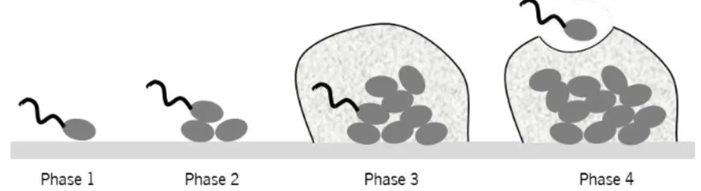
The initial attachment of microorganisms to the surface (Phase 1) is driven by hydrophobic and/or electrostatic interactions as well as specific bacterial surface molecules. The next step is the formation of micro- and then macro-colonies (Phase 2) together with the formation of the polymeric matrix (Phase 3). Lastly, when maturation occurs, the enlarged biofilm shows focal dissolution and begins to release planktonic bacterial cells (Phase 4), which can spread to other locations and develop other biofilms. A schematic representation of biofilm formation phases are shown in figure below.
Figure 3 Stages of biofilm formation. Initial attachment of microorganisms to the surface (Phase 1); formation of micro-colonies (Phase 2); formation of macro-colonies with the formation of a self-produced polymeric matrix (Phase 3); maturation of biofilm, with the release of planktonic cells.
Biofilms are often resistant against antimicrobial agents (such as antibiotics), due to several mechanisms (e.g.: the presence of the EPS, presence of dormant cells and multicellular resistance strategies).
17 Biofilm structure itself acts as a barrier to host defenses, because it reduces the mobility of immune cells and restricts antibiotic diffusion, contributing for the chronicity of the disease [106, 107]. Mechanisms of biofilms resistance against antimicrobials agents include:
Presence of EPS: A biofilm is a permanent source of infection and confers protection to the microorganisms towards antibiotic therapy due to the presence of the EPS matrix [108]. The EPS of biofilms contains polysaccharides, proteins, and DNA (eDNA) that form a glue-like substance for adhesion to the surface and for the three-dimensional biofilm architecture [109]. The EPS function as a barrier providing protection to the cells in the biofilm, and is considered one of the causes associated to antimicrobial resistance, where the antimicrobial agents may be prevented from penetrating the biofilm if they bind to components of the biofilm matrix or to microorganism membranes [110]. Alginate is an polysaccharide of EPS in a mucoid P. aeruginosa biofilm and has been shown to increase the tolerance to aminoglycosides [111].
Multicellular strategies: additional to the protective matrix, biofilm resistance depends on different multicellular strategies from exchanged plasmids, transposons and mutations in genes encoding microbial resistance [107, 112]. The high densities of microorganisms within the biofilm under pressure of antimicrobials enhance horizontal gene transfer and the frequency of mutation [113]. Additionally, delay of antimicrobial penetration through EPS can also induce the expression of genes mediating resistance in the biofilm [114].
Altered metabolism: the expression of distinct metabolic pathways based on the local environmental circumstances in the biofilm is controlled by various genotypes and phenotypes coexist within the biofilm population. Studies have shown that biofilms feature chemical patterns that correspond to gradient of antimicrobials with differences in concentration from outside to inside the biofilm. Due this fact he metabolic activity of microorganisms is higher in the external part of the biofilm and lower in the internal part leading to a reduced susceptibility to antimicrobials [110, 115] In this case the metabolism is adapted due to external factors.
18
Slow growth: biofilm is a metabolic heterogeneous population induced by multiple microcolonies part of such communities. Within the biofilms are created zones characterized by an poor nutrition and can be developed stationary phase-like dormant-cells which can lead for antimicrobials resistance [110, 116] by restricting the access for these antimicrobials that contribute to the general resistance seen in biofilms [117].
Persister cells: some cells of microorganisms can survive after prolonged exposure to antimicrobials which are denominated persister cells. This subpopulation of microorganisms that differentiate into a dormant and protected state. One aspect of chronic characteristics of bacterial infectious disease is the presence of these dormant cells, which are able to resist to the action of most antibiotics. These dormant bacteria, which colonizes specifically the deeper parts of the biofilms, can suppress their metabolism, including cell membrane formation, protein synthesis, and DNA replication [118, 119]. Dormant bacteria can survive to antibiotic exposure because their antibiotic target sites are deactivated, which means that they tolerate sublethal concentrations of antibiotics [120, 121].
Oxygen condition: the oxygen tension in the depth of biofilm is low such as been described for P. aeruginosa and these low tension (hypoxia) change the composition of multidrug efflux pumps with consequently antimicrobial resistance like response to stress [122]. Aditionlly, the ability of this bacterium to adapt to the oxygen-limited environments is associated with a drastic physiological change in P. aeruginosa (e.g. increased alginate production; alterations in the outer membrane; biofilm development), which contributes to an increased antibiotic tolerance [123]. In addition, the anaerobic environment within biofilms will most likely affect aminoglycoside antimicrobial activity due to the downregulation of energy metabolism genes [124] and by triggering changes in gene expression [125].
Swarming: the microorganisms with the ability to swarm reflect a social multicellular behavior and its reflets in decrease the effectiveness of the agents against infections [126].
19 QS: it is complex system, which regulates the behaviour with others cells, control of
microbial population density and expression of VF [127].
Biofilms are seen as a major problem in medical settings and it has been estimated that up to 80% of all infections worldwide are biofilm-related [128].
Therefore, microbial adhesion and biofilm formation on medical devices (Figure 4) often leads to deterioration, blockage and loss of function and removal of the devices is often the unique solution [129].
Figure 4 Photomicrographs captured by scanning electron microscopy of biofilms developed on the VAP ETT [16].
Biofilms are typically formed on the inner surface and in case of the VAP infection in ETT [130] which contribute to the development of the infection, allowing the contact and persistence of pathogens within the host [131]. In ventilated patients the biofilms forms on ETT very quickly after intubation and its act as a significant source of inoculation of the lungs by bacteria [132]. The tube appears to be a point of access of microorganisms to the lower respiratory tract (e.g.: via microaspiration during intubation itself, development of biofilm within the tube) [10, 11].
1.5 VAP therapy
VAP patients still receive inadequate initial antibiotics treatment even if it is well known that the incidence of MDR pathogen infections is on the rise in ICU. VAP is one of the major sites for emergence of MDR pathogens because subtherapeutic antibiotic concentrations in the lung require longer duration of therapy, thereby favoring selection of resistant bacteria [16].
20
When VAP is suspected, empirical antibiotics should be administered immediately. The accurate identification of etiologic pathogens might improve therapy procedures and the control of the infection to avoid complications for the patient. Although bacteriological sampling is important, it should not significantly delay the start of treatment [14]. Even if the bacteriological test is not available, the therapy should be initiated. The most relevant information to start treatment, until there is no specific clinical information on the cause of the infection agent, it's whether it comes to early onset VAP or late onset VAP. Usually, the antibiotic therapy is initiated with a broad-spectrum antibiotic [16]. It is still necessary to know the whole clinical history of the patient with infection to choose the appropriate treatment.
Approximately 50 % of all antibiotics administered in ICUs are for treatment of VAP [8].
The usual duration of treatment for early onset VAP is eight days and longer in the case of late-onset VAP or if MDR organisms are suspected or identified [133, 134]. Late late-onset VAP requires broad spectrum antibiotic whereas early onset VAP can be treated with limited spectrum antibiotics [4].
1.5.1 Conventional therapy
There is a general agreement that rapid initiation of appropriate antimicrobial therapy improves the outcome of the disease [14]. In VAP, conventional therapy typically includes the administration of empirical antibiotics in an attempt to cure the infection. However, due to certain resistance mechanisms, specific to each microorganism, the recommended treatment varies. The therapy is started according to the time of onset (Table 3).
Table 3 Comparison of recommended initial empiric therapy for VAP according to time of onset [4, 133]
Early onset VAP Late onset VAP
Second or third generation cephalosporin: e. g., ceftriaxone: 2 g daily;
cefuroxime: 1.5 g every 8 hours; cefotaxime: 2 g every 8 hours OR
Fluoroquinolones
e. g., levofloxacin: 750 mg daily;
Cephalosporin
e. g., cefepime: 1–2 g every 8 hours; ceftazidime 2 g every 8 hours OR
Carbepenem
e. g., imipenem + cilastin: 500 mg every 6 hours or 1 g every 8 hours;
21
Table 3 (Continued) Comparison of recommended initial empiric therapy for VAP according to time of onset [4, 133]
e. g., levofloxacin: 750 mg daily; moxifloxacin: 400 mg daily OR
Aminopenicillin + beta-lactamase inhibitor e. g., ampicillin + sulbactam: 3 g
every 8 hours OR
Ertapenem 1 g daily
meropenem: 1 g every 8 hours OR
Beta-lactam/beta-lactamase inhibitor
e. g.: piperacillin + tazobactam: 4.5 g every 6 hours PLUS Aminoglycoside e. g.: amikacin: 20 mg/kg/day; gentamicin: 7 mg/kg/day; tobramycin: 7 mg/kg/day OR Antipseudomonal fluoroquinolone e. g.: ciprofloxacin 400 mg every 8 hours; levofloxacin 750 mg daily
PLUS
Coverage for MRSA
e. g.: vancomycin: 15 mg/kg every 12 hours OR
linezolid: 600 mg every 12 hours
The therapy can be initiated based on the information about the causative microorganism (Table 4).
Owing to the high rate of resistance to monotherapy observed with P. aeruginosa, combination therapy is always recommended [135].
Table 4 Recommended therapy for suspected or confirmed multidrug resistant organisms and fungal VAP [4, 133]
Pathogen Treatment
Methicillin-resistant Staphylococcus aureus See Table 3
22
Table 4 (Continued) Recommended therapy for suspected or confirmed multidrug resistant organisms and fungal VAP [4, 133]
Acinetobacter species Carbapenem
e. g.: imipenem + cilastin; 1 g every 8 hours; meropenem 1 g every 8 hours
OR
Beta-Lactam/beta-lactamase inhibitor e. g., ampicillin + sulbactam: 3 g every 8 hours OR
Tigecycline: 100 mg loading dose, then 50 mg every 12 hours
Extended-spectrum beta-lactamase (ESBL) positive enterobacteriaceae
Carbepenem
e. g.: imipenem + cilastin: 1 g every 8 hours; meropenem: 1 g every 8 hours
Fungi Fluconazole: 800 mg every 12 hours;
caspofungin: 70 mg loading dose, then 50 mg daily; voriconazole (for aspergillus species): 4 mg/kg every 12 hours
Legionella Macrolides (e. g.: azithromycin)
OR
Fluoroquinolones (e. g.: levofloxacin)
Several antibiotics have re-emerged as alternatives to treat P. aeruginosa. Polymyxins are used in cases of MDR P. aeruginosa. Polymyxin B (PolyB) is an antibiotic primarily used for resistant Gram-negative infections and frequently used to control pulmonary infections caused by P. aeruginosa [136]. For treatment of C. albicans, amphotericin B (AmB) was considered the best choice to treat serious and invasive Candida infections [137].
1.5.2 Alternative approaches in biofilm control: the QS inhibitors
Due to the frequent failures of antibiotherapy towards biofilms new therapeutic modalities to treat the infection are required. Additionally, empirical antimicrobial regimen leads to the overuse of antibiotics and, thus, emergence of antimicrobial resistance. This is a major problem associated with the use of antibiotics and a constant concern as it leads to increased mortality. The formation
23 of biofilm in the VAP ETT is also a concern, because this is usually impervious to systemic antibiotics, making the treatment of infection more complicated [106].
For P. aeruginosa, high percentages of isolates resistant to aminoglycosides, ceftazidime, fluoroquinolones, piperacillin/tazobactam, and carbapenems were reported from several countries in Europe in 2011 [138].
To improve the quality of care and consequent decrease in mortality associated with VAP, new challenges are placed in the field of treatment of infection. The alternatives to conventional therapies are related by using new products that may interact and interfere more effectively with microorganisms causing the disease, leading to more efficient therapy. The new strategies generally aim to interfere with exoproducts produced by microorganisms, inhibiting cellular communication and reducing biofilm production. Possible alternatives to synthetic antimicrobials include QSI, antimicrobial peptides, biofilm degradation enzymes and bacteriophages.
Antimicrobial peptides: antimicrobial peptides (AMPs) are part of the inmate immune response found among all classes of life. Yours characteristics hydrophobic and cationic allow them pouring and fragment the citoplasmatic membrane [139]. Recently was described that certain AMPs are able to destabilize EPS by binding to the eDNA with consequent disrupt of biofilm [140].
Enzymes: new approaches to biofilm control also include the use of biofilm matrix-degrading enzymes in order to detach the cells of biofilms and make them more susceptible to antimicrobial. DNase I is one of these enzymes which degrades the eDNA of EPS. Dispersin B (DspB) and α-amylase act likewise DNase on the biofilms. A combination of the enzymes with antibiotics is promising in clinical context [141].
Bacteriophages: also known as phages, are viruses that infect bacteria. Thus, phages are candidates to prevent and control biofilm since they are able infect and lyse cells in single and polymicrobial species biofilms [142, 143]. However bacteria can escape to phage infection by increasing your biofilm formation ability [144].
QS inhibitors (QSI): in this regard, advances in the QS field have been made. In recent years, a number of biotechnology companies that aim specifically at developing
anti-24
QS and anti-biofilm drugs have emerged [145]. The great diversity of QS signal generators, receivers, carriers, regulators and the signals themselves, represent multiple targets for inhibition of QS. This strategy of inhibition of QS pathways by interfering with signal generation, signal relay, signal transduction or destruction of the signal entirely is also known as “quorum quenching” [146]. QS inhibition strategies may have targeted species-specific QS molecules. Many studies have been done and many compounds studied with potential QSI were proposed such as furanone [147] and patulin [148], two compounds of natural origin. Some antibiotics (such as the macrolide azithromycin (AZT), the β-lactam ceftazidime and the fluoroquinolone ciprofloxacin (CIP)) have QSI activity in addition to their conventional antibiotic activity [149]. QSI are compounds able to quench the action of and usually are considered as safe. Additionally, it is not expectable any development of resistance since QSI not impose any selective pressure to bacteria, as seen in classical antibiotic treatment [150].
REFERENCES
1. Ventilação Mecânica (1a parte)
[http://estudmed.com.sapo.pt/anestesiologia/ventilacao_mecanica_1.htm]
2. Overview of Mechanical Ventilation - Critical Care Medicine - Merck Manual Professional Edition [https://www.merckmanuals.com/professional/critical-care-medicine/respiratory-failure-and-mechanical-ventilation/overview-of-mechanical-ventilation]
3. Brochard L: Mechanical ventilation: invasive versus noninvasive. Eur Respir J 2003, 22(47 suppl).
4. Guidelines for the management of adults with hospital-acquired, ventilator-associated, and healthcare-associated pneumonia. Am J Respir Crit Care Med 2005, 171:388–416.
5. Mechanical Ventilation - Lung and Airway Disorders - Merck Manual Home Edition
[https://www.merckmanuals.com/home/lung-and-airway-disorders/respiratory-failure-and-acute-respiratory-distress-syndrome/mechanical-ventilation]
6. Pneumatikos IA, Dragoumanis CK, Bouros DE: Ventilator-associated pneumonia or endotracheal tube-associated pneumonia? An approach to the pathogenesis and preventive strategies emphasizing the importance of endotracheal tube. Anesthesiology 2009, 110:673–80.
7. Zolfaghari PS, Wyncoll DLA: The tracheal tube: gateway to ventilator-associated pneumonia.
25 8. Hunter JD: Ventilator associated pneumonia. BMJ 2012, 344:e3325–e3325.
9. Pinciroli R, Mietto C, Berra L: Respiratory therapy device modifications to prevent ventilator-associated pneumonia. Curr Opin Infect Dis 2013, 26:175–83.
10. Grgurich PE, Hudcova J, Lei Y, Sarwar A, Craven DE: Diagnosis of ventilator-associated pneumonia: controversies and working toward a gold standard. Curr Opin Infect Dis 2013, 26:140–50.
11. Mietto C, Pinciroli R, Patel N, Berra L: Ventilator associated pneumonia: evolving definitions and preventive strategies. Respir Care 2013, 58:990–1007.
12. Safdar N, Crnich CJ, Maki DG: The pathogenesis of ventilator-associated pneumonia: its relevance to developing effective strategies for prevention. Respir Care 2005, 50:725–39; discussion 739–41.
13. Joseph NM, Sistla S, Dutta TK, Badhe AS, Parija SC: Ventilator-associated pneumonia: a review.
Eur J Intern Med 2010, 21:360–8.
14. Hunter JD: Ventilator associated pneumonia. Postgrad Med J 2006, 82:172–8.
15. Afshari A, Pagani L, Harbarth S: Year in review 2011: Critical Care - infection. Crit Care 2012, 16:242.
16. Piazza O, Wang X: A translational approach to ventilator associated pneumonia. Clin Transl Med 2014, 3:26.
17. Kalanuria AA, Zai W, Mirski M: Ventilator-associated pneumonia in the ICU. Crit Care 2014, 18:208.
18. Chastre J, Fagon J-Y: Ventilator-associated pneumonia. Am J Respir Crit Care Med 2002, 165:867–903.
19. Hoffman LR, Déziel E, D’Argenio DA, Lépine F, Emerson J, McNamara S, Gibson RL, Ramsey BW, Miller SI: Selection for Staphylococcus aureus small-colony variants due to growth in the presence of Pseudomonas aeruginosa. Proc Natl Acad Sci U S A 2006, 103:19890–5.
20. Lambert PA: Mechanisms of antibiotic resistance in Pseudomonas aeruginosa. J R Soc Med
2002, 95 Suppl 4:22–6.
21. Nseir S, Jozefowicz E, Cavestri B, Sendid B, Di Pompeo C, Dewavrin F, Favory R, Roussel-Delvallez M, Durocher A: Impact of antifungal treatment on Candida-Pseudomonas interaction: a preliminary retrospective case-control study. Intensive Care Med 2007, 33:137–42.
22. Pier GB: Pulmonary disease associated with Pseudomonas aeruginosa in cystic fibrosis: current status of the host-bacterium interaction. J Infect Dis 1985, 151:575–80.
23. Tan MW, Mahajan-Miklos S, Ausubel FM: Killing of Caenorhabditis elegans by Pseudomonas aeruginosa used to model mammalian bacterial pathogenesis. Proc Natl Acad Sci U S A 1999, 96:715–20.
26
24. Kaleli I, Cevahir N, Demir M, Yildirim U, Sahin R: Anticandidal activity of Pseudomonas aeruginosa strains isolated from clinical specimens. Mycoses 2007, 50:74–8.
25. Berman J, Sudbery PE: Candida Albicans: a molecular revolution built on lessons from budding yeast. Nat Rev Genet 2002, 3:918–30.
26. Morales DK, Hogan DA: Candida albicans interactions with bacteria in the context of human health and disease. PLoS Pathog 2010, 6:e1000886.
27. Sandven P: Epidemiology of candidemia. Rev Iberoam Micol 2000, 17:73–81.
28. Peleg AY, Hogan DA, Mylonakis E: Medically important bacterial-fungal interactions. Nat Rev
Microbiol 2010, 8:340–9.
29. Cugini C, Calfee MW, Farrow JM, Morales DK, Pesci EC, Hogan DA: Farnesol, a common sesquiterpene, inhibits PQS production in Pseudomonas aeruginosa. Mol Microbiol 2007, 65:896–906.
30. Kaufmann GF, Park J, Janda KD: Bacterial quorum sensing: a new target for anti-infective immunotherapy. Expert Opin Biol Ther 2008, 8:719–24.
31. Li Y-H, Tian X: Quorum sensing and bacterial social interactions in biofilms. Sensors (Basel)
2012, 12:2519–38.
32. Bassler BL, Losick R: Bacterially speaking. Cell 2006, 125:237–46.
33. Antunes LCM, Ferreira RBR: Intercellular communication in bacteria. Crit Rev Microbiol 2009, 35:69–80.
34. Fuqua WC, Winans SC, Greenberg EP: Quorum sensing in bacteria: the LuxR-LuxI family of cell density-responsive transcriptional regulators. J Bacteriol 1994, 176:269–75.
35. Tegmark K, Morfeldt E, Arvidson S: Regulation of agr-dependent virulence genes in Staphylococcus aureus by RNAIII from coagulase-negative staphylococci. J Bacteriol 1998, 180:3181–6.
36. Ayala VI, Teijaro JR, Farber DL, Dorsey SG, Carbonetti NH: Bordetella pertussis infection exacerbates influenza virus infection through pertussis toxin-mediated suppression of innate immunity. PLoS One 2011, 6:e19016.
37. Thalanayar PM, Holguin F: Rash, disseminated intravascular coagulation and legionella: Episode 10 and a rewind into the past. Respir Med Case Reports 2015, 15:95–100.
38. Roux D, Gaudry S, Dreyfuss D, El-Benna J, de Prost N, Denamur E, Saumon G, Ricard J-D: Candida albicans impairs macrophage function and facilitates Pseudomonas aeruginosa pneumonia in rat. Crit Care Med 2009, 37:1062–7.
39. Chen AI, Dolben EF, Okegbe C, Harty CE, Golub Y, Thao S, Ha DG, Willger SD, O’Toole GA, Harwood CS, Dietrich LEP, Hogan DA: Candida albicans ethanol stimulates Pseudomonas aeruginosa


![Figure 2 Molecular mechanisms of the interactions between P. aeruginosa and C. albicans [28] a| P](https://thumb-eu.123doks.com/thumbv2/123dok_br/17579310.818633/39.892.277.606.389.878/figure-molecular-mechanisms-interactions-p-aeruginosa-c-albicans.webp)
![Figure 4 Photomicrographs captured by scanning electron microscopy of biofilms developed on the VAP ETT [16]](https://thumb-eu.123doks.com/thumbv2/123dok_br/17579310.818633/43.892.173.719.418.615/figure-photomicrographs-captured-scanning-electron-microscopy-biofilms-developed.webp)