UNIVERSIDADE DA BEIRA INTERIOR
Ciências
Optimization of a gellan gum support by
experimental design for recombinant proteins
partition
Filipe dos Santos Frias
Dissertação para obtenção do Grau de Mestre em
Bioquímica
(2º ciclo de estudos)
Orientadora: Prof. Doutora Ângela Maria Almeida de Sousa
Co-orientador: Prof. Doutor Luís António Paulino Passarinha
Acknowledgments
First of all, I would like to express my sincere gratitude to my parents and brother for all their sacrifices, encouragement, support and unconditional love.
Furthermore, I would also like to give a special thanks to my supervisors Professor Doctor Ângela Sousa and Professor Doctor Luís Passarinha for all their availability, guidance, expertise, patience and trust. Their help, efforts, vast knowledge, criticism and suggestions were crucial for the development of this project. It was a privilege to work and learn with them.
I would also like to acknowledge the University of Beira Interior, in particular the Health Sciences Research Center, where all the work was developed.
I am also grateful to all people involved in the Health Sciences Research Center of the University of Beira Interior, particularly to my colleagues in the Biotechnology and Biomolecular Sciences group whose advice and companionship made my work much more easy and pleasant.
Finally, I am extremely thankful to all my friends. Their constant availability, support, belief, motivation and love through all this time was crucial to surpass the most difficult times.
Resumo alargado
A cromatografia é um dos métodos mais usados para a separação e purificação de diferentes biomoléculas terapêuticas. É uma técnica que tem sido muito explorada, ao longo dos últimos anos, nas áreas da indústria farmacêutica e biotecnológica para a obtenção de proteínas e ácidos nucleicos com elevado grau de pureza.
Os suportes cromatográficos têm sido alvo de grandes desenvolvimentos a fim de encontrar uma matriz que reúna as características ideais como porosidade, estabilidade química e física, elevada eficiência de transferência de massa, baixo custo de produção associado, biocompatibilidade, hidrofilicidade, boa capacidade de manutenção do fluxo e capacidade de reutilização após vários ensaios cromatográficos.
A goma de gelana é um polímero polissacárido natural linear que tem uma vasta gama de aplicações em diversas áreas, desde a indústria alimentar (agente espessante e gelificante), cosmética (loções, cremes, maquilhagem, etc), indústria biotecnológica (como um substituinte do agar), indústria farmacêutica (sistema de entrega direcionada de fármacos e microencapsulações) e medicina (construção de “scaffolds” para regeneração de tecidos). Devido às suas propriedades como versatilidade, biocompatibilidade, biodegradabilidade e elevada estabilidade, a gelana tem atraído grande interesse por parte do mercado e tem ganho crescente importância entre as diferentes indústrias.
A gelana possui a capacidade de, em determinadas condições e na presença de catiões divalentes, sofrer uma alteração conformacional formando uma forte rede tridimensional devido às interações entre a gelana, os iões e as moléculas de água dando origem a um gel termoreversível, resistente a altas temperaturas e a extremas condições ácidas.
Tendo em conta as características apresentadas, o presente trabalho teve como objetivo otimizar a formulação de uma matriz cromatográfica pela técnica de emulsão água-em-óleo a fim de ser usada em diversos processos cromatográficos. A inovação deste projeto tem como base a produção de microesferas através de um método de emulsão água-em-óleo, usando dois líquidos imiscíveis, a água e um óleo alimentar vegetal. Trata-se de uma técnica que não requer o uso de instrumentos complexos, é de fácil manuseamento, tem baixo custo associado e permite o fácil controlo dos parâmetros associados a este processo. A preparação das esferas foi feita de acordo com os seguintes parâmetros: concentração da goma de gelana (1 % - 2,5 %), velocidade de agitação (250 rpm – 750 rpm) e temperatura (20 ºC – 100 ºC). Considerando que todos estes fatores afetam a estabilidade e estrutura das microesferas, foram feitas várias formulações utilizando um desenho experimental, estratégia que permitiu obter as condições ótimas no sentido de se produzirem micropartículas com o mínimo diâmetro possível. As condições ótimas fornecidas pelo desenho foram 1,41 % de concentração de gelana, 749,47 rpm de velocidade de agitação e 99,20 ºC para obter
cromatográfico, foi necessário a presença de catiões divalentes numa solução de reforço, para onde as esferas foram transferidas a fim de aumentar a estabilidade. O catião escolhido foi o bário, que devido ao seu grande tamanho iónico é considerado bastante eficaz na gelificação da gelana, pois contribui largamente para a formação da estável e rígida rede tridimensional. Além disso, o bário também permitiu a formação de esferas uniformes de tamanho reduzido com um pequeno ratio de inchaço.
Para a validação do modelo fornecido pelo software do desenho experimental, foi necessário efetuar cinco réplicas de dois pontos previstos dados pelo programa. A visualização das esferas produzidas foi feita através de microscopia ótica que permitiu a medição dos diâmetros das micropartículas e através de microscopia eletrónica de varrimento, que tornou possível a análise estrutural das esferas num estado liofilizado, bem como a análise topográfica e morfológica da sua superfície. Além disso, esta última metodologia também permitiu avaliar a porosidade das esferas, de onde foi constatado a ausência de canais interiores, sugerindo que qualquer interação estabelecida com a matriz de goma de gelana se dê apenas na sua superfície.
Aproveitando a natureza aniónica do polímero de gelana e o facto de não ter que ser funcionalizada com ligandos como os suportes cromatográficos comerciais, foi possível explorar diferentes interações com as proteínas modelo albumina sérica bovina (BSA), α-quimotripsina e lisozima, bem como com um extracto parcialmente purificado da proteína catecol-O-metiltransferase humana na isoforma solúvel (hSCOMT). Nos ensaios com as proteínas modelo, o tampão usado foi o MES a pH 6.2, o que conferiu carga negativa à BSA e cargas positivas à α-quimotripsina e lisozima devido aos seus pontos isoelétricos. Assim, a BSA não ligou à matriz tendo sido eluída após a injeção da amostra na coluna, ainda com a passagem do tampão sem sal, enquanto as outras duas proteínas interagiram devido à oposição de cargas, tendo sido eluídas com o aumento da força iónica. Quanto à amostra pré-purificada de SCOMT, foi usado um tampão de pH 4.0 nas condições de equilíbrio, o que conferiu carga positiva a esta proteína. Esta condição permitiu a interação desta proteina com a coluna, tendo sido maioritariamente eluída através da manipulação do pH (alterando o pH do tampão para 6.4), permitindo assim a eliminação de algumas proteínas contaminantes presentes na amostra.
A fim de se caracterizar melhor esta nova matriz cromatográfica, foi determinada a capacidade dinâmica de ligação utilizando uma estratégia de saturação da coluna com uma solução de lisozima 0,5 mg/mL a 1 mL/min. Os valores obtidos na capacidade de ligação das microesferas de goma de gelana a 10 % e 50 % da curva de saturação foram 2,43 mg/mL e 4,73 mg/mL, respetivamente. Comparando com outras matrizes cromatográficas comerciais e tendo em conta que apenas a área de superfície das microesferas de gelana é funcional, estes valores estão dentro do esperado.
Estes estudos permitiram concluir que a estabilidade da matriz cromatográfica de gelana foi incrementada com o desenvolvimento deste projecto de mestrado e que permitiu a interação
com proteínas de várias naturezas, através de estratégias de eluição com manipulação de força iónica e pH.
Em suma, os dados apresentados manifestam uma versatilidade da gelana em interagir com diferentes biomoléculas e, devido à sua capacidade de gelificação, foi possível a elaboração deste inovador e promissor suporte cromatográfico para a cromatografia de troca catiónica, a partir de microesferas da goma de gelana.
Palavras-chave
Cromatografia de troca iónica; Matriz cromatográfica; Microesferas de gelana; Purificação de proteínas; Técnica de emulsão água-em-óleo
Abstract
Chromatography is one of the most studied methods, due to its simplicity, versatility and high reproducibility, to separate and purify molecules that can have therapeutic, industrial and biotechnological interest. In recent years, the development of new chromatographic matrices has been continuously increased in order to afford rapid and efficient separations and decrease the use of resources. Gellan gum is a natural anionic exopolysaccharide and, in the presence of divalent cations, has the ability to form thermos-reversible strong gels resistant to temperature and extreme acidic conditions.
In this work, it was proposed the preparation of gellan gum microbeads to be used as a stable chromatographic stationary phase. In order to produce the matrix, a low-cost water-in-oil emulsion technique was adopted. To obtain optimal conditions to the bead formulation, experimental design was applied, which allowed the optimization of the experimental conditions and to produce microbeads with the smallest diameter possible. Due to the negative charge of gellan gum, it was possible to study the interactions established with three model proteins (BSA, α-chymotrypsin and lysozyme) and with a therapeutic complex protein, SCOMT. In the model protein assays, MES buffer with pH 6.2 was used, which conferred negative charge to BSA and positive charge to α-chymotrypsin and lysozyme, due to its isoelectric points. Thus, BSA did not bind to the matrix while the other two proteins were retained to the gellan gum, being eluted with an increase of ionic strength. Regarding to pre-purified SCOMT sample, a buffer with pH 4.0 was used under equilibrium conditions, conferring positive charge to the protein, thus, it also interacted with the column and was majorly eluted by pH manipulation (by changing the buffer pH to 6.4), allowing the elimination of some protein contaminants.
Dynamic binding capacity assay of the gellan gum microbeads was made, in order to characterize this support as a novel chromatographic matrix. The values of DBC of the gellan gum stationary phase to 10 % and 50 % of breakthrough were 2.43 mg/mL and 4.73 mg/mL, respectively. These DBC values are satisfactory when compared to commercial resins used in affinity chromatography, taking into account that protein interaction only occurred at the gellan bead surface. These results indicated that gellan gum microbeads obtained by water-in-oil emulsion technique can be used as an innovative and promising chromatographic support due to its gelling ability and versatility to interact with different biomolecules.
Keywords
Contents
Chapter I - Introduction 1
1 Gellan gum 1
1.1 Gellan gum as a microbial polysaccharide 1
1.2 Molecular structure 2
1.3 Chemical, physical and gelling properties 2
1.4 Production of gellan gum 5
1.4.1 Fermentation process 5 1.4.2 Biosynthetic pathway 7 1.5 Gellan applications 10 2 Chromatography 15 2.1 Chromatographic process 15 2.2 Preparative chromatography 16 2.3 Chromatographic methods 16
2.3.1 Size-exclusion chromatography (SEC) 18 2.3.2 Ion-exchange chromatography (IEC) 20 2.3.3 Hydrophobic interaction chromatography (HIC) 22 2.3.4 Reversed-phase chromatography (RPC) 24
2.3.5 Affinity chromatography (AC) 25
2.3.5.1 Immobilized metal-ion affinity chromatography (IMAC) 26
2.4 Matrices and ligands 27
2.4.1 Chemical features of chromatographic supports 28 2.4.2 Physical and structural features of chromatographic supports 30 2.4.3 Ligand features on chromatographic supports 32
2.5 Dynamic binding capacity (DBC) 33
2.6 Chromatographic supports for protein purification 34 2.7 Preparation of microbeads by water-in-oil emulsion 36
2.8 Experimental design 38
3 Catechol-O-methyltransferase (COMT) 41
3.1 General overview of catechol-O-methyltransferase 41 3.2 Physiological functions of COMT in catecholamine metabolism 43 3.3 Parkinson´s disease and COMT inhibitors 44 3.4 hSCOMT biosynthesis and purification 45
Chapter II – Objectives 47
Chapter III – Materials and methods 49
2.1 Bead formation with water-in-oil emulsion 49 2.2 Optimization of the bead formulation with DoE 50 2.3 Microscopic bead visualization and scanning electron microscopy (SEM) 52
2.4 Ion exchange chromatography 52
2.5 Electrophoretic and dot-blot analysis 54
2.6 Dynamic binding capacity 54
Chapter IV – Results and Discussion 55
1 Gellan gum beads production 55
2 Optimization of the bead formation with DoE 57
2.1 Goodness of fit 59
2.2 Model validation 61
2.3 Influence of one-factor and interaction plots on gellan gum beads
production 66
2.4 Response surface methodology 71
2.5 Scanning electron microscopy (SEM) 73 3 Applicability of gellan gum beads in chromatography 75 3.1 Chromatographic assays with isolated model proteins 75 3.2 Chromatographic assays with combined model proteins 77 3.3 Chromatographic assays with catechol-O-methyltransferase (COMT) 83
4 Dynamic binding capacity (DBC) 89
Chapter V – Conclusion 93
Chapter VI – Future perspectives 95
List of figures
Figure 1. Repeating unit of the exopolysaccharide gellan produced by S.
paucimobilis ATCC 31416. 2
Figure 2. Schematic model of the conformational transitions and gelation of
low acyl gellan gum through temperature changes with and without added
cations. 3
Figure 3. Chemical structure of native and deacetylated gellan gum. 4
Figure 4. (a) Typical fermentation process for gellan production and its
purification at a laboratory scale. (b) Time of course of growth and gellan production by S. elodea ATCC 31416 at 30 ºC during a batch growth in a defined
medium with 20 g/l glucose at initial pH of 8.0. 6
Figure 5. Proposed pathway leading to the nucleotide sugar precursors,
UDP-D-glucose, UDP-D-glucuronic acid and dTDP-L-Rhamnose, involved in gellan gum
biosynthesis with glucose as the substrate. 8
Figure 6. (a) Organization of the gellan biosynthetic gel clusters (from S.
elodea ATCC 31461. (b) A model for biosynthesis and assembly of gellan. 10
Figure 7. Selective protein properties. 17
Figure 8. In SEC, large molecules run through the space between media with a
shorter pathway, while the smaller molecules run through inside the packing
particles with a longer pathway. 19
Figure 9. Net charge of a protein as function of pH, showing the pH ranges in
which the protein is positively or negatively charged. 21
Figure 10. Types of ion exchangers. 21
Figure 11. (a) Highly ordered water shells surround the hydrophobic surfaces of
ligands and proteins. (b) The equilibrium of the hydrophobic interaction is
controlled predominantly by salt concentration. 23
Figure 12. Proteins and peptides bind to an RCP medium under aqueous
conditions and elute as the eluent becomes more hydrophobic. 24
Figure 13. An affinity matrix binding to its target protein. 25
Figure 14. A bead of a typical IMAC matrix with an attached chelator that
coordinates a metal. 27
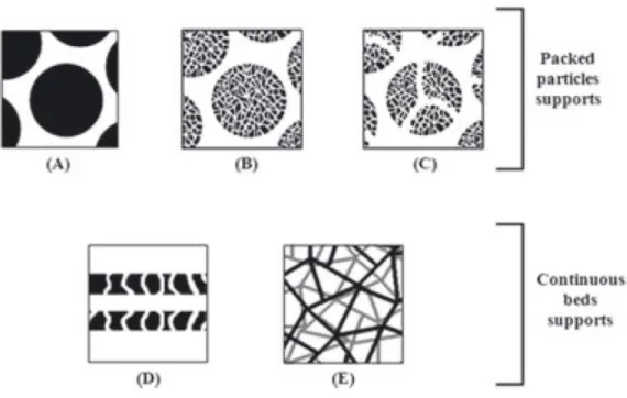
Figure 15. Schematic representation of the physical and structural properties
of the constituent materials of different chromatographic supports. 30
Figure 16. Photomicrograph of a polydisperse oil-in-water emulsion consisting
of oil droplets in a continuous aqueous phase. 37
Figure 18. (a) A three dimensional response surface plot showing the expected
performance (η) as a function between x1 and x2. (b) A contour plot of
variables interaction of a response surface. 40
Figure 19. Reaction catalyzed by catechol-O-methyltransferase (COMT). 42
Figure 20. Schematic representation of the three-dimensional structure of
COMT. 43
Figure 21. Dopaminergic transmission in prefrontal cortex (PFC). 44
Figure 22. Chemical structures of tolcapone and entacapone,
second-generation COMT inhibitors. 45
Figure 23. Scheme design of water-in-oil emulsion. 56
Figure 24. Microscopic visualization with different magnification lens of several
sizes of the formed beads from different runs in a hydrated state. 59
Figure 25. Predicted values versus observed values plot. 61
Figure 26. (a) Diameter sizes for the validation of the DoE model given by
Design Expert software version 7.0 in a confidence interval of 95 % and (b)
considering all the sizes obtained from all the experiments. 62
Figure 27. Contour plots for the predicted response (predicted bead diameter)
of the defined optimal points. 66
Figure 28. Interactions between stirring velocity and temperature, when
assuming gellan concentration as a constant parameter on the beads diameter. 67
Figure 29. Interactions between gellan gum concentration and temperature,
when assuming stirring velocity as a constant parameter on the beads diameter. 68
Figure 30. Interactions between gellan gum concentration and stirring velocity,
when assuming temperature as a constant parameter on the beads diameter. 69
Figure 31. The effect of temperature, when assuming gellan gum concentration
and stirring velocity as constant parameters, on the beads diameter. 70
Figure 32. Sum of squares and p-values of the different variables of the
proposed model. 71
Figure 33. Response surface methodology. 72
Figure 34. Representative pictures of lyophilized gellan gum beads by scanning
electron microscopy (SEM). 74
Figure 35. Chromatographic profile obtained for the isolated model proteins
assay. 76
Figure 36. Chromatographic profile obtained for the combined model proteins
assay. 78
Figure 37. SDS-PAGE electrophoretic analysis of the peak fractions collected in
Figure 38. Chromatographic profile obtained for the combined model proteins
assay. 82
Figure 39. SDS-PAGE electrophoretic analysis of the peak fractions collected in
the chromatographic assay of combined model proteins, BSA + α-chymotrypsin +
lysozyme. 83
Figure 40. Chromatographic profile obtained for the pre-purified SCOMT
protein assay. 84
Figure 41. (a) SDS-PAGE electrophoretic analysis with silver staining of the
peak fractions collected in the chromatographic assay of pre-purified SCOMT. (b) Dot-blotting analysis of the peak fractions collected in the chromatographic
assay of pre-purified SCOMT. 85
Figure 42. Chromatographic profile obtained for the pre-purified SCOMT
protein assay and respective dot-blot analysis. 86
Figure 43. SDS-PAGE electrophoretic analysis with silver staining of the peak
fractions collected in the chromatographic assay of pre-purified SCOMT. 86
Figure 44. Chromatographic profile obtained for the pre-purified SCOMT
protein assay and respective dot-blot analysis. 88
Figure 45. SDS-PAGE electrophoretic analysis with silver staining of the peak
fractions collected in the chromatographic assay of pre-purified SCOMT. 88
Figure 46. Dynamic binding capacity of gellan gum beads for 0.5 mg/mL
List of tables
Table 1. Chemical composition of different types of gellan gum. 5
Table 2. Specifications of gellan gum. 12
Table 3. Several areas highlighting the utility of gellan gum as a polymer. 14
Table 4. Action principles in protein purification. 18
Table 5. Examples of different basic material constituents of chromatographic
supports applied in purification. 29
Table 6. Features of media suited for protein chromatography. 31
Table 7. Chromatographic supports used in IEC. 35
Table 8. Levels of variables used in the central composite design. 50
Table 9. Assays obtained by the conjugation of the different variables from the
experimental plan for optimization of the gellan gum beads. 51
Table 10. Assays obtained by the conjugation of the different variables from
the experimental plan for optimization of the gellan gum beads with the
respective bead diameter as output. 58
Table 11. Statistical coefficients of the model. 60
Table 12. Predicted points given by the program in order to obtain the
minimum diameter bead. 61
Table 13. Average bead diameter established by the visualization of five
replicas from each optimal point. 62
Table 14. Range of bead size values in order to the model be validated in the
95 % confidence interval for each predicted point by the software. 64
Table 15. % of BIAS and CV obtained from the calculation of the error of the
values from the second 95 % confidence interval obtained. 65
Table 16. Dynamic binding capacity of cation-exchange resins and heparin for
List of acronyms
3,5-DNC 3,5-dinitrocatechol
AC Affinity chromatography
ANOVA+ Analysis of variance
BaCl2 Barium chloride
BSA Bovine serum albumin
CCD Central composite design
CG Gas chromatography
CI Confidence interval
COMT Catechol-O-methyltransferase
CV Coefficient of variation
DBC Dynamic binding capacity
D-Glc D-glucose
D-GlcA D-glucuronic acid
DoE Design of experiments
EPS Extracellular polysaccharides
FDA Food and Drug Administration
G1P Glucose-1-phosphate
G6P Glucose-6-phosphate
GSLs Glycosphingolipids
HA-gellan High acyl gellan
HCl Hydroclorich acid
HIC Hydrophobic interaction chromatography
hMBCOMT Human membrane catechol-O-methyltransferase
hSCOMT Human soluble catechol-O-methyltransferase
IDA Imino diacetate
IEC Ion-exchange chromatography
IMAC Immobilized metal-chelate chromatography
JECFA Joint Expert Committee on Food Additives
LA-gellan Low acyl gellan
LC Liquid chromatography
L-Rha L-rhamnose
MES 4-Morpholineethanesulfonic acid
NaCl Sodium chloride
NPC Normal phase chromatography
OAc O-acetyl
OGl O-glyceryl
OMA Outer membrane auxiliary
OVAT One-variable-at-a-time PCPs Polysaccharide co-polymerases PD Parkinson´s disease PFC Prefrontal cortex PgmG Phosphoglucomutase pI Isoelectric point
PSA Ammonium persulfate
PVDF Polyvinylidene fluoride
RmlA TDP-glucose pyrophosphorylase
RmlB dTDP-D-glucose-4,6-dehydratase
RmlC dTDP-6-deoxy-D-glucose-3,5-epimerase
RmlD dTDP-6-deoxy-L-mannose dehydrogenase
RPC Reverse phase chromatography
RSM Response surface methodology
SAM S-adenosyl-L-methionine
SCOMT Soluble catechol-O-methyltransferase
SDS Sodium dodecyl sulfate
SDS-PAGE Sodium dodecyl sulfate polyacrylamide gel electrophoresis
SEC Size-exclusion chromatography
SEM Scanning electron microscopy
TBS Tris-buffered saline
TED Tris(carboxylmethyl) ethylene diamine
TEMED N,N,N’,N’-tetramethylethylenediamine
Tris Tris(hydroxymethyl)aminomethane
UgdG UDP-glucose dehydrogenase
UgpG UDPglucose pyrophosphorylase
Chapter I
Introduction
1 Gellan gum
1.1 Gellan gum as a microbial polysaccharide
In recent years, the use of microbial polysaccharides in different industrial sectors like food, pharmaceuticals, chemicals, cosmetics, oil drilling, and paper manufacturing has strongly increased because of its several physicochemical and rheological properties. Moreover, it has gained much importance due to its novel property of forming thermo-reversible gels when heated and cooled [1,2].
Microbial extracellular polysaccharides, produced by a wide range of bacteria, are high molecular weight biopolymers, presenting an extreme diversity in terms of chemical structure and composition [3]. Some of the applications of the biopolymers include their utility as emulsifiers, stabilizers, binders, gelling agents, coagulants, lubrificants, film formers, thickening agents, and have additionally potential use in catalysis, separation of materials, tissue engineering and drug delivery in the form of tablets, capsules, beads and hydrogels [4]. Among the extracellular polysaccharides (EPS), gellan is a multifunctional gelling agent produced in high yields. Gellan gum is the generic name for the anionic water-soluble extracellular heteropolysaccharide produced by bacterium Pseudomonas elodea. It was previously referred to by codename S-60 and PS-60. The gellan gum producing microorganism was isolated from the elodea plant tissue [5]. In 1994, it was discovered that the gellan gum producing bacterium was a new non-pathogenic bacterial strain of species Sphingomonas
paucimobilis ATCC 31461, and classified in α-4 subclass of the proteobacteria [6]. Sphingomonas is a group of gram negative, rod shaped chemoheterotrophic with a single
flagellum, strictly aerobic bacteria that produce yellow pigmented colonies and contains glycosphingolipids (GSLs) in their outer membranes instead of lipopolysaccharides [7].
1.2 Molecular structure
The native exopolysaccharide product gellan from Sphingomonas paucimobilis is composed of a linear repeating tetrasaccharide of D-glucose (D-Glc), D-glucuronic acid (D-GlcA) and L-rhamnose (L-Rha) in the ratio of 2:1:1 (Figures 1 and 4 a A6). It has acetyl (OAc) and O-glyceryl (OGl) moieties on the D-glucosyl residue adjacent to the D-glucuronyl residue as the side chain [8]. The native gellan is partially esterified: the 1,3-DGlc residue can be linked to L-glycerate at C-2 and/or to acetate at C-6 (Figure 1) and there is 1 mol of glycerate and 0.5 mol of acetate per repeating unit [9].
Figure 1. Repeating unit of the exopolysaccharide gellan produced by S. paucimobilis ATCC 31416. In the native polymer, O-acetyl (OAc) is present at 0.5 mol per repeating unit and O-glyceryl (OGl) at 1 mol per repeating unit. D-Glc, D-glucose; D-GlcA, D-glucuronic acid; L-Rha, L-rhamnose [9].
1.3 Chemical, physical and gelling properties
Gellan gum, an anionic heteropolysaccharide because of the presence of free carboxylate groups, has an average molecular mass of about 500 kDa. It has been shown to dissolve readily in water, adopting a disordered conformation (random coil) at higher temperatures (>~ 40 ºC) which subsequently undergoes a disordered-to-ordered transition on cooling. X-ray diffraction studies on gellan gum have shown that the ordered conformation is a threefold, left-handed, parallel double helix [10]. The double-helical structure is stabilized by interchain hydrogen bonding occurring regularly along the helix between hydromethyl groups of the 4-linked glucosyl units in one chain and carboxylate groups in the other. The aggregation of the gellan gum helices occurs in aqueous solutions and is strongly influenced not only by the polymer concentration but also by the temperature and the presence of cations [11]. While helical ordering at low temperatures may impart weak gels characteristics, the formation of a true hydrogel network is achieved through cation-mediated association of helices (Figure 2). This association can be facilitated through either monovalent or divalent cations, although divalent cations produce stronger gels. The mechanism of gelation involves the formation of double helical junction zones followed by
aggregation of double helical segments to form a three-dimensional network by complexation with cations and hydrogen bonding with water [10].
Figure 2. Schematic model of the conformational transitions and gelation of low acyl gellan gum through temperature changes with and without added cations [12].
Increasing the ion or polysaccharide concentration in the medium raises the gelling and melting point of the polysaccharide gels. The presence of the cations increases the number of junction zones and decreases the rotational freedom. The structures are rendered more heat resistant and the elastic modulus of the gel is increased [13]. It has been suggested that the O-acetyl group on the native polysaccharide has only a weak effect on aggregation of gellan molecules, whereas the L-glyceryl residue seems to be detrimental in crystal packing [14]. The organization of double helices in the unit cells portrays the junction zone implicated in the gelation process. In the presence of monovalent cations, such as K+, pairs of gellan helices are aligned in an antiparallel fashion with their helix axes apart and connected by strong carboxylate---K+---water---K+---carboxylate interactions. On the other hand, when divalent ions, such as Ca2+, are used for gelation, each K+---water---K+ bridge in this arrangement is expected to be replaced by a single Ca2+ ion so that gellan molecules are linked by obviously stronger carboxylate---Ca2+---carboxylate interactions. This rearrengement of the junction zone provides a convincing structural explanation for the ability of calcium to make gellan gels of a desired strength and texture at a significantly lower ionic strength than that of potassium or other monovalent ion. Divalent cations act as direct bridges by site binding between pairs of carboxyl groups, while monovalent cations induce aggregation by suppressing electrostatic repulsions [10]. For that reason, we can say that divalent counter ions promote greater gelation compared to monovalent ones [15].
The physical properties of gellan vary considerably and are functions of EPS concentration, temperature, aqueous environment, and the presence of monovalent and divalent cations in
which offers a solution to many problems encountered in the current gelling agents, because it can form a transparent gel in the presence of multivalent cations, which is resistant to heat and acid. Therefore, gellan gum is one of the most intensively studied polysaccharides and an appropriate model in order to study thermo-reversible sol-gel transitions [16].
The gellan gum, in its native form, contains two acyl substituents, L-glycerol and acetyl, being known as high acyl gellan (HA-gellan). The substituents may be removed by alkaline hydrolysis to give deacetylated gellan, also called low acyl gellan (LA-gellan). The acyl substituents drastically affect the rheology of the gels formed with various cations. HA-gellan usually produces elastic, soft (weak), non-brittle, and opaque gels while LA-gellan enables the formation of non-elastic, hard (firmer, strong), brittle, and transparent gels under optimal gelling conditions [17]. Therefore, a wide range of structures, with varied rheological properties and appealing textures may be designed by controlling the acyl content. Structure of gellan gum along with native and deacetylated gellan gum, as well the chemical composition of the different types of gellan are illustrated in Figure 3 and Table 1, respectively [18].
Table 1. Chemical composition of different types of gellan gum (modified from [18]).
Gellan gum Neutral sugars
Glc/Rha=6/4 Uronic acid %
Acetyl
group % Protein % Ash %
Native 69 11 3 10 7
Deacetylated 62 13 0 17 8
Deacetylated
and clarified 66.5 22 0 2 9.5
The approximated chemical composition of gellan gum is glucose 60 %, rhamnose 20 % and glucuronic acid 20 %. In addition to these, it contains considerable amounts of non-polysaccharide material such as cell protein and ash, which can be removed by filtration or centrifugation [20,21].
The clarified gellan gum results from filtration of hot and deacetylated gellan gum and is used for enhanced removal of cell proteins. Clarification of gellan gum is of value especially when the gum is to be used as an agar substitute [22].
1.4 Production of gellan gum
1.4.1 Fermentation process
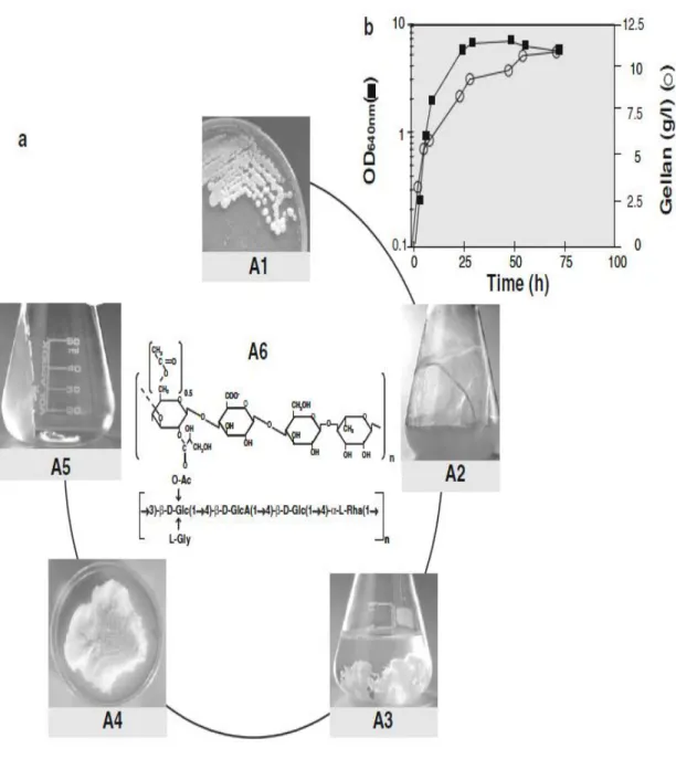
Figure 4 shows a fermentation process for gellan gum production and its purification at a laboratory scale. Gellan production is a growth-associated process with a maximum production of 12 g/l (Figure 4 b). The viscosity of the culture medium increases during the exponential and stationary phases and reaches a very high value at the end of the process. This characteristic creates great difficulty in terms of separating gellan from the cells, and the dilution of culture broth prior to isopropyl alcohol precipitation is required. After repetitive isopropyl alcohol precipitation steps, gellan is resuspended in water, followed by dialysis and lyophilization. Finally, gellan can be resuspended in water to produce a gel (Figure 4 a) [23].
Figure 4. (a) Typical fermentation process for gellan production and its purification at a laboratory scale. A1 Mucoid phenotype of the gellan producing strain S.elodea ATCC 31416, A2 culture broth after 72 h of growth, A3 gellan precipitated with isopropyl alcohol, A4 lyophilized gellan, A5 gellan gel (1 g/l), A6 molecular structure of the repeat unit of gellan, Glc glucose, GlcA glucoronic acid, Rha L-rhamnose, Ac acetate, Gly glycerate. (b) Time of course of growth and gellan production by S. elodea
ATCC 31416 at 30 ºC during a batch growth in a defined medium with 20 g/l glucose at initial pH of 8.0.
The procedure adopted to recover gellan from the culture broth involves a dilution (with saline solution) in order to reduce its viscosity, followed by centrifugation to separate the cells. The supernatant is precipitated with cold isopropyl alcohol and the dried EPS quantified. The viscosity of the broth increased significantly during growth [23].
The pH plays an important role in the production of gellan gum by S. paucimobilis, as it significantly influences both cell growth and product formation. The optimal pH value for bacterial polysaccharide production is higher that of fungal glucan production. The recommended pH value for gellan production ranges from 6.5 to 7.0 [24,25]. More acidic or more alkaline environment reduces the cell growth and, consequently, gellan production [26].
1.4.2 Biosynthetic pathway
The gellan biosynthetic pathway is a multi-step process that can be divided into three sequential steps: (a) the intracellular synthesis of the sugar-activated precursors (Figure 5);
(b) the assembly of the tetrasaccharide repeat units linked to the inner membrane (Figure 6); (c) translocation of the repeat unit to the periplasmic space followed by their polymerization
and exportation of the polymer through the outer membrane (Figure 6) [23]. The gellan biosynthesis starts with the cytosolic formation of the nucleotide-sugar precursors, UDP-D-glucose, UDP-D-glucuronic acid, and dTDP-L-rhamnose. The enzymes required for the synthesis of these sugar nucleotides are phosphoglucomutase (PgmG), UDPglucose pyrophosphorylase (UgpG), UDP-glucose dehydrogenase (UgdG), TDP-glucose pyrophosphorylase (RmlA), dTDP-D-glucose-4,6-dehydratase (RmlB), dTDP-6-deoxy-D-glucose-3,5-epimerase (RmlC), and dTDP-6-deoxy-L-mannose dehydrogenase (RmlD) [9].
Glucose-1-phosphate occupies a key position from which two routes derive, one leading to UDP-D-glucose and UDP-D-glucuronic acid and the other leading to dTDP-L-rhamnose. The identification and biochemical characterization of genes/enzymes involved in the formation of the following gellan precursors were carried out: the pgmG gene, encoding a phosphoglucomutase (EC 5.4.2.2), which catalyses the reversible conversion of glucose-6-phosphate into glucose-1-glucose-6-phosphate [27]; the ugpG gene, encoding a glucose-1- glucose-6-phosphate uridilyltransferase (or UDP-glucose pyrophosphorylase; EC 2.7.7.9), which catalyses the reversible conversion of glucose-1-phosphate and UTP into UDP-D-glucose and diphosphate [28]; the ugdG gene, encoding a UDP-glucose dehydrogenase, which converts UDP-D-glucose into UDP-D-glucuronic acid [29]; and rmlA, the first gene of the 4-gene rml cluster, which encodes a glucose-1-phosphate thymidilyltransferase (or TDP-glucose pyrophosphorylase; EC 2.7.7.24) that converts glucose-1-phosphate and TTP into TDP-D-glucose, necessary for the formation of dTDP-L-rhamnose [30]. Among the gellan biosynthetic enzymes, the PgmG protein plays a pivotal role, presumably being an ideal target for metabolic engineering. Indeed, this enzyme catalyses a step representing a branch point in carbohydrate metabolism; G6P enters catabolic processes to yield energy and reducing power and G1P is a precursor of sugar nucleotides that are used by cells in the synthesis of various polysaccharides [23].
Figure 5. Proposed pathway leading to the nucleotide sugar precursors, glucose, UDP-D-glucuronic acid and dTDP-L-Rhamnose, involved in gellan gum biosynthesis with glucose as the substrate [9].
The synthesis of the sugar precursors is followed by the formation of the repeat units by sequential transfer of the sugar donors to an activated lipid carrier by committed glycosyltransferases. Then, gellan gum polymerization and exportation (Figure 6) are the next steps. Most of these gellan-specific processes are catalyzed by enzymes encoded by the gel cluster of genes that contain at least 22 genes (Figure 6 a) [9,31].
Gellan tetrasaccharide repeat units are assembled on a lipid carrier (Figure 6 step 2). The priming glycosyltransferase is encoded by the gene gelB, which is homologous to spsB from
Sphingomonas sp. ATCC 31554 [31]. SpsB was demonstrated to be a
glycosyl-isoprenylphosphatate transferase that transfers glucose-1-phosphatase from UDP-glucose to the lipid carrier [32]. The addition of the second sugar, glucuronic acid, from UDP-glucuronic acid into the glucosyl-α-pyrophosphorylpolyprenol intermediate is catalyzed by GelK [27]. Pollock and co-workers proposed the gene spsL for encoding a glucosyl-(β1–4)-glucuronosyl transferase. Due to the strong homology between spsL and gelL genes, it is possible that the glycosyltransferase GelL catalyses the addition of the third sugar (glucose) to the repeat unit. Finally, the enzyme that adds the last sugar to the tetrasaccharide repeat unit, rhamnose, is putatively encoded by the remaining gene with homology to glycosyltransferases, gelQ [9,31]. The assembly of the repeat unit will only be complete after the addition of the substituent groups, acetate and glycerate, to the first glucose residue. These reactions are catalyzed by two enzymes having acetyltransferase and glyceryltransferase activities, respectively [31].
The next step in polysaccharide biosynthesis is the polymerization of the repeat units to form longer chains. The genes involved in this process of gellan polymerization and the export to the cell surface have been identified and located in the gel cluster region IV and are named
gelS and gelG, respectively. GelS is the translocase of the repeat units and GelG is the
polymerase (Figure 6 b, step 3). The determination of length distribution of the synthesized polysaccharide chains is controlled by a family of proteins termed polysaccharide co-polymerases (PCPs), which interact both with the polymerase enzyme and the secretion machinery. The PCP enzyme in S. elodea is encoded by the genes gelC and gelE. GelE also plays an important role by regulating GelC activity, in fact, GelE seems to have ATPbinding activity. GelC shows the typical PCP N- and C-terminal transmembrane helices separated by a segment with a predicted coiled-coil region located in the periplasm. A computational analysis of GelE structure reveals a potential amphipathic helix at the C-terminal region, which may be involved in the association of GelE to the plasma membrane and, therefore, interacting with GelC [33].
The next step, the exportation of the polysaccharide, is mediated by integral outer membrane proteins from the outer membrane auxiliary (OMA) family, which forms a protein channel through the membrane that allows the polysaccharide to gain access to the cell surface. This exportation process is mediated by the OMA protein homologue encoded by the gene gelD in S. elodea [34,35].
Figure 6. (a) Organization of the gellan biosynthetic gel clusters (from S. elodea ATCC 31461. (b) A model for biosynthesis and assembly of gellan. Activated sugar nucleotides are sequentially added to a lipid carrier to form repeat units of the polysaccharide. The undecaprenyl pyrophosphate (und-PP)-linked tetrasaccharide repeat unit is assembled at the interface between the cytoplasm and the inner membrane by sequential activity of GelB, GelK, GelL, and GelQ glycosyltransferases. These newly synthesized und-PP-linked repeat units are then translocated across the membrane in a process requiring GelS. These repeat units provide the substrate for GelG polymerization and also require GelC/GelE proteins, possibly by forming oligomers that connect polymerization to export and regulate polysaccharide chain length. Gellan chains are then exported by GelD, which acts as a channel. IM Inner membrane, OM outer membrane [23].
1.5 Gellan applications
Most polysaccharide´s applications are related to their behavior in aqueous media. Their physical and chemical characteristics, such as water binding capacity, high average molecular weight, polyelectrolyte behavior, molecular structure, and the possibility of being chemically modified, enables this type of molecules to present diverse functional properties like thickening, film forming, gelling, emulsion stabilizing, flocculating and nano/microstructures production abilities [36]. Furthermore, in the case fucose and rhamnose-rich bacterial polysaccharides, they also present interesting biological activities that render these
molecules to be used in a wide range of applications, particularly in added value products like cosmetics, pharmaceuticals, medical devices, and functional food products [37,38]. As examples, rhamnose is commonly used as precursor for the productions of aroma and flavors, and together with fucose has attracted more attraction since it has been found to counteract with several of the mechanism involved in skin aging [39].
In 1992, successful toxicity trials were completed and approval has been granted by the Food and Drug Administration (FDA) in the USA. Gellan gum was accepted to be used as a food additive [40]), and specifications, as summarized in Table 2, were prepared at the 46th Joint Expert Committee on Food Additives (JECFA) in 1996 [18]. Since then, gellan has been used in the food industry as additive that functions, not only as versatile gelling agent, but also as texturizing, stabilizing, suspending, film-forming, and structuring agent. Types of food products that contain gellan gum include bakery fillings, dairy products, low-fat spreads, frostings, icing and glazes, desserts gels, puddings, jams and jellies, sauces, structured foods and toppings [41].
Table 2. Specifications of gellan gum (modified from [18]).
Property Value
Definition
Gellan gum is a high molecular weight polysaccharide produced by a pure culture fermentation of carbohydrates by Sphingomonas
elodea, purified by recovery with isopropyl alcohol, dried and milled
Molecular mass Approximately 500 kDa Description Off-white powder
Functional uses Thickening agent, gelling agent, stabilizer, etc
Solubility Soluble in water, forming a viscous solution, insoluble in ethanol Loss on drying Not more than 15 % (105 ºC, 2.5 h)
Lead Not more than 2 mg/kg Nitrogen Not more than 3 %
Gel test with calcium ion Add 1 g of sample, 0.5 g sodium chloride, heat to 80 ºC for 1 min. Allow solution to cool. A firm gel is produced
Gel test with sodium ion To 1 % solution of sample, add 0.5 g sodium chloride, heat to 80 ºC and stir for 1 min. Allow solution to cool. A firm gel is formed Isopropyl alcohol Not more than 750 mg/kg
Microbial criteria:
1. Total plate count Not more than 10,000 colonies/gm 2. E. coli Negative by test
3. Salmonella Negative by test 4. Yeasts and molds Not more than 400 colonies/gm
Gellan is commercially available in three forms: no, low, and high acyl content with the respective denominations of Gelrite®, Kelcogel® F, and Kelcogel® LT100. Kelgogels are food-grade gellans used as gelling agents in food and personal care applications (lotions, creams, sunscreens, make-up, face masks, hair care products, air freshener and toothpastes) [42].
In the biotechnology industry, Gelrite® is used as a substituent of agar in plant tissue culture media and it is particularly useful for the culture of thermophilic bacterial species, as the gels are thermostables and can withstand prolonged incubations at high temperatures. In these microbiological media applications, the high purity of gellan gum and the water-like clarity of the gels are distinct additional advantages [43]. It also has potential environmental applications like the biodegradation of gasoline [44] and the transportation of gel-encapsulated cells protected from biotic and abiotic stress, such as predation by protozoa and bacteriophages, and the inhibitory effect of toxic compounds, respectively [45].
In the medicine field, the use of gellan has been investigated for tissue engineering (construction of 3D scaffolds, cartilage reconstruction and guided bone regeneration) and wound healing (in wound dressings to inhibit postsurgical adhesion and scar formation). Gellan sulfate derivatives are promising materials for rheumatoid arthritis, as they have a tendency for selective binding of fibronectin molecules, and for the development of cell-hybrid materials for artificial veins design due to the anticoagulant activity of such derivatives [46].
In pharmaceuticals technology, gellan gum can be used to produce easy-to-swallow solid dosage forms, such as hydrogels, beads and coated tablets, and to modify the rate of release of active ingredients from tablets and capsules. Gellan gum is also conveniently used for controlled or sustained release of various drugs and also for microencapsulating preparation. Formulations based on gellan gum for oral, ophthalmic, gastric, and nasal applications have also been developed [47].
Gells of gellan gum are also used as solid matrix for separation of DNA fragments onto electrophoresis and DNA isolation [48] and, more recently, gellan gum has been applied in the development of a new gel matrix for ionic exchange chromatographic approaches [49]. In summary, we can argue that gellan gum has gained importance among several industries and attracted a considerable market interest, as we can see in Table 3. Due to its unique properties, versatility, stability, biocompatibility, and biodegradability, the large variety of applications as well as the high and steadily increasing number of patents filing, it is suggested that gellan gum has been adopted as one of the most important commercialized bacterial exopolysaccharides and, due to its potential, it will be even more significant in the future [18].
Table 3. Several areas highlighting the utility of gellan gum as a polymer (modified from [18]).
Drug used Formulation Action Application
Clarithromycin In situ floating gel Anti-bacterial Gastric ulcers
Levofloxacin
hemihydrates In situ floating gel Anti-bacterial
Helicobacter pyroli infections, peptic ulcers
Naproxen In situ floating gel Anti-pyretic and NSAID Rheumatic arthritis, inflammation
Cimetidine In situ floating gel H2 receptor antagonist Peptic ulcer
Acetohydroxamic
acid Floating bead Anti-bacteria
Helicobacter pyroli infections
Mometasone
furoate Nasal in situ gel Corticosteroid Allergic rhinitis
Metoclopramide HCl
Intranasal
microspheres Anti-emetic
Cancer therapy, nausea, vomiting, pregnancy migraine
Glipizide Gellan gum beads Hypoglycaemic agent Diabetes
Gellan gum and beverage or food component
Spherical flavored gel
bead Flavourant Food industry
Carvedilol Hydrogel
microspheres Anti-hypertensive
Hypertension, Angina pectoris
Gatifloxacin Ocular inserts Fluoroquinolone
antibiotic Bacterial conjunctivitis
Indomethacin Ophthalmic in situ gel NSAID Uveitis, inflammation of eyes
Metformin Gum cordial/gellan
beads Hypoglycaemic agent Diabetes
Propranolol Gellan beads B-blocke Hypertension
Paracetamol Oral in situ gel - -
Ascorbic acid Gellan gum films Nutritional and
2 Chromatography
2.1 Chromatographic process
The development of techniques and methods for protein separation and purification has been essential for many of the recent advancements in biotechnology research. The chromatographic technology is undoubtedly one of the most diverse and powerful purification methods for downstream process applications. The diversity and quantity of biomolecules present in crude extracts as well the similarities between impurities and the target biomolecule are considered the critical challenges in the extraction and purification steps. Thus, the global aim of a protein purification process in not only the removal of unwanted contaminants, but also the concentration of the desired protein and their transfer to an environment where it is stable, biologically active, with a high purity degree and in a form ready for the intended application [50].
Chromatography is widely used due to its simplicity, robustness, high resolving power, versatility, high reproducibility and can be applied in the purification of a large variety of biomolecules [51]. Depending on the physical and chemical characteristics, such as charge, molecular size, hydrophobicity and polarity of each target biomolecule and matrices, several chromatographic techniques ca be used, by exploiting different interactions between the solute in the mobile phase with the stationary phase. There are two mechanisms used for chromatographic separation of proteins: adsorption (ion exchange, hydrophobic interaction, reverse phase and affinity chromatography) and molecular filter chromatography (size exclusion chromatography) [47,50].
Ideally, a chromatographic stationary phase should be selective, macroporous, incompressible, reusable, mechanical and chemically stable with low unspecific adsorption and high binding capacity, present an adequate mass transfer and maintain good flow properties [52].
The solutes can stay in the mobile phase or be distributed in the stationary phase by promoting specific interactions. The stationary phase is packed into a vertical column and should be insoluble in the buffer whereas the mobile phase is pumped through the column. The differential separation occurs due to continuous addition of mobile phase, as known as elution, resulting from alterations on ionic strength and pH or by addition of a competitive agent, causing a sequence of separated peaks that reflect the concentration of biomolecules versus time or volume of eluent at the column exit, which is tipically represented in a chromatogram [53].
followed by selective elution or by binding impurities, allowing the biomolecule to pass through the column without being retained (negative chromatography) [54].
2.2 Preparative chromatography
Chromatography can be applied in a preparative or an analytical scale [55]. Analytical chromatography aims at separating complex mixtures to identify and quantify the components of mixtures, simple or complex of a small sample. Once the required signals have been acquired for a component, the analyte is discarded. The purpose remains the rapid determination of the component structure, through the direct acquisition of the proper information, and the calculation of its concentration, through calibration of the detector signal. The preparative chromatography aims to isolate and purify the target molecules from impurities using a large amount of sample to be applied in a further goal. However, some concerns remains, like the need to produce as concentrated a fraction as possible, to collect and transfer it without pollution, and to do the separation as quickly and cheaply as possible [56].
In the preparative chromatography, maximum throughput at a high purity is the principal consideration. Therefore, an adsorption/desorption mode is preferred. The process can be divided into several steps: equilibration (adjusting mobile phase and stationary phase to binding conditions); sample application/loading (loading of the sample solution onto the column); washing (removal of unbound material, often with several steps); elution (desorption of the desired compound by stepwise or continuous change of the mobile phase composition); regeneration/cleaning (desorption of sticky impurities, colors, endotoxins and lipids); preservation/sanitation (change of mobile phase to avoid microbial contamination during storage) [55].
2.3 Chromatographic methods
General properties of proteins are used to isolate them from other proteins or non-protein contaminants. Minor differences between various proteins, such as size, charge, hydrophobicity and biospecific interactions (Figure 7) are used to purify the target protein with the application of several chromatographic methods [57].
Figure 7. Selective protein properties. Example of properties that are used to separate one protein from another [57].
Currently, several different action principles are employed for chromatography of biomolecules (Table 4). These action principles are gel filtration or size-exclusion chromatography (SEC), ion-exchange chromatography (IEC), hydrophobic interaction chromatography (HIC), reverse phase chromatography (RPC) and affinity chromatography (AC) [58].
Table 4. Action principles in protein purification (modified from [58]).
Name Action principle Separation by
Size-exclusion chromatography Size exclusion Molecular size and shape
Ion-exchange chromatography Ionic binding Surface charge
Hydrophobic interaction
chromatography Hydrophobic complex formation
Hydrophobicity and hydrophobic patches
Reversed-phase chromatography Hydrophobic complex formation Hydrophobicity
Affinity chromatography Biospecific adsorption/desorption Molecular structure and chemical structure
2.3.1 Size-exclusion chromatography (SEC)
Size-exclusion chromatography, also designated as gel filtration, is an entropically controlled separation technique that depends on the relative size or hydrodynamic volume and shape of a macromolecule with respect to the average pore size of the packing. With a properly calibrated SEC column, all the statistical average molecular weights of a polymer can be determined, as well as its molecular weight distribution. For protein separations, composite packings particles, such as the Superdex series, are increasing in popularity because of their high pore volume, inertness and small particle size availability [59].
The separation process depends on the different ability of various proteins to enter all, some or none of the channels in the porous beads. Molecules running through a SEC column have to solve a maze which becomes more complex to smaller the molecule is, because small molecules have more potential channels that they can access. On the other hand, larger molecules are, for steric reasons, excluded from the channels, and pass quickly between the beads. The detour through the channels will thus retard smaller molecules in comparison to larger proteins (Figure 8). Although the separation in SEC is generally assumed to be according to its molecular weight, it is more accurate to claim that SEC separation is achieved by the differential exclusion or inclusion within porous particles. The ease of diffusion is dependent on the hydrodynamic volume, which is the volume created by the movement of the molecule in water. The difference between hydrodynamic volume and molecular weight is the shape. Proteins tend to be globular molecules while DNA or polysaccharides tend to be linear molecules. Linear molecules have much larger hydrodynamic volume than globular molecules, so a 10.000 MW DNA molecule will elute much earlier than a 10.000 MW protein [57].
Figure 8. In SEC, large molecules run through the space between media with a shorter pathway, while the smaller molecules run through inside the packing particles with a longer pathway [60].
Sample components are eluted isocratically (single buffer, no gradient) and separation can be performed within a broad pH, ionic strength, and temperature range. The medium accepts a variety of additives: co-factors, protein stabilizers, detergents, urea and guanidine hydrochloride. The buffer composition does not usually affect resolution, although including a low salt concentration, for example 25 to 150 mM NaCl, is recommended to eliminate any weak electrostatic interactions between proteins and the SEC matrix. The buffer conditions are selected to suit the sample type and to maintain target protein activity, because the proteins are transferred to the buffer used for equilibration of the column. Equilibration buffer can thus be selected according to the conditions required for further purification, analysis, storage or use [54].
SEC is a powerful method for purification of proteins that have passed one or several initial purifications steps. After those steps, the material has been concentrated and bulk impurities have been removed. Gel filtration can now be used to remove remaining impurities and it will also remove oligomers or aggregates of the target protein [54]. Therefore, the SEC separation method gives the least resolution with the lowest capacity and largest dilution of the sample with respect to all other forms of chromatography. Still, this technique is frequently used due
advantage of SEC is its gentle non-interaction with the sample, enabling high preservation of biological activity. Moreover, since the separation is not dependent on any adsorptive property of the molecule, SEC provides a method for separating multimers that are not easily distinguished by other chromatographic methods.
SEC is a non-binding method, which means that no concentration of the sample components take place. Since separation starts directly as the sample is applied on the column, the cross section must be large enough to cope with the desired sample volume. In fact, the sample zone is broadened during the passage through the column resulting in dilution of the sample [54]. However, the length of the column is also significant since it affects both resolution and process time, thus a relationship between the quantity of injected sample and the column volume (maximum 5 %) must be complied in order to guarantee an efficient separation between the molecules. Consequently, SEC is preferably used as a final polishing step when sample volumes have been reduced [57].
2.3.2 Ion–exchange chromatography (IEC)
Ion-exchange chromatography separates biomolecules according to differences in their surface charge to give separation with high-resolution and high sample loading capacity. The net charge of a protein depends on its amino acid sequence (proteins can have both positive and negative charges) and on the pH of the buffer in which it has been solubilized. At its isoelectric point (pI), the net charge of a protein is neutral. At pH values above this pI, the protein is negatively charged being considered an anion. Therefore, if the matrix has positively charged groups (an anion exchanger) it can be used to purify the protein if the buffer pH is above the pI of the protein. On the other hand, at pH values below the pI, the protein is positively charged, hence, a cation exchange may bind this protein (Figures 9 and 10) [53]. The pH interval in which ion exchange chromatography is carried out is restricted by the pH range in which the protein is stable. To achieve good adsorption, the pH of the buffer chosen should be at least one pH unit above or below the isoelectric point of the analytes to be separated [57].
Figure 9. Net charge of a protein as function of pH, showing the pH ranges in which the protein is positively or negatively charged [61].
Figure 10. Types of ion exchangers (modified from [62]).
Proteins bind as they are loaded onto a column at low ionic strength and then, the ionic conditions are altered so that bound substances are desorbed differentially. Elution is usually performed by increasing salt concentration or changing pH, in a linear or stepwise gradient.
The most common salt is NaCl, but other salts can also be used to modulate separation, for example, K+, Ca2+, Mg2+, CH
3COO-, SO42-, I-, Br- [54].
IEC is one the more powerful protein purification methodology available and probably the most frequently used chromatographic technique for the separation of proteins, polypeptides, nucleic acids, polynucleotides, and other charged biomolecules. An advantage of this method is that elution normally takes place under mild conditions, so that the protein can maintain its native conformation during the chromatographic process. In general, ion exchangers are more densely substituted than other adsorbents used in protein chromatography and its capacity for protein binding is very high. Its broad specificity also allows the removal of significant impurities such as deamidated forms, endotoxins and unwanted glycoforms. Still, non-specific interactions with proteins due to hydrophobic or other non-ionic interactions are low. Additional reasons for the success of IEC are the straightforward separation principle and ease of performance and controllability of the method. Moreover, ion exchanger resins are very robust and can be sanitized in place and used for hundreds of cycles. The main disadvantage of IEC is its limitation in selectivity [57].
2.3.3 Hydrophobic interaction chromatography (HIC)
Hydrophobic interaction chromatography takes advantage of the hydrophobicity of proteins promoting its separation on the basis of reversible hydrophobic interactions between immobilized hydrophobic ligands and non-polar regions on the protein surface. The adsorption increases with high salt concentrations in the mobile phase and the elution is achieved by decreasing the salt concentration of the eluent (Figure 11) [50]. Therefore, the term “salt-promoted adsorption” could be used for this type of chromatography [63]. The different types of elution conditions can be used for purification of complex mixtures of proteins that would be difficult to separate using other chromatographic techniques. In fact, HIC has been successfully used for separation purposes as it displays binding characteristics complementary to other protein chromatographic techniques [64].
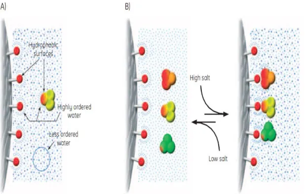
Figure 11. A) Highly ordered water shells surround the hydrophobic surfaces of ligands and proteins. Hydrophobic substances are forced to merge to minimize the total area of such shells (maximize entropy). Salts enhance the hydrophobic interaction. B) The equilibrium of the hydrophobic interaction is controlled predominantly by salt concentration [65].
The hydrophobic ligands on HIC media can interact with the hydrophobic surfaces of proteins. In pure water, any hydrophobic effect is too weak to cause interaction between ligand and proteins or between the proteins themselves. However, certain salts enhance hydrophobic interactions because they remove the hydration water, leaving the hydrophobic groups available to interact, thus, adding such salts, brings about binding (adsorption) to HIC media. For selective elution (desorption), the salt concentration is lowered gradually and the sample components elute in order of hydrophobicity (Figure 11 B) [65].
Hydrophobic interaction chromatography can be used for capture, intermediate to other techniques or as polishing steps in a purification protocol. It allows the protein separation that differs in one amino acid residue, separating a native protein from incorrectly folded proteins. The structural damage to the biomolecule is minimal, certainly due to the stabilizing influence of salts. Still, recoveries are often high. Thus, HIC combines the non-denaturating characteristics of salt precipitation and the precision of chromatography to yield activity recoveries. Therefore, at laboratorial and industrial scales, the biomolecule purification such as serum proteins, nuclear proteins, recombinant proteins, membrane proteins, enzymes, and hormones is extremely performed by HIC [66].
![Figure 5. Proposed pathway leading to the nucleotide sugar precursors, UDP-D-glucose, UDP-D- UDP-D-glucuronic acid and dTDP-L-Rhamnose, involved in gellan gum biosynthesis with glucose as the substrate [9]](https://thumb-eu.123doks.com/thumbv2/123dok_br/18859583.930252/32.892.133.742.112.489/proposed-nucleotide-precursors-glucuronic-rhamnose-involved-biosynthesis-substrate.webp)
![Table 3. Several areas highlighting the utility of gellan gum as a polymer (modified from [18])](https://thumb-eu.123doks.com/thumbv2/123dok_br/18859583.930252/38.892.95.762.134.1128/table-areas-highlighting-utility-gellan-gum-polymer-modified.webp)
![Figure 7. Selective protein properties. Example of properties that are used to separate one protein from another [57]](https://thumb-eu.123doks.com/thumbv2/123dok_br/18859583.930252/41.892.272.664.108.557/figure-selective-protein-properties-example-properties-separate-protein.webp)
![Figure 12. Proteins and peptides bind to an RCP medium under aqueous conditions and elute as the eluent becomes more hydrophobic [54]](https://thumb-eu.123doks.com/thumbv2/123dok_br/18859583.930252/48.892.124.749.468.706/figure-proteins-peptides-medium-aqueous-conditions-eluent-hydrophobic.webp)
![Figure 13. An affinity matrix binding to its target protein. A - the bead; B - the space arm; C - the ligand; D - the target protein [57]](https://thumb-eu.123doks.com/thumbv2/123dok_br/18859583.930252/49.892.258.681.751.938/figure-affinity-matrix-binding-target-protein-ligand-protein.webp)
![Table 5. Examples of different basic material constituents of chromatographic supports applied in purification (modified from [51])](https://thumb-eu.123doks.com/thumbv2/123dok_br/18859583.930252/53.892.142.788.153.781/examples-different-material-constituents-chromatographic-supports-purification-modified.webp)