LAMP technology: Rapid identification of
Brucella
and
Mycobacterium avium
subsp.
paratuberculosis
Marcos D. Trangoni
1, Andrea K. Gioffré
1, María E. Cerón Cucchi
2, Karina C. Caimi
1,
Paula Ruybal
1, Martín J. Zumárraga
1, Silvio L. Cravero
11
Institute of Biotechnology, Centre of Agronomy and Veterinary Sciences, National Agricultural Technology Institute, Buenos Aires, Argentina. 2
Institute of Pathobiology, Centre of Agronomy and Veterinary Sciences, National Agricultural Technology Institute, Buenos Aires, Argentina.
Submitted: November 13, 2013; Approved: September 14, 2014.
Abstract
In this study, we developed new sets of primers to detect Brucella spp. and M. avium subsp. paratuberculosis(MAP) through isothermal amplification. We selected a previously well-charac-terized target gene,bscp31, specific forBrucellaspp. and IS900for MAP. The limits of detection us-ing the loop-mediated isothermal amplification (LAMP) protocols described herein were similar to those of conventional PCR targeting the same sequences. Hydroxynaphtol blue and SYBR GreenTM allowed direct naked-eye detection with identical sensitivity as agarose gel electrophoresis. We in-cluded the LAMP-based protocol in a rapid identification scheme of the respective pathogens, and all tested isolates were correctly identified within 2 to 3 h. In addition, both protocols were suitable for specifically identifying the respective pathogens; in the case ofBrucella, it also allowed the identifi-cation of all the biovars tested. We conclude that LAMP is a suitable rapid molecular typing tool that could help to shorten the time required to identify insidious bacteria in low-complexity laboratories, mainly in developing countries.
Key words: loop-mediated isothermal amplification, molecular typing, brucellosis, paratuber-culosis.
Introduction
Brucellosis and paratuberculosis are diseases caused bacterial pathogens of veterinary concern (Manning and Collins, 2001; Samartino and Enright, 1993). Brucellosis is clearly defined as a zoonotic disease. In Santa Fe province, which accounts for 32% of milk production in Argentina, the cumulative incidence rate of zoonoses in rural veteri-narians (1964-2008) was reported to be 34.1%, with a brucellosis frequency of 29.1% (Molineri et al., 2013). However, the role of Mycobacterium avium subsp. paratuberculosis, the causal agent of paratuberculosis, in Crohn’s disease in humans is currently under discussion (Das and Seril, 2012; Kuenstner, 2012).
Brucellosis is caused by facultative intracellular pathogens of theBrucellagenus, and domestic and wild
an-imals are considered natural reservoirs of the disease. Brucella melitensis, Brucella abortus and Brucella suis also induce human disease, and rare but persisting cases of human brucellosis caused byBrucella canisandBrucella species of marine mammals have also been recognized (Pappaset al., 2005).
M. aviumsubsp.paratuberculosis(MAP) belongs to theM. avium-intracellularecomplex (MAC), comprising two species,M. intracellulareandM. avium, and the sub-species M. avium subsp. avium, M. avium subsp. hominisuiss,M. aviumsubsp.silvaticumand MAP (Mijset al., 2002).MAC members possess properties that enable them to grow in natural biotopes without losing their patho-genicity for certain hosts (Bietet al., 2005). MAP causes chronic progressive enteritis in ruminants, which is known
Send correspondence to: S.L. Cravero. Institute of Biotechnology, Centre of Agronomy and Veterinary Sciences, National Agricultural Technology Insti-tute, N. Repetto y de Los Reseros s/n. B1712 WAA Hurlingham, Buenos Aires, Argentina. E-mail: [email protected].
as paratuberculosis or Johne’s disease (Chiodini et al., 1984; Larsenet al., 1975).
The traditional methods for detecting these pathogens are largely based on phenotypic traits, and the diagnosis of brucellosis and paratuberculosis involves bacteriological culture, histopathology and serological tests such as en-zyme-linked immunosorbent assay (ELISA)-based tech-niques and agglutination tests (Gallet al., 2008; Manning and Collins, 2001). However, the isolation of the pathogen is required to confirm the diagnosis, a process that is time consuming, especially for MAP, which requires long peri-ods (up to two months) to develop in culture media. Molec-ular biology techniques have allowed the sensitive diagno-sis of different bacteria through the application of nucleic acid amplification, which minimizes the requirement of biosafety conditions. In addition to contributing to the diag-nosis, nucleic acid amplification provides an accurate mo-lecular tool for identification at the species or subspecies level. The polymerase chain reaction (PCR) is the main nu-cleic acid amplification method currently used, and it is ex-pected that this technique will eventually supersede many of the classical direct methods of infectious agent detection (OIE, 2008). Indeed, high sensitivity, specificity and rapid-ity are the major advantages of PCR over other nucleic acid-based techniques. Nonetheless, PCR requires basic equipment, such as thermocyclers, electrophoretic systems and PCR-product detection systems, and the lack of such equipment often limits its use in developing countries.
The loop-mediated isothermal amplification (LAMP) technique is characterized by its simplicity because the en-tire process of amplification and detection is performed in a single step in which the reaction components are subjected to isothermal conditions (Nagamineet al., 2002; Notomiet al., 2000), which requires less specialized equipment than conventional PCR technologies. Therefore, LAMP is ac-cessible for laboratories in developing countries.
The LAMP method is based on the isothermal strand-displacement activity of the Bacillus subtilis- de-rivedBstDNA polymerase. This enzyme when combined with four target-specific primers renders the single-tempe-rature amplification of a highly specific fragment from a DNA template at amounts greater than those of an equiva-lent PCR (Nagamineet al., 2002; Notomiet al., 2000). Fur-thermore, this higher amplification efficiency allows straightforward visual detection by colorimetric methods (Gotoet al., 2009; Paridaet al., 2008).
Many studies have referred to LAMP as a successful and promissory alternative for the sensitive and specific de-tection of human and veterinary pathogens (Barkwayet al., 2011; Dukeset al., 2006; Savanet al., 2004; Sirichaisin-thopet al., 2011; Wanget al., 2012; Zhanget al., 2013). The main objective of the present study was to develop and apply a LAMP strategy for the specific detection of Brucellaspp. and MAP, important bacterial pathogens in Argentina, to simplify diagnosis. The purpose of this
strat-egy focuses on lowering the reaction time and equipment costs for bacterial detection.
Materials and Methods
Bacterial strains and growth conditions
To standardize the LAMP protocols, we used a B. abortusS2308 strain and a wild-type MAP isolate, which was previously typed by conventional methods in our labo-ratory.
TheB. abortusS2308 colonies were obtained from tryptose agar plates and grown in 2.5 mL tryptic soy broth (Difco BD, USA) at 200 rpm for 48 h at 37 °C.
The MAP isolate was first confirmed by insertion se-quence900(IS900) and F57 PCR and then grown in 7H9 liquid medium (Difco, BD, USA) supplemented with 0.2% mycobactin J (Allied Monitor, Fayette, MO USA).
To evaluate the specificity of the LAMP assay and to test the performance of the LAMP protocol in crude lysates, we employed strains from the different hosts and sources listed in Results section.Ochrobactrum anthropiDNA was also evaluated as a negative control. All of the isolates be-long to the INTA strain collection.
DNA extraction from reference strain cultures and wild-type isolates
High-quality DNA extraction from reference strains
Chromosomal DNA of high quality was obtained to test the detection limit. DNA extraction fromB. abortus S2308 was performed as follows.A 2.5 mL aliquot of cul-ture was lysed with 30 mL of 25 mg/mL proteinase K
(Promega, WI, USA) and 126mL of 10% sodium dodecyl
sulfate (SDS) for 2 h at 60 °C. The DNA was precipitated by adding 0.1 volumes of 3 M sodium acetate (pH 5.3) and 0.6 volumes of isopropanol and then removed with a sterile loop. The DNA was washed twice in 70% ethanol and sus-pended in 600 mL of Tris-EDTA (TE) buffer (10 mM
Tris-HCl pH 8, 1 mM EDTA). A second step of precipita-tion and purificaprecipita-tion was performed. Finally, the DNA was suspended in 100mL of TE. DNA extraction from the MAP
culture was performed as previously described (van Embdenet al., 1992).The DNA integrity was assessed by 0.8% agarose gel electrophoresis and then quantitated us-ing a Nanodrop ND-1000 spectrophotometer (Thermo Fisher Scientific, DE, USA). Ten-fold serial DNA dilutions were performed with sterile distilled water, and 1mL of
each dilution was used as the template for amplification.
Rapid DNA extraction from wild-type isolates
Cell lysis was performed by physical treatment. A loopful of growth from a solid medium or a 0.5-1 mL aliquot devoid of medium by centrifugation was suspended in 200mL of distilled water. The sample was boiled and
sample was subjected to a brief centrifugation at 12,000 xg, and 5mL of the supernatant was used as the template for
amplification.
Target genes and primer design
Target sequences that are traditionally used to iden-tify Brucella spp. and MAP were selected. The LAMP primers were designed to target the bscp31 gene from Brucellaspp. (Baily et al., 1992) and IS900 from MAP (Greenet al., 1989). Complete LAMP primer sets, includ-ing both loop primers for each selected sequence, were
de-signed using Primer Explorer V4 software
(http://primerexplorer.jp/elamp4.0.0/index.html). The pri-mer sequences are listed in Table 1.
LAMP reaction and detection
LAMP assays for Brucella spp. and MAP (Bru-LAMP and MAP-(Bru-LAMP, respectively) were performed in a final reaction volume of 25 mL with 1.4 mM dNTPs
(Promega, WI, USA), 8 mM SO4Mg, 0.8 M betaine (Sigma-Aldrich, MO, USA) and 8 U ofBstDNA Polymer-ase (New England Biolabs, MA, USA). The LAMP reactions also contained 1.6mM of FIP and BIP primers,
0.16mM of F3 and B3 primers, 0.8mM of LF and LR
prim-ers and the corresponding DNA as the template. The tem-plates consisted of 1mL of high-quality DNA or 5mL of the
supernatant of the cell lysate obtained by the rapid DNA ex-traction method. The reaction tubes were incubated in dif-ferent equipment (MyCycler thermocycler (Bio-Rad, CA, USA) and a thermal bath) to evaluate the robustness of the amplification method. For the MyCycler thermocycler, the tubes were incubated for 60 min at 60 °C and then for 5 min
at 80 °C. The incubation using the thermal bath was for 60 min at 60 °C. For MAP-LAMP, the incubation tempera-ture was 65 °C instead of 60 °C due to the characteristic high GC content of mycobacterial genomes. The LAMP amplicons were visualized by different strategies: a) 2% agarose gel electrophoresis, staining with 0.5 mg/mL of
ethidium bromide and visualization under ultraviolet (UV) light; b) naked-eye inspection by colorimetric methods such as SYBR GreenTMstaining and hydroxy naphthol blue (HNB). For SYBR GreenTMstaining, 1mL of 1/10 SYBR
GreenTMI (Invitrogen, CA, USA) solution was added di-rectly to each reaction tube after incubation, and the DNA was visualized under UV light. For HNB (JT Baker, USA) staining, a final concentration of 120 mM was utilized.
HNB was added prior to amplification (Gotoet al., 2009).
PCR amplification
The reactions were performed with primers B4/B5 for Brucella spp. (B4/B5 PCR) and S204/S749 for MAP (IS900PCR), as previously described (Bailyet al., 1992; Englund et al., 1999). The amplification reactions were performed in a final volume of 25mL with 1.25 U of Taq
DNA Polymerase (Promega, WI, USA), 200mM dNTPs
(Promega, WI, USA), 0.5mM of each primer and 1mL of
high-quality DNA or 5mL of the supernatant of the cell
lysates obtained during the rapid DNA extraction method. The reactions consisted of an initial denaturation step at 95 °C for 5 min, followed by 35 amplification cycles and a final extension step at 72 °C for 10 min. The amplification cycles comprised a first step at 94 °C for 1 min, an anneal-ing step at 60 °C forBrucellaspp. or 59 °C for MAP for 1 min and an extension step at 72 °C for 1 min. The PCRs
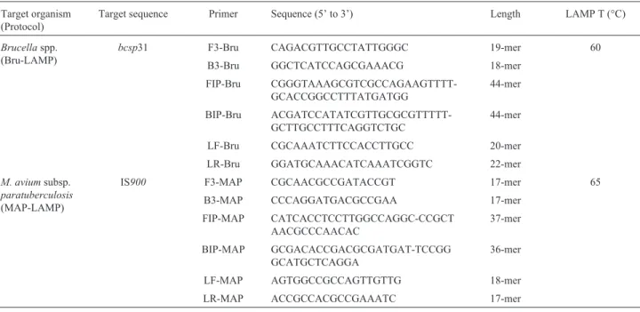
Table 1- LAMP primers designed in this study.
Target organism (Protocol)
Target sequence Primer Sequence (5’ to 3’) Length LAMP T (°C)
Brucellaspp. (Bru-LAMP)
bcsp31 F3-Bru CAGACGTTGCCTATTGGGC 19-mer 60
B3-Bru GGCTCATCCAGCGAAACG 18-mer
FIP-Bru CGGGTAAAGCGTCGCCAGAAGTTTT-GCACCGGCCTTTATGATGG
44-mer
BIP-Bru ACGATCCATATCGTTGCGCGTTTTT-GCTTGCCTTTCAGGTCTGC
44-mer
LF-Bru CGCAAATCTTCCACCTTGCC 20-mer
LR-Bru GGATGCAAACATCAAATCGGTC 22-mer
M. aviumsubsp.
paratuberculosis
(MAP-LAMP)
IS900 F3-MAP CGCAACGCCGATACCGT 17-mer 65
B3-MAP CCCAGGATGACGCCGAA 17-mer
FIP-MAP CATCACCTCCTTGGCCAGGC-CCGCT AACGCCCAACAC
37-mer
BIP-MAP GCGACACCGACGCGATGAT-TCCGG GCATGCTCAGGA
36-mer
LF-MAP AGTGGCCGCCAGTTGTTG 18-mer
were performed in a MyCycler thermocycler (Bio-Rad, CA, USA). The sizes of the PCR products (223 bp and 563 bp, respectively) were determined by comparison with a molecular weight marker using 1.5% agarose gel electro-phoresis, ethidium bromide staining (0.5mg/mL) and UV
light visualization.
Results
Detection limit and specificity of LAMPvs.PCR
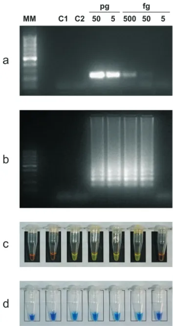
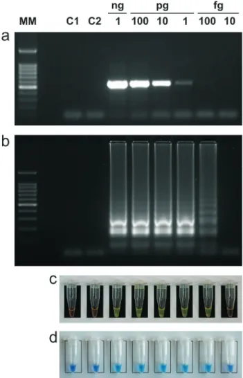
After confirming amplification using the novel sets of primers, we determined the detection limit by 10-fold serial dilutions of purified genomic DNA. Positive LAMP reac-tions were confirmed by the appearance of a ladder-like pattern on agarose gels stained with ethidium bromide; pos-itive PCRs were confirmed by specific size amplicon visu-alization. The detection limit of Bru-LAMP and MAP-LAMP were 50 fg and 100 fg per reaction, respectively (Figures 1b and 2b). The sensitivities reached by all the LAMP protocols were in accordance with those obtained with PCR targeting the same genes; however, the last point detected by PCR was barely observed (Figures 1a and 2a). The specificity of the Bru-LAMP protocol was deter-mined using DNA fromOchrobactrum anthropi, a phylo-genetically related bacterium, as the control. No evidence of cross-reactivity was detected with the control DNA tested by PCR (Figure 1a) or LAMP (Figure 1b).
The specificity of the MAP-LAMP protocol was eva-luated using cell lysates fromM. bovis, M. aviumsubsp. avium, M. gordonae, M. scrofulaceum, M. porcinum,and two phylogenetically related bacteria,Nocardia farcinica andNocardia testacea. Interestingly, a specific amplifica-tion of MAP was obtained when using the MAP-LAMP protocol designed in this study (Table 2).
End-point detection of LAMP products by single staining
The end-point detection of the products of LAMP amplification was performed by naked-eye inspection by fluorescent staining for nucleic acid detection. For SYBR GreenTM, a dilution of the original orange color indicates a negative result, whereas a fluorescent green color indicates a positive amplification. For HNB, a violet or sky-blue color indicates a negative or a positive result, respectively. All the direct end-point detections showed the same sensi-tivity (Figures 1c-d and 2c-d).
Usefulness of LAMP in molecular typing schemes
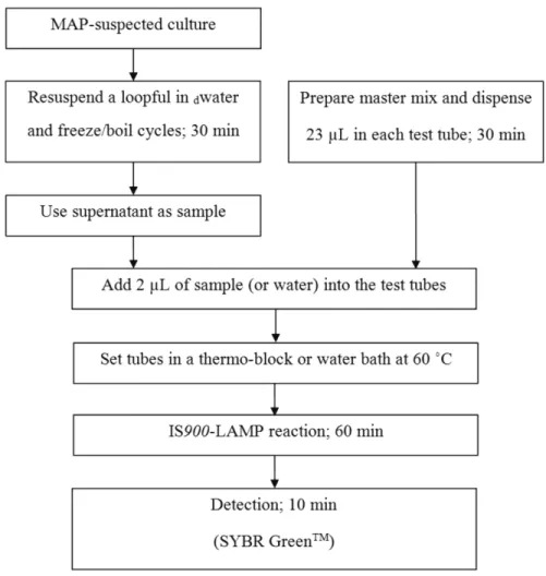
Next, we evaluated the potential of LAMP as part of a rapid molecular typing scheme using non-purified DNA as the template. For this purpose, we tested 16 MAP field iso-lates and 20Brucellaspp. isolates along with 17 reference strains. As an example, Figure 3 shows how the MAP cul-tures were processed.
All the samples tested by the MAP-LAMP protocol were identified as MAP according to IS900PCR (Table 2). Additionally, cell lysates ofNocardiaspp. andM. avium subsp.avium, an atypical mycobacteria, were also asses-sed; as expected, the results were negative (Table 2). The results obtained for all the samples were the same, regard-less of the equipment employed (thermostatic water bath or thermocycler) (data not shown). The same protocol was adapted forBrucella, and the correct identification of the genus was achieved, regardless of the species or serovar tested (Table 2). Thus, a correct identification of the isolate
Figure 1- Comparative analytical sensitivity of Bru-LAMP and PCR. Agarose gel electrophoresis and direct naked-eye detection. 1a) B4/B5 PCR, 1.5% agarose gel. 1b) Bru-LAMP, 2% agarose gel. 1c) Bru-LAMP SYBR GreenTM. 1d) Bru-LAMP, HNB. Serial DNA dilution ofBrucella
could be obtained in less than 3 h, which demonstrates the suitability of applying LAMP as a routine protocol.
Discussion
We aimed to develop LAMP-based protocols for the specific and sensitive detection of two bacterial pathogens. To this end, we designed primers and tested LAMP sensi-tivity using three different methods for end-point determi-nation: agarose gel electrophoresis, SYBR GreenTM and hydroxynaphthol blue.
In our study, we specifically designed LAMP primers for the identification ofBrucellaspp. usingbcsp31 as the target sequence. This gene encodes the 31 kDaBrucella cell surface salt-extractable protein (BCSP) and is highly conserved in the genus Brucella (Baily et al., 1992; Bricker, 2002). Ohtsukiet al.(2008) first reported the
iden-tification ofBrucellaspp. using two LAMP primer sets tar-geting the same gene. The detection limit obtained in our study with theBrucellagenus-specific LAMP was similar to that obtained by Ohtsukiet al.(2008). Another LAMP for the identification of members of this genus with a higher sensitivity has been previously described; however, a dif-ferent gene, which codes for the 25 kDa outer membrane protein (Omp25), was selected as the target (Lin et al., 2011).
Primer design is the most important factor affecting the performance of LAMP. However, further optimization of the protocol may be needed to improve the sensitivity of the test. Taking into account that the LAMP reaction com-bines 6 to 8 different regions, a highly specific amplifica-tion product is expected.
We obtained a detection limit with the MAP-LAMP protocol that was similar to that obtained by Enosawaet al. (2003), who targeted the same sequence. In the present study, we tested the specificity of the MAP primer set with DNA from closely related mycobacteria, such asM. avium subsp.avium, orM. bovis, another important mycobacterial pathogen of veterinary concern. Although the MAP-LAMP developed in our study proved to be discriminative for sub-species determination, some nonspecific amplification with M. scrofulaceum strains has been reported using LAMP targeting IS900(Enosawaet al., 2003). We herein demonstrated that the MAP-LAMP protocol was suitable, even for a panel of atypical mycobacteria, including M. scrofulaceum, orNocardiaspp., a closely related genus.
The comparative results between the PCR and novel LAMP protocols reported here demonstrate that isothermal amplification can achieve the same sensitivity as conven-tional PCR, regardless of the pathogen. This is consistent with previous comparative analyses in which LAMP reached the same sensitivity, or even higher levels, as nested-PCR and real-time PCR (Enosawaet al., 2003; Lin et al., 2009). Hence, the high processivity of the isothermal amplification yields a detectable product faster than con-ventional PCR, which is a hallmark of this method.
It is important to note that the methodology used for product detection could bias the sensitivity. The PCR de-tection limit is established by the presence of specific bands on agarose gel electrophoresis, which depends not only on the concentration of the amplicon obtained but on several other factors. For instance, the concentration of ethidium bromide, the sensitivity of the detection system and image processing (if available), among other factors, may alter the results. However, as we showed in this study, the threshold at which the LAMP reaction changes from positive to nega-tive is abrupt and, as a consequence, the end-point determi-nation is accurate.
LAMP not only leads to the isothermal amplification of DNA in a stoichiometric reaction (Notomiet al., 2000) but also to the variation of by-products. For instance, the in-creased formation of magnesium pyrophosphate (Moriet Figure 2- Comparative analytical sensitivity of MAP-LAMP and PCR.
Agarose gel electrophoresis and direct naked-eye detection. 1a) IS900
PCR, 1.5% agarose gel. 1b) LAMP, 2% agarose gel. 1c) MAP-LAMP, SYBR GreenTM. 1d) MAP-LAMP, HNB. Serial DNA dilution of
M. aviumsubsp.paratuberculosis(from 1 ng to 10 fg); C1,M. avium
al., 2001) and the subsequent reduction in the concentration of magnesium cations can be titrated by HNB (Gotoet al., 2009). Although the visualization of magnesium pyrophos-phate precipitate is a simple end-point detection strategy (Moriet al., 2001), the sensitivity of this technique is lower than that of fluorometric and colorimetric methods and of-ten requires the use of a centrifugation step to facilitate the visualization of the precipitate or trained technicians.
Considering that an ideal detection method must fit certain criteria, such as sensitivity, reproducibility and an accessible cost, it is important to know the performance of the different methods available in the laboratory. As the use of SYBR GreenTMor HNB shows better reproducibility, we selected this method to compare the relative sensitivity to that of agarose gel electrophoresis. The comparative sensi-tivity achieved by both methods was similar in all the LAMP protocols tested. The direct determination of the end-point, however, was easier when the SYBR GreenTM method was performed, even without an UV-transillumi-nator. Although a method of choice, SYBR GreenTM re-quires the opening of the reaction tubes after amplification; which can result in carry-over contamination, and the risk of amplicon contamination limits its application to those laboratory settings in which LAMP is used as a non-routine practice. A lower color contrast between positive and nega-tive amplification was observed when the HNB method was used; however, the sensitivity obtained in each assay
was the same as the other studied methods. Therefore, the HNB method is more suitable for laboratories in which LAMP is frequently used due to its low risk of contamina-tion.
To achieve a scheme that allows the rapid identifica-tion of bacterial pathogens, we coupled the LAMP protocol to a simple step of cell lysis that enabled us to easily release DNA from cultures. This approach allowed a significant re-duction in the time required for identification compared to the traditionally time-consuming protocols that involve DNA extraction and conventional PCR. Importantly, the reduction in the complexity of the protocols could also help to expand the use of molecular biology techniques to labo-ratories that have not yet adopted DNA-based techniques.
The evaluation of pathogen detection directly from clinical samples certainly constitutes a challenge, and a LAMP method tested using a wide panel of field samples could be a useful tool to diagnose diseases that impact pro-duction systems. Finally, LAMP is a simple, rapid, low-cost genetic testing technology that is specific and sensi-tive. This technology can be coupled to schemes with typ-ing purposes and could contribute to the conventional methods used for the identification ofBrucellaspp. and MAP in non-sophisticated laboratories, especially in devel-oping countries.
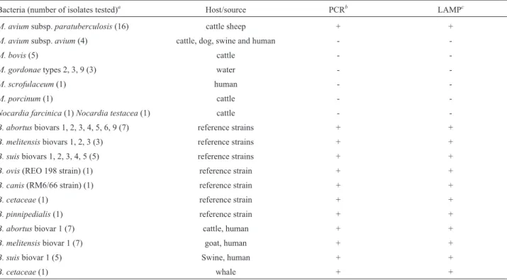
Table 2- LAMP performance and specificity evaluated using cell lysate samples from cultures of different bacteria.
Bacteria (number of isolates tested)a Host/source PCRb LAMPc
M. aviumsubsp.paratuberculosis(16) cattle sheep + +
M. aviumsubsp.avium(4) cattle, dog, swine and human -
-M. bovis(5) cattle -
-M. gordonaetypes 2, 3, 9 (3) water -
-M. scrofulaceum(1) human -
-M. porcinum(1) cattle -
-Nocardia farcinica(1)Nocardia testacea(1) cattle -
-B. abortusbiovars 1, 2, 3, 4, 5, 6, 9 (7) reference strains + +
B. melitensisbiovars 1, 2, 3 (3) reference strains + +
B. suisbiovars 1, 2, 3, 4, 5 (5) reference strains + +
B. ovis(REO 198 strain) (1) reference strain + +
B. canis(RM6/66 strain) (1) reference strain + +
B. cetaceae(1) reference strain + +
B. pinnipedialis(1) reference strain + +
B. abortusbiovar 1 (7) cattle, human + +
B. melitensisbiovar 1 (7) goat, human + +
B. suisbiovar 1 (5) Swine, human + +
B. cetaceae(1) whale + +
a71 samples were evaluated. b
Mycobacteriumspp. andNocardiaspp. isolates were processed by IS900PCR.Brucellaspp. were processed by B4/B5 PCR.
Acknowledgments
The authors would like to thank Bernardo Sioya for his excellent technical assistance and Dr. Julia Sabio y García for critical reading of the manuscript. MT holds a doctoral fellowship from INTA. AG, KC and PR are career members of CONICET. This study was supported by Na-tional Institute of Agricultural Technology (INTA) grants AEGR2412 and AERG 52-232121.
References
Baily GG, Krahn JB, Drasar BS et al. (1992) Detection of
Brucella melitensisandBrucella abortusby DNA
amplifi-cation. J Trop Med Hyg 95:271-275.
Barkway CP, Pocock RL, Vrba Vet al.(2011) Loop-mediated isothermal amplification (LAMP) assays for the species-specific detection of Eimeria that infect chickens. BMC Vet Res 7:67.
Biet F, Boschiroli ML, Thorel MFet al.(2005) Zoonotic aspects
of Mycobacterium bovis and Mycobacterium
avium-intracellularecomplex (MAC). Vet Res 36:411-436.
Bricker BJ (2002) PCR as a diagnostic tool for brucellosis. Veteri-nary Microbiol 90:435-446.
Chiodini RJ, Van Kruiningen HJ, Merkal RS (1984) Ruminant paratuberculosis (Johne’s disease): the current status and fu-ture prospects. Cornell Vet 74:218-262.
Das KM, Seril DN (2012) Mycobacterium avium subspecies paratuberculosisin Crohn’s disease: the puzzle continues. J Clin Gastroenterol 46:627-628.
Dukes JP, King DP, Alexandersen S (2006) Novel reverse tran-scription loop-mediated isothermal amplification for rapid detection of foot-and-mouth disease virus. Arch Virol 151:1093-1106.
Englund S, Ballagi-Pordany A, Bolske Get al.(1999) Single PCR and nested PCR with a mimic molecule for detection of
My-cobacterium aviumsubsp.paratuberculosis. Diagn
Micro-biol Infect Dis 33:163-171.
Enosawa M, Kageyama S, Sawai Ket al.(2003) Use of loop-mediated isothermal amplification of the IS900sequence for rapid detection of cultured Mycobacterium avium subsp. paratuberculosis.J Clin Microbiol 41:4359-4365. Gall D, Nielsen K, Nicola Aet al.(2008) A proficiency testing
method for detecting antibodies againstBrucella abortusin quantitative and qualitative serological tests. Rev Sci Tech 27:819-828.
Goto M, Honda E, Ogura Aet al.(2009) Colorimetric detection of loop-mediated isothermal amplification reaction by using hydroxy naphthol blue. Bio Techniques 46:167-172.
Green EP, Tizard ML, Moss MTet al.(1989) Sequence and char-acteristics of IS900, an insertion element identified in a hu-man Crohn’s disease isolate of Mycobacterium paratuberculosis. Nucleic Acids Res 17:9063-9073.
Kuenstner JT (2012)Mycobacterium avium paratuberculosisand Crohn’s Disease: an association requiring more research. J Crohn’s Colitis 6:393.
Larsen AB, Merkal RS, Cutlip RC (1975) Age of cattle as related to resistance to infection with Mycobacterium paratuberculosis. Am J Vet Res 36:255-257.
Lin GZ, Zheng FY, Zhou JZet al.(2011) Loop-mediated isother-mal amplification assay targeting theomp25 gene for rapid detection ofBrucellaspp. Mol Cell Probes 25:126-129.
Manning EJ, Collins MT (2001)Mycobacterium aviumsubsp.
paratuberculosis: pathogen, pathogenesis and diagnosis.
Rev Sci Tech 20:133-150.
Mijs W, de Haas P, Rossau Ret al.(2002) Molecular evidence to support a proposal to reserve the designation
Mycobacte-rium avium subsp. avium for bird-type isolates and ‘M.
aviumsubsp.hominissuis‘ for the human/porcine type ofM. avium. Int J Syst Evol Microbiol 52:1505-1518.
Molineri A, Signorini ML, Perez Let al.(2013) Zoonoses in rural veterinarians in the central region of Argentina. Aust J Rural Health 21:285-290.
Mori Y, Nagamine K, Tomita Net al.(2001) Detection of loop-mediated isothermal amplification reaction by turbidity de-rived from magnesium pyrophosphate formation. Biochem Biophys Res Commun 289:150-154.
Nagamine K, Hase T, Notomi T (2002) Accelerated reaction by loop-mediated isothermal amplification using loop primers. Mol Cell Probes 16:223-229.
Notomi T, Okayama H, Masubuchi H et al. (2000) Loop-mediated isothermal amplification of DNA. Nucleic Acids Res 28:E63.
Ohtsuki R, Kawamoto K, Kato Yet al.(2008) Rapid detection of Brucellaspp. by the loop-mediated isothermal amplification method. J Appl Microbiol 104:1815-1823.
OIE (2008) Validation and quality control of PCR methods for the diagnosis of infectious disease. In: Manual of diagnostic tests and vaccines for terrestrial animals. Commission OBS (ed). pp 46-55.
Pappas G, Akritidis N, Bosilkovski Met al.(2005) Brucellosis. N Engl J Med 352:2325-2336.
Parida M, Sannarangaiah S, Dash PKet al.(2008) Loop mediated isothermal amplification (LAMP): a new generation of inno-vative gene amplification technique; perspectives in clinical diagnosis of infectious diseases. Rev Med Virol 18:407-421. Samartino LE, Enright FM (1993) Pathogenesis of abortion of bo-vine brucellosis. Comp Immunol Microbiol Infect Dis 16:95-101.
Savan R, Igarashi A, Matsuoka Set al.(2004) Sensitive and rapid detection of edwardsiellosis in fish by a loop-mediated iso-thermal amplification method. Appl Environ Microbiol 70:621-624.
Sirichaisinthop J, Buates S, Watanabe Ret al.(2011) Evaluation of loop-mediated isothermal amplification (LAMP) for ma-laria diagnosis in a field setting. Am J Trop Med Hyg 85:594-596.
van Embden JD, van Soolingen D, Small PMet al.(1992) Genetic markers for the epidemiology of tuberculosis. Res Microbiol 143:385-391.
Wang F, Jiang L, Ge B (2012) Loop-mediated isothermal amplifi-cation assays for detecting shiga toxin-producing
Esche-richia coli in ground beef and human stools. J Clin
Microbiol 50:91-97.
Zhang W, Meng X, Wang J (2013) Sensitive and rapid detection
of Trueperella pyogenes using loop-mediated isothermal
amplification method. J Microbiol Methods 93:124-126.
Associate Editor: Rodrigo da Silva Galhardo