Universidade Nova de Lisboa
Instituto de Higiene e Medicina Tropical
Population diversity and transmission dynamics of
Plasmodium
sp.
Dissertação apresentada para cumprimento dos requisitos necessários à obtenção do grau de Doutor no Ramo de Ciências Biomédicas, Especialidade em Parasitologia, realizada sob
orientação científica da Invª. Doutora Ana Paula Arez
Instituto de Higiene e Medicina Tropical
Population diversity and transmission dynamics of
Plasmodium sp.
Cristina Isabel Rodrigues Mendes
Licenciada em Química Aplicada pela Universidade Nova de Lisboa
Mestre em Biotecnologia pela Universidade Nova de Lisboa
Dissertação apresentada para cumprimento dos requisitos necessários à obtenção do grau de Doutor no Ramo de Ciências Biomédicas, Especialidade em Parasitologia, realizada sob orientação científica da Invª. Doutora Ana Paula Arez
Orientador: Invª. Doutora Ana Paula Arez
Unidade de Parasitologia Médica
Instituto de Higiene e Medicina Tropical
Co-Orientador: Inv. Doutor Pedro Berzosa
Centro Nacional de Medicina Tropical Instituto de Salud Carlos III
Comissão Tutorial: Prof.Doutor Bruno de Sousa
Faculdade de Psicologia e de Ciências da Educação Universidade de Coimbra
O trabalho foi financiado pela Fundação para a Ciência e Tecnologia, através da bolsa de
doutoramento SRFH/BD/41473/2007, pelo PEst-OE/SAU/LA0018/2011 - Proj.
Estratégico LA0018 2011/2012 e pelo projeto de investigação PTDC/SAU-EPI/113326/2009.
Results presented in this thesis have been published or are in preparation for
publication:
Published:
Mendes C., Dias F., Figueiredo J., Mora V.G., Cano J., de Sousa B., do Rosário V.E.,
Benito A., Berzosa P., Arez A.P. (2011) Duffy negative antigen is no longer a barrier to
Plasmodium vivax – molecular evidences from the African West Coast (Angola and Equatorial Guinea). PLos Neglected Tropical Disease; 5(6):e1192.
Mendes C., Salgueiro P., Gonzalez V., Berzosa P., Benito A., do Rosário V.E., de Sousa
B., Cano J., Arez A.P. (2013) Genetic diversity and signatures of selection of drug
resistance in Plasmodium populations from both human and mosquito hosts in continental Equatorial Guinea. Malaria Journal; 12:114
In preparation:
Mendes C., Custódio A., Pinto J., Arez AP., Silveira H. Molecular evidence of positive
Aos meus pequenotes,
O presente trabalho não teria sido possível sem a colaboração de várias
instituições e pessoas, às quais eu gostaria de agradecer:
Ao Instituto de Higiene e Medicina Tropical e ao Centro de Malária e outras
Doenças Tropicais, de Lisboa, que me deram todas as condições necessárias para que eu
conseguisse desenvolver o meu trabalho. Queria agradecer à “antiga” Unidade de
Malária, onde desenvolvi o meu trabalho e especialmente ao Professor Virgílio E. do
Rosário por todo o apoio dado como diretor desta unidade, durante os anos de
doutoramento.
Al Centro Nacional de Medicina Tropical del Instituto Carlos III, de Madrid, por
acogerme como estudiante y haberme permitido desarrollar la parte inicial de mi trabajo
experimental, la cual incluyó la extracción de muestras biológicas, la detección e
identificación de Plasmodium y el estudio de las mutaciones asociadas a la resistencia a distintos antimálaricos.
À Fundação para a Ciência e Tecnologia (FCT) pela concessão da bolsa de
doutoramento (SRFH/BD/41473/2007), ao PEst-OE/SAU/LA0018/2011 - Proj.
Estratégico LA0018 2011/2012, aos fundos concedidos no âmbito do projeto
PTDC/SAU-EPI/113326/2009 e finalmente ao Instituto de Salud Carlos III, Ministério
da Ciência e Inovação, Madrid, que tornaram possível a realização deste trabalho.
A todas as pessoas da Guiné-Equatorial e Angola que aceitaram participar neste
estudo.
À Invª Doutora Ana Paula Arez, por ter aceite ser minha orientadora. Agradeço
toda a orientação, ideias e discussões que foram trocadas ao longo destes anos, que me
deram a oportunidade de crescer como investigadora. Agradeço também toda a
confiança depositada em mim.
Al Doctor Pedro Berzosa Diaz, mi codirector, por aceptarme como estudiante y
y favorecer mi formación como investigadora.
Ao Prof. Doutor Bruno Sousa, por ter aceite fazer parte da minha comissão
tutorial. Por ter estado sempre disponível para discutirmos todos os pormenores do
tratamento estatístico.
Ao Prof. Doutor Henrique Silveira, por apesar de não ser meu orientador, ter
estado sempre presente e por ter demonstrado sempre uma grande disponibilidade para
me receber, trocar ideias e discutir resultados. Um muito obrigado pelo apoio e
incentivo.
A mis colegas de trabajo en ISCIII-Madrid, especialmente a Vicen, por su
preciosa ayuda en el laboratorio; a Paco y Carlos por ofrecerme su amistad y apoyo en
todo momento - !Muchísimas Gracias!; a Patricia, Carmen, Aida, Maria, Pilar, Laura
por todo el apoyo y por largas conversaciones en la comida
A Jorge Cano y a todo el personal del proyecto de Guinea por realizar la
colección de muestras y porque sin ellos este trabajo no se hubiera podido llevar a cabo.
A todos os meus colegas do IHMT, especialmente à Rute Félix e à Patrícia
Machado por terem estado sempre presentes, por termos partilhado alegrias, tristezas,
bons resultados e algumas frustrações. Pat obrigada pela leitura da tese. Vão fazer
sempre parte da minha vida. Adoro-vos!!
À Ana Catarina Alves, Dinora Lopes, outras duas grandes amigas! Obrigada
pela amizade e boa disposição. Mesmo nas alturas mais difíceis foi sempre importante
saber que estavam lá. Mostraram que também é possível fazer amizades no trabalho.
Obrigada!!
À Cláudia Gomes, Joana Figueiredo e Mónica Guerra, as minhas “meninas” e
companheiras de grupo Mixinfect. Obrigada pela vossa boa disposição e pela ajuda no laboratório. Foi um prazer ter trabalhado convosco!
À Patricia Salgueiro, Ana Afonso, Ana Custódio, Lara Borges e à Martinha
À Celeste Figueiredo, por ser das pessoas mais eficientes que conheço. Por me
ter ajudado sempre a tratar de toda a burocracia e também por ser uma boa amiga.
Obrigada pelas conversas, punham-me sempre animada.
Aos meus amigos de sempre pela amizade. Por estarem sempre presentes mesmo quando não estamos perto… Obrigada Cata e António pelas preciosas orientações no castelhano .
Aos meus pais e irmã que são o meu pilar e que ao longo de todos estes anos têm
estado sempre lá para mim. Agradeço o apoio, a coragem, a força e amor
incondicional… Não poderia ter uma família melhor! Adoro-vos do fundo meu coração!
Ao Zé, por ter uma paciência infinita para mim. Por ter estado sempre ao meu
lado, por me apoiar incondicionalmente e por nunca ter deixado de acreditar em mim.
Obrigada por seres um marido espectacular e um pai simplesmente fantástico. Sem ti,
nunca teria chegado até aqui. Amo-te muito!
Diversidade populacional e dinâmica de transmissão de
Plasmodium
sp.
Cristina Mendes
PALAVRAS-CHAVE: Malária, Anopheles gambiae, Plasmodium sp., Plasmodium vivax, antigénio Duffy, resistência a antimaláricos, hospedeiro humano.
Apesar dos esforços desenvolvidos nas últimas décadas, a malária continua a ser um dos maiores problemas de saúde pública no mundo, sendo a principal causa de morbilidade e mortalidade principalmente na África Subsaariana.
Fazer uma análise global, que integre todos os intervenientes deste sistema complexo, que engloba três entidades biológicas, fatores socioeconómicos e ambientais, não é fácil, mas pensamos ser um ponto fulcral para um maior conhecimento sobre esta doença. Neste estudo, utilizando um conjunto completo de amostras – sangue periférico e mosquitos – pretendeu-se analisar este complexo sistema de forma abrangente.
Deste modo, este trabalho teve como principais objetivos: 1) Caraterizar as populações parasitárias circulantes nos dois hospedeiros – humano e mosquito vetor - através da identificação das espécies de Plasmodium presentes; marcadores moleculares de diversidade (Pfmsp2) e marcadores moleculares associados a resistência a fármacos (mutações pontuais nos genes Pfdhps, Pfdhfr, Pfcrt e Pfmdr1); 2) Analisar as pressões seletivas atuantes sobre os genes associados a resistência a fármacos e 3) Analisar a diversidade de genes do mosquito vetor - AgTG1 e AgTG2 - tentando relacioná-los com a presença/ausência de infeção.
As amostras biológicas utilizadas para este trabalho foram recolhidas em três países diferentes: na Guiné Equatorial continental foram colhidas amostras de sangue e mosquitos adultos em duas localidades, Miyobo e Ngonamanga. Em Angola obtiveram-se as amostras de sangue em quatro localidades diferentes (Gabela, Porto Amboim, Kissala – Sumbe e Praia – Sumbe) e foram ainda usados neste estudo mosquitos adultos provenientes de Antula, Guiné-Bissau.
Em relação ao primeiro e segundo objetivos deste trabalho, foi possível constatar
a presença das quatro espécies de Plasmodium em ambos os hospedeiros, com
prevalências superiores às reportadas oficialmente, incluindo P. vivax, espécie que ainda não tinha sido detetada na Guiné Equatorial continental. Detetou-se igualmente indivíduos Duffy negativos infetados com duas estirpes diferentes de Plasmodium vivax
encontra-patogénicos, e por conseguinte envolvidos numa resposta contra a infeção.
Por fim, este trabalho permitiu concluir que na Guiné Equatorial continental existem as quatro espécies de Plasmodium, incluindo a espécie P. vivax que até à data não estava descrita no país. Foi encontrada uma elevada prevalência de mutações associadas à resistência à sulfadoxina-pirimetamina, pelo que se recomenda uma contínua monitorização destas mutações. Por fim constatou-se que os genes AgTG1 e
Population diversity and transmission dynamics of
Plasmodium
sp.
Cristina Mendes
KEYWORDS: Malaria, Anopheles gambiae, Plasmodium sp., Plasmodium vivax,
Duffy antigen, antimalarial-resistance associated genes, human host
Despite all efforts made over the past decades, malaria remains a major public health problem in the world, affecting mainly the Sub-Saharan Africa.
A comprehensive analysis that integrates all factors in this complex system, which consists of three biological entities, socio-economic and environmental factors, is not easy, but it is crucial for a better understanding of this disease. In this study, using a complete set of peripheral blood samples and mosquitoes, we intended to analyse this complex system.
So, the main objectives of this study were to: 1) Characterise the circulating parasite populations in the two hosts – human and mosquito vector - through the identification of Plasmodium species; molecular marker diversity (Pfmsp2) and drug resistance-associated markers (mutations in the genes Pfdhps , Pfdhfr , Pfcrt and
Pfmdr1; 2) analyse the selective pressures acting on genes associated with drug resistance and 3 ) analyse the diversity of genes in the mosquito vector - AgTG1 and
AgTG2 - trying to relate them to the presence / absence of infection.
The biological samples used in this study were collected in three different countries: blood samples were collected in mainland Equatorial Guinea (in two villages: Miyobo and Ngonamanga) and in Angola (in four different villages: Gabela, Porto Amboim, Kissala - Sumbe and Praia - Sumbe); mosquitoes were collected, also, in the two villages of Equatorial Guinea and in Antula, Guinea Bissau.
Regarding the first and second objectives of this study, it was possible to detect the presence of the four Plasmodium species in both hosts, with prevalence higher than officially reported, including Plasmodium vivax, a species that had not been previously described in this country. Duffy negative individuals infected with two different strains of P. vivax (VK247 and classic strains) were also found.
This study revealed that the four Plasmodium species are present in mainland Equatorial Guinea, including P. vivax, specie that had not been described yet in this country. High prevalence of mutation in genes associated with resistance to the sulphadoxine-pyrimethamine combination were found, so it is recommended a close and continuous monitoring of these mutations frequency, since there is the danger of an eventual reduction in the efficacy of combined therapy. Finally it was found that the
A. Anopheles
ACT Artemisinin-based Combination Therapies
AgTG1 Transglutaminase 1 of Anopheles gambiae AgTG2 Transglutaminase 2 of Anopheles gambiae
AMPs Antimicrobial peptides
AS Artesunate
AS/AQ Artesunate/Amodiaquine
BIMCP Bioko Island Malaria Control Project
Ca2+ Calcium ion
CEC1 Cecropin 1
CLIPs Clip-domain serine proteases
CQ Chloroquine
CSP Circumsporozoite Protein
CTLs C-type lectins
DARC Duffy antigen / receptor for chemokines
DDT Dichloro Dipehnyl Trichlroethane
DEF1 Defensin 1
G6PD Glucose-6-Phophate Dehydrogenase
GAM Gambicin
Glurp Glutamate-rich Protein
GNBPs Gram-Negative Bacteria-binding Proteins
GTP Guanosine TriPhosphate
HbS Haemoglobin S
He Heterozygosity
Imd Immune Deficiency pathway
IPT Intermittent preventive treatment
IRS Indoor Residual Spraying
ITNs Insecticide-Treated Nets
LLINs Long-Lasting Insecticidal Nets
LRIMs Leucine-Rich Immune Proteins
MAGs Male Accessory Glands
MOI Multiplicity of Infections
MSP-1 Merozoite Surface Protein 1
MSP-2 Merozoite Surface Protein 2
MSP-3 Merozoite Surface Protein 3 alpha
NF-kB Nuclear Factor kB
P. Plasmodium
PAMPs Pathogen-Associated Molecular patterns
Pfdhfr Plasmodium falciparum dihydrofolate reductase
Pfdhps Plasmodium falciparum dihydropteroate synthase
Pfmdr1 Plasmodium falciparum multidrug resistance 1 gene
PGRPs Peptidoglycan Recognition Proteins
PK Pyruvate Kinase
PO Phenoloxidase
PPO Prophenoloxidase
PRR Pattern Recognition Receptor
PYR Pyrimethamine
RBC Red Blood Cell
RDT Rapid Diagnostic Test
RT-PCR Real-Time PCR
s.l. sensu lato
s.s. sensu stricto
SCRs Scavenger Receptors
SFX Sulphadoxine
SP Sulphadoxine-Pyrimethamine
SRPNs Serpins
STAT Signal Transducers and Activators of Transcription
STR Short Tandem Repeat
TEPs Thioester-containing Proteins
TG Transglutaminase
Scientific articles ... iii
Agradecimentos ... vii
Resumo ... xi
Abstract ... xiii
Abbreviations ... xv
Table of Contents ... xvii
List of Figures ... xxiii
List of Tables ... xxv
Chapter 1 – ... 27
General Introduction ... 27
I. Malaria ... 28
I.1. Geographic distribution of malaria ... 28
I.1.1. Equatorial Guinea ... 29
I.1.2. Guinea - Bissau ... 30
I.1.3. Angola ... 31
I.2. Malaria control: antimalarials and insecticides ... 32
I.2.1. Malaria vector control ... 32
I.2.1.1. Insecticide resistance ... 33
I.2.2. Malaria control ... 34
I.2.2.1. Intermittent preventive treatment ... 34
I.2.2.2. Diagnosis of malaria ... 34
I.2.2.3. Malaria treatment ... 35
I.2.2.4. Antimalarial resistance ... 36
II. Parasite ... 37
II.1. Taxonomic classification ... 37
II.2. Plasmodium life cycle ... 38
II.3. Infection dynamics ... 40
II.3.1. Plasmodium falciparum ... 42
II.3.2. Plasmodium vivax ... 43
II.3.3. Plasmodium malariae ... 44
II.3.4. Plasmodium ovale ... 45
II.4. Mixed Infections ... 45
II.5.1.2. STRs ... 49
II.5.2. Plasmodium vivax ... 49
II.5.2.1. Genetic markers – csp ... 50
III. Human Host ... 50
III.1. Human factors associated with susceptibility / resistance to malaria ... 50
III.1.1. Duffy antigen ... 51
IV. Mosquito vector ... 53
IV.1. Anopheles life cycle ... 54
IV.2. Malaria vectors ... 56
IV.2.1. Anopheles gambiae s.l. ... 56
IV.2.2. Anopheles funestus ... 56
IV.2.3. Anopheles carnevalei ... 57
IV.2.4. Anopheles moucheti moucheti ... 58
IV.3. Mosquito immunity ... 58
IV.3.1. Signal modulation and transduction ... 62
IV.3.2. Effector mechanisms ... 63
IV.3.2.1. Antimicrobial peptides ... 63
IV.3.2.2. Melanisation ... 64
IV.3.2.3. Phagocytosis ... 64
IV.3.3. Coagulation ... 65
IV.3.3.1. Transglutaminases ... 66
References ... 71
Aims of this thesis ... 87
Specific objectives ... 88
Chapter 2 – Genetic diversity and signatures of selection of drug resistance in Plasmodium populations from both humans and mosquito host in continental Equatorial Guinea ... 89
Abstract ... 91
Background... 91
Background ... 92
Methods ... 92
Sample collection ... 92
Detection and identification of Plasmodium species ... 94
Plasmodium falciparum genetic diversity ... 94
Drug resistance associated genes ... 95
Genetic diversity and linkage disequilibrium ... 97
Pfdhfr and Pfdhps haplotype characterization ... 97
Discussion ... 97
Plasmodium species diversity ... 97
Plasmodium falciparum genetic diversity ... 97
Anti-malarial resistance evolution ... 100
Conclusions ... 101
Competing interests ... 102
Author Contributions ... 102
Acknowledgments ... 102
Author details ... 102
References ... 102
Additional file 1 ... 104
Additional file 2 ... 105
Additional file 3 ... 106
Additional file 4 ... 108
Additional file 5 ... 109
Chapter 3- Duffy negative antigen is no longer a barrier to Plasmodium vivax – Molecular evidences from the African West Coast(Angola and Equatorial Guinea) ... 111
Abstract ... 113
Background: ... 113
Methodology / Findings: ... 113
Conclusions: ... 113
Introduction ... 113
Author Summary ... 114
Methods ... 114
Ethics statement ... 114
Sampling ... 114
Genotyping of Duffy blood group ... 115
Results ... 116
Detection and identification of Plasmodium species ... 116
Genotyping of Pvcsp genes ... 116
Genotyping of Duffy blood group ... 116
Discussion ... 116
Supporting Information ... 117
Acknowledgments ... 117
Author Contributions ... 117
References ... 117
Chapter 4 - Molecular evidence of positive selection in the transglutaminases genes in Anopheles gambiae ... 119
Molecular evidence of positive selection in transglutaminases of Anopheles gambiae ... 121
Keywords ... 121
Abstract ... 121
Introduction ... 122
Methods ... 123
Mosquito sampling ... 123
DNA extraction and species identification ... 124
Transglutaminase sequencing ... 125
Data analysis ... 125
Protein diversity ... 126
Results ... 126
Polymorphism and diversity ... 127
Population differentiation and Phylogeny ... 131
Selection tests ... 133
Protein analysis ... 136
Discussion ... 138
Authors' contributions ... 142
Acknowledgements ... 142
References ... 142
Additional file 4-table S4 ... 151
Chapter 1 – Introduction
Figure 1 – Trends in malaria incidence, 2000 - 2012..……….. .29
Figure 2 – Plasmodium life cycle ………..39
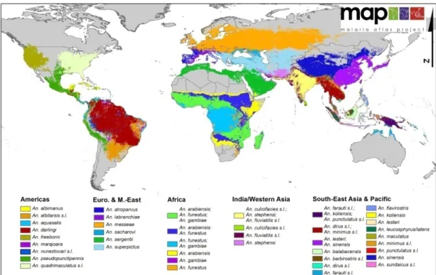
Figure 3 – The global distribution of 34 dominant vector species and / or species complexes ………...54
Figure 4 – The life-cycle of Anopheles ………..55
Figure 5 – Parasite numbers during critical steps of transformation of gametes to ookinetes, to oocysts, and through the migration of sporozoites from the midgut
epithelium to salivary glands ………..59
Figure 6 – Schematic representation of the mechanisms of defence in A. gambiae
against Plasmodium ………....60
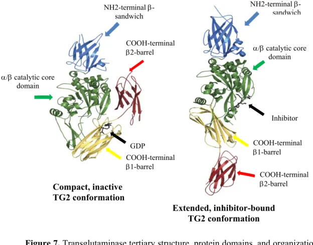
Figure 7 – Transglutaminase tertiary structure, protein domains, and organization ….67
Chapter 2 - Genetic diversity and signatures of selection of drug resistance in
Plasmodium populations from both human and mosquito hosts in continental
Equatorial Guinea
Figure 1 – Total prevalence of mutations in the eight codons of Pfdhfr and Pfdhps
genes ………...96
Chapter 3 - Duffy Negative Antigen Is No Longer a Barrier to Plasmodium vivax – Molecular Evidences from the African West Coast (Angola and Equatorial Guinea)
Figure 1 – Map of the five collection places in Equatorial Guinea and Angola …….115
Figure 1 – Phylogenetic trees for the complete DNA sequence of AgTG1 (A) and
AgTG2 (B) genes ………..132
Figure 2 – Values of Ka/Ks ratio for the coding region of: A-AgTG1 gene – comparison between infected and non-infected mosquitoes; B - AgTG2 gene – comparison between infected and non-infected mosquitoes; C - AgTG1 gene – comparison between M- and S- forms and D - AgTG2 gene – comparison between M- and S- forms ………..135
Figure 3 – Distribution of the different variants of the AgTG1 (A) and AgTG2 (B) proteins in the non-infected and infected mosquitoes ………..136
Figure 4 – Structural model of AgTG1 protein (A) and AgTG2 protein (B). Three-dimensional (3D) structural localization of mutated amino acids represented in solid
Chapter 1 – Introduction
Table 1 – Some of the most important infection characteristics of the five species of
Plasmodium that infect Human ………..41
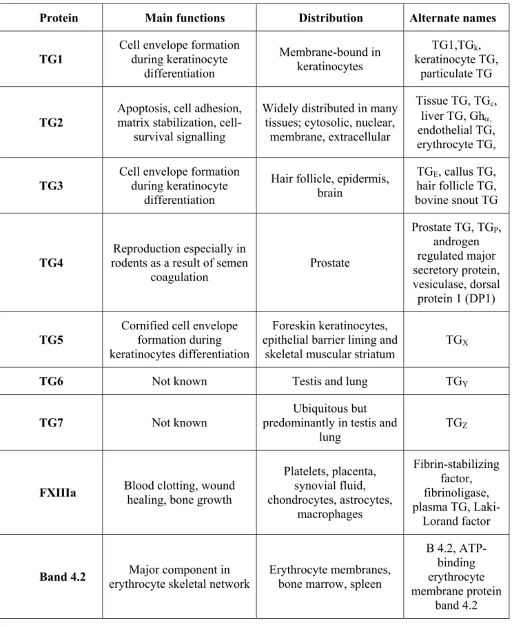
Table 2 – Main characteristics of the nine human TG ………...69
Chapter 2 - Genetic diversity and signatures of selection of drug resistance in Plasmodium populations from both human and mosquito hosts in continental Equatorial Guinea
Table 1 – Neutral microsatellite diversity of Plasmodium falciparum populations from Ngonamanga and Miyobo in humans and mosquitoes ………...95
Table 2 – Statistics of the 15 STR loci of Plasmodium falciparum-positive individuals: mutants to PYR ………...98
Table 3 – Statistics of the 15 STR loci of Plasmodium falciparum-positive individuals: mutants to SFX ………...99
Additional file 1 – Prevalence of Plasmodium infections in humans, in two villages of mainland Equatorial Guinea ……….104
Additional file 2 – Prevalence of Plasmodium infections in mosquitoes, in two villages of mainland Equatorial Guinea ……….105
Additional file 3 – Characterization of mutations in Pfcrt, Pfmdr1,Pfdhps and Pfdhfr
genes, in humans and mosquitoes ……….106
Additional file 4 – Pfdhfr point mutations and their respective STRhaplotypes in allele size ………108
Guinea)
Table 1 – Prevalence of infection in both humans and mosquitoes, in Angola and Equatorial Guinea ……….116
Chapter 4 – Molecular evidence of positive selection in transglutaminases of Anopheles gambiae
Table 1 – Intraspecific polymorphism for A. gambiaeAgTG1and AgTG2genes …. 128
Table 2 – Neutrality tests for the two transglutaminases genes AgTG1 and AgTG2 in Infected mosquitoes, Non-Infected mosquitoes, A. gambiae M-form and A. gambiae S-form ………..134
Additional file 1-table S1 – Sequences and annealing temperatures of primers used to amplify the AgTG1 and AgTG2genes of A. gambiae⁄⁄⁄⁄⁄⁄⁄⁄⁄⁄⁄⁄..148
Additional file 2-table S2 – Matrix of pairwise comparisons of Fst for the two populations and for the two groups of infection studied ⁄⁄⁄⁄⁄⁄⁄⁄⁄⁄⁄..149
Additional file 3-table S3 – Hierarchical analysis of molecular variance (AMOVA)
among the infected and non-infected groups ⁄⁄⁄⁄⁄⁄⁄⁄⁄⁄⁄⁄⁄⁄⁄⁄150
Chapter 1 –
I.
Malaria
The understanding of malaria and its complex life cycle has increased
enormously in the last years, but despite decades of research and efforts to combat it,
malaria continues to be one of the main public health problems in the world, affecting
mainly the poorest areas of the planet. According to the latest World Health
Organization (WHO) report, this mosquito-borne disease was responsible in 2012 for
207 million clinical cases, of which approximately 81% were in the African Region,
resulting in approximately 627 000 deaths, affecting primarily children under five years
old and pregnant women living in sub-Saharan Africa (WHO, 2013).
Malaria is caused by a protozoan parasite from the genus Plasmodium, and it is known that there are five species that may affect humans – Plasmodium falciparum, Plasmodium vivax, Plasmodium malariae, Plasmodium ovale and Plasmodium knowlesi. These parasites are transmitted to humans when female mosquitoes of the genus Anopheles feed on human blood. There are more than 30 anopheline species that transmit malaria to humans (WHO, 2013).
I.1. Geographic distribution of malaria
During the past decade, multiple organizations – as World Bank, Global Fund,
Affordable Medicine Facility-malaria, The US President’s Malaria Initiative, Bill &
Melinda Gates Foundation and others – have concerted efforts to combat malaria all
over the world (WHO, 2013; Murray et al., 2012). The measures undertaken had great
impact especially in countries with high malaria transmission and it is estimated that 3.3
million lives have been saved during this period (WHO, 2013).
Despite all these efforts, malaria remained endemic in 103 countries, causing
approximately 207 million clinical cases (range 135–287 million) and 627 000 deaths
(range 473 000–627 000) in 2012. Figure 1 shows that the most affected areas are the
tropical and subtropical regions of the world, specially the sub-Saharan Africa, Central
Figure 1. Trends in malaria incidence, 2000 - 2012 (from http://www.who.int/gho/malaria/en/, accessed in March 21st, 2014).
The World Health Assembly and Roll Back Malaria have as main objective to
achieve a 75% reduction in malaria cases by 2015, when compared to levels in 2000.
This objective, as well as all the progresses made until now, can be compromised since
the international funding for malaria control has levelled off due to a reduction in the
funding sources (WHO, 2013).
I.1.1. Equatorial Guinea
Equatorial Guinea is located in West Central Africa and has an area of 28 051
Km2 and according to the most recent reports, the country has a population of about 704
000 inhabitants
(https://www.cia.gov/library/publications/the-world-factbook/geos/ek.html, accessed in October 16th, 2013). This country is divided in three
main regions, one continental – Rio Muni - and two islands – Bioko and Annobon. The
continental area is bordered by Cameroon and Gabon; the island of Bioko is located 32
Km of Cameroon coast and is where the capital city (Malabo) is situated. The Annobon
Equatorial Guinea has a tropical climate with distinct wet and dry seasons. The
continental area presents two dry seasons - from December to March and July to
September - and two rainy seasons - one stronger from September to November and
other from March until late June.
In Equatorial Guinea, malaria remains the major endemic disease and the
leading cause of child mortality and morbidity, being characterized as hyper- and
holoendemic (Rehman et al., 2013). It is important to distinguish the insular from the
continental region regarding epidemiological characteristics of malaria, since control
measures have been different in different regions of the country. In 2004, the Bioko
Island Malaria Control Project (BIMCP) was launched, consisting mainly in the indoor
residual spraying (IRS) programme, and aiming to eliminate malaria infection in the
island. The prevalence of infection has been significantly reduced (from 42%
pre-intervention, to 18% in 2008) on the insular region (Pardo et al., 2006; Kleinschmidt et
al., 2009) whilst the prevalence of infection remained above 50% in children under five
years old in mainland region (Kleinschmidt et al., 2009).
Four species of Plasmodium – P. falciparum, P. vivax, P. malariae and P. ovale
– were present in the insular region, however in the mainland, P. vivax was described for the first time only quite recently (Mendes et al., 2011). In both regions, P. falciparum was the most prevalent species, being responsible for approximately 90% of the cases.
Concerning the mosquito vector, multiple Anopheline species were found in the
continental region, Anopheles melas and A. gambiae s.s. being considered the main vectors (Moreno et al., 2004). In addition to these species, it is still possible to find
Anopheles moucheti moucheti, Anopheles carnevalei and Anopheles funestus (Molina et al., 1993; Cano et al., 2006).
I.1.2. Guinea - Bissau
Guinea-Bissau is a country located in Western Africa, bordered by the North
Atlantic Ocean, between Guinea and Senegal. With a total area of 36 125 Km2 and a
population of 1.628 603 people
two distinct seasons: a rainy season (from June to November) and a dry season (from
December to May).
In Guinea-Bissau, malaria remains a serious health problem, presenting
approximately 55 000 clinical cases for year (WHO, 2013), being considered as
mesoendemic-to-holoendemic with intense and seasonal transmission during the rainy
season. Plasmodium falciparum is the most prevalent species, responsible for almost 100% of the cases although there are reports of the presence of P. malariae and P. ovale
in this country (Arez et al., 2003). Anopheles gambiae s.s. and A. melas are the main malaria vectors in the area.
I.1.3. Angola
Angola, a country with 18.056 072 inhabitants and a total area of 1.246 700 Km2
(https://www.cia.gov/library/publications/the-world-factbook/geos/ao.html, accessed in
October 16th, 2013), faces several problems resulting from 27 years of civil war. Angola
has shown in recent years a high growth rate due to its oil production, however much of
the country's infrastructure is still damaged or undeveloped, since it is estimated that
80% of hospitals and health centres have been damaged / destroyed during the war,
restricting the access to health systems to less than 30% of the population. These
problems associated with many other factors as the lack of basic sanitation and
difficulties in access to health centres and hospitals, allowed the spread of many
diseases, including malaria.
According to the latest report, the prevalence of malaria has dropped 50% over
the last five years as a result of control efforts. Nevertheless this disease remains one of
the major’s public health problems in Angola, being responsible for 91 deaths per 1 000
live births (President’s Malaria Initiative, 2013).
Malaria is endemic all over the country, being classified as hyperendemic in the
north and along the Atlantic coast. In the central and southern areas is classified as
mesoendemic unstable. There are two transmission peaks, one occurring between
March and May and the other between October and November. The most prevalent
species of Plasmodium is P. falciparum, but the other three - P. vivax, P. malariae and
There are numerous species of mosquitoes responsible for transmitting malaria
parasites, A. gambiae and A. funestus being considered primary vectors (WHO, 2013).
I.2. Malaria control: antimalarials and insecticides
Despite the high number of clinical cases and deaths occurring all over the world
due to malaria, this is a preventable and treatable disease. According to the WHO
(2013) is essential to act at the level of transmission of the parasite by the mosquito
vector, but also in the development of illness and severe disease.
I.2.1. Malaria vector control
Historically, vector control has been an important tool to reduce and even eradicate malaria in some parts of the world. Nowadays there are different options available to vector control that include chemical, biological, natural plant products, and environmental management (Raghavendra et al., 2011).
For WHO, the interventions with higher impact are insecticide-treated nets (ITNs), that include the long-lasting insecticidal nets (LLINs) and the conventional nets that are later treated with an insecticide; and IRS.
The ITNs forms a physical barrier between the infected mosquitoes and man and to be effective need to have high coverage rates. Today it is estimated that a total of 88
countries, including 39 in Africa, distribute ITNs free of charge. In fact, the percentage
of households owning at least one ITN in sub-Saharan Africa is estimated to have risen
from 3% in 2000 to 56% in 2012, but declined slightly to 54% in 2013, and the last data
indicates that approximately 86% of the population with access to an ITN actually uses
it, suggesting that efforts to encourage ITN use have been successful (WHO, 2013).
IRS with insecticides continues to be one of the main pillars for malaria control.
malaria control. In the last years, the proportion of at-risk population that was protected
arise from less than 5% in 2005 to 11% in 2010 but fell to 8% in 2012, with 58 million
people benefiting from the intervention, only in the African Region (WHO, 2013).
Another important measure is the larval control of malaria vector Anopheles
mosquitoes. The principle of chemical larviciding is to eliminate or reduce the vector population by killing the larvae. This preventive method has been neglected, in spite of some authors thinking that should be taken into account in the new malaria control programs (Walker & Lynch, 2007). The WHO recommends larviciding only in settings where mosquito breeding sites are few, fixed, findable and easy to identify, map and
treat (WHO, 2013).
I.2.1.1. Insecticide resistance
Until the early 19th century, the application of insecticides was the primary
control tool in the vector control programs (Breman, 2001). Several insecticides have
been used so far, but in the 20th century, after the discovery of the insecticidal potential of dichlorodipehnyltrichlroethane (DDT), a new era of insect control began. DDT was the first synthetic organic insecticide used for effective vector control; it was cheap and very efficient. However, with the extensive use of this and other insecticides, the insecticide resistances start to appear and spread in many Anopheles species
(Raghavendra et al., 2011).
Today the insecticide resistance is one of the major problems for vector control
programs and according the last report, mosquito resistance to at least one insecticide
used for malaria control has already been identified in 64 countries. The one that rise
bigger concern is the resistance to pyrethroids, especially in Africa.
To try to overcome this problem, WHO made a series of recommendations such
as: 1) Resistance management measures should be part of every vector control program
and deployed preventively, without waiting for signs of the presence of resistance or of
control failure; 2) A substantial intensification of resistance monitoring is needed; 3)
Using the same insecticide for multiple successive IRS cycles is not recommended; 4)
In areas with high LLIN coverage, pyrethroids should not be used for IRS (WHO,
I.2.2. Malaria control
To control malaria one should take into account several aspects: first it is needed
appropriate preventive measures; a good and reliable diagnostic and finally an effective
treatment.
I.2.2.1. Intermittent preventive treatment
Intermittent preventive treatment (IPT) is recommended for pregnant women
and for children less than five years old living in malaria endemic countries. It is
estimated that a total of 36 of 45 sub-Saharan African countries had adopted IPT as
national policy by the end of 2011, using sulfadoxine-pyrimethamine (SP) as the drug
of reference (WHO, 2013). Several studies reported the importance of the use IPTs and
ITNs during pregnancy, since leads to a reduction in stillbirths, improvements in birth
weight of babies (since malaria infection during the pregnancy it is one of the main
causes of low birth weight) and a reduction in the prevalence of parasitaemia and
anaemia in pregnant women (Gamble et al., 2009; Eisele et al., 2012; Singh et al.,
2013).
I.2.2.2. Diagnosis of malaria
Current recommendations of effective, yet expensive artemisinin-based
combination therapies (ACT) for malaria in Sub-Saharan Africa have increased the
importance of laboratory-confirmed diagnosis.
In the majority of malaria endemic countries many fever cases are treated
presumptively with antimalarials without parasitological diagnosis; further, not all
confirmed malaria cases receive appropriate treatment. It is therefore important the
implementation of a universal diagnostic test, which allows to obtain reliable results.
The current reference method for malaria diagnosis is direct optical microscopic
visualization of parasites on thick and/or thin blood smears (Kyabayinze et al., 2008),
but unfortunately, this technique is influenced by many aspects as: the experience of the
laboratory technicians, the quality of the microscopes and the lack of quality control
(RDTs) for malaria have substantial potential to help solve these questions, especially in
poor areas (Reyburn et al., 2007), but it must be ensured that RDTs are highly sensitive
and specific for Plasmodium species detection (Wongsrichanalai et al., 2007). In fact, the number of patients tested by microscopic examination increased to a peak of 188
million in 2012, whereas the number of RDTs supplied by manufacturers increased
from 88 million in 2010 to 205 million in 2012 (WHO, 2013).
I.2.2.3. Malaria treatment
The first drug used to fight the high fevers caused by malaria was quinine, a
medicinal plant isolated from Chinchona tree. In the 20th century, started to appear the first organic compounds that intend to substitute the quinine, among them were the
pamaquine, quinacrine and ultimately chloroquine (CQ) (Thompson et al., 1972).
Chloroquine quickly became the drug of choice to combat malaria, since it was a
cheap and effective drug. In 1955, WHO launched a campaign for malaria eradication
with the wide distribution of CQ together with DDT, which produced some regional
successes (Wellems & Plowe, 2001). During the late 1950s, the first cases of resistant
P. falciparum were detected in Colombia and at the Cambodia-Thailand border (Payne et al., 1987).
Later, other antimalarial drugs were developed, such as SP that already presents
some cases of resistance, particularly in the south-western Asia and South America, but
still is widely used in Africa mainly for IPT; mefloquine; amodiaquine and more
recently the ACTs.
Today and according to the WHO, uncomplicated P. falciparum infectionshould be treated with an ACT and currently there are 5 recommended: artemether plus
lumefantrine, artesunate plus amodiaquine, artesunate plus mefloquine, artesunate plus
SP and dihydroartemisinin plus piperaquine. The choice of the ACT should be based on
the therapeutic efficacy of the combination in the country or area of intended use. For P. vivax infections, the guidelines are that it should be treated with chloroquine in areas where this drug is effective or with an appropriate ACT (not artesunate plus SP) in areas
I.2.2.4. Antimalarial resistance
Antimalarial drug resistance is a major public health problem which delays the
malaria control. Today, parasites have already developed resistance to all the drugs
available for malaria control, somewhere in the world. According to the WHO,
antimalarial resistance has been defined as the “ability of a parasite strain to survive
and/or multiply despite the administration and absorption of a drug given in doses equal
to or higher than those usually recommended but within tolerance of the subject”. This
definition was later modified to specify that the drug in question must “gain access to
the parasite or the infected red blood cell for the duration of the time necessary for its
normal action” (Bloland, 2001).
Chloroquine
The first cases of P. falciparum resistance were detected in Colombia and at the Cambodia-Thailand border during the late 1950s (Payne et al., 1987) and spread
gradually through South America, Southeast Asia, and India in the 1960s and 1970s. In
Africa the first resistance reports only appeared in the late 1970s, in Kenya and
Tanzania (reviewed in Wellems & Plowe, 2001).
It is thought that CQ efficacy lies in its ability to interrupt haematin, which is
released in large amounts as the parasite consumes and digests haemoglobin in its
digestive food vacuole a process of detoxification as malaria parasites grow within their
host’s red blood cells (RBCs) (Dorn et al., 1998).
Plasmodium falciparum chloroquine resistance transporter (Pfcrt) is a predicted transporter that is localized in the digestive vacuole membrane and may be involved in
drug efflux and/or pH regulation. Several point mutations in this gene seemed to be
associated with the CQ resistance, being the N75E, K76T the most frequent in Africa
and considered as the best markers (Wellems & Plowe, 2001; Le Bras et al., 2003; Bray
et al., 2005).
Sulfadoxine-Pyrimethamine
After the appearance of the CQ resistance, the antifolate combination of SP has
increasingly become the drug of choice for the treatment of uncomplicated P.
falciparum malaria. However, SP resistance has developed quickly and the first report of resistance of P. falciparum to pyrimethamine was in the north-east Tanzania in 1954 (Clyde, 1954).Despite the resistance reports this drug still have some efficacy in some
countries of Africa and is used in the IPT.
The major cause of resistance to antifolate drugs are point mutations in P. falciparum dihydrofolate reductase (Pfdhfr) and P. falciparum dihydropteroate synthase (Pfdhps) genes that rapidly diminished their clinical effectiveness. The Pfdhfr
is a key enzyme in the redox cycle for production of tetrahydrofolate, and the Pfdhps is an enzyme involved in the biosynthesis of folate (Cowman et al., 1988; Brooks et al.,
1994).
Several studies showed that there are four main point mutations in the Pfdhfr
gene that are associated to resistance to pyrimethamine (N51I, C59R, S108N and
I164L) (Basco et al., 1995a; Curtis et al., 1996). The S108N mutant exhibits a low level
of resistance, the N51I/S108N or the C59R/S108N double mutants, intermediate levels
of resistance, and the N51I/C59R/S108N triple mutant has a higher level of resistance to
this drug. Similarly, resistance to sulfadoxine is due to four mutations in the Pfdhps
(S436F, A437G, K540E, A581G) (Brooks et al., 1994). Each successive mutation
causes a decrease in the susceptibility to these drugs. In fact, the association between
the mutations in the two genes, originating the quintuple mutant (Pfdhfr: N51I, C59R, S108N and Pfdhps: A437G, K540E) is associated with the clinical failure of the SP (Kublin et al., 2002; Talisuna et al., 2004).
II.
Parasite
II.1. Taxonomic classification
The human malaria parasites are classified as belonging to the Eukaryota
an apical intracellular complex (visible with electronic microscope) and by not having
cilia or flagella, except for microgametes (Ayala et al., 1998). They belong to the
Hematozoa class (characterized by organisms that parasitise erythrocytes); to the
Haemosporida order (that present a mobile zygote – ookinete) and to the Plasmodiidae
family (with two different types of multiplication in their life cycle: sexual and asexual
phases). This parasite belongs to Plasmodium genus (characterised by having an asexual phase in cells other than erythrocytes - hepatocytes) (Knell, 1991; Ayala et al., 1998)
and to Plasmodium and Laverania sub-genus. Finally, they are classified in the following species: P. falciparum, P. vivax, P. malariae, P. ovale and P. knowlesi
(Antinori et al., 2012).
All species of human malaria parasites present a similar and complex life-cycle
requiring two different hosts - the human and the female Anopheles mosquito - and showing three types of genomes: a) a nuclear genome with 14 linear chromosomes; b) a
linear mitochondrial genome and finally c) a 35kb circular plastid genome that is
housed in the apicoplast (Antinori et al., 2012).
II.2.
Plasmodium
life cycle
All species of Plasmodium that infect humans show a similar life cycle (Figure 2) which is characterised by a sexual phase, named sporogony, that take place in the
mosquito vector; and an asexual phase, named schizogony, that occurs in the human
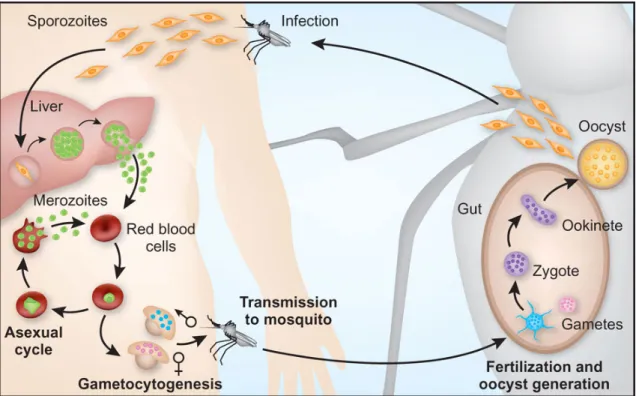
Figure 2.Plasmodium life cycle (adapted from Pasvol, 2010).
The Plasmodium life cycle starts when a female anopheline mosquito feeds on infected blood, and the gametocytes (sexual cells) began their development in the
mosquito. This phase is called fertilization and corresponds to the sexual phase. The
gametocytes suffer differentiation, forming the female and male gametes. Fertilization
occurs between these two gametes originating a zygote (the unique diploid form of all
life cycle), which undergoes meiosis and differentiation into motile ookinetes – invasive
forms in the next few hours.
After approximately 24 hours, the ookinetes pass across the midgut epithelium
and lodged under the basal lamina forming vegetative oocysts. These forms mature, and
after several rounds of mitosis, sporozoites are formed – haploid forms. After the
oocysts rupture, sporozoites are released into the hemolymph and travel through the
mosquito haemacoel until reach salivary glands. When a new blood meal is taken by the
mosquito female, the sporozoites are injected into the bloodstream of a new host. A few
minutes later, they are already starting the invasion of the liver cells, starting the
The newly arrived sporozoites enter the liver hepatocytes leading to liver
schizonts. When they mature, merozoites are released into the peripheral blood – new
invasive forms – where they will invade erythrocytes. In the case of P. vivax and P. ovale, the sporozoites can differentiate into hypnozoites, stages that can remain dormant in the liver for long periods of time.
In the erythrocytes, the merozoites begin to differentiate into trophozoites. This
period is called prepatent period and its extension is characteristic of each species. After
two or three days of mitotic divisions erythrocytic schizonts are formed. When
erythrocytes disrupt, merozoites are released and will infect other RBCs, starting a new
cycle in the blood. This phase is responsible for symptoms of malaria illness, and as the
number of parasites increases (parasitaemia), the infected person becomes more
severely affected (Knell, 1991; Antinori et al., 2012).
II.3. Infection dynamics
The five species of Plasmodium infecting humans exhibit different biological and infection characteristics. Table 1 shows some of the most important differences
Table 1: Some of the most important infection characteristics of the five species of Plasmodium that infect humans (adapted from Antinori et al., 2012).
Characteristics P. falciparum P. knowlesi P. malariae P. ovale P. vivax
Pre-erythrocytic
stage (days) 5-7 8-9 14-16 9 6-8
Pre-patent period
(days) 9-10 9-12 15-16 10-14 11-13
Erythrocytic cycle
(hours) 48 24 72 50 48
Red blood cells
affected All All
Mature
erythrocytes Reticulocytes Reticulocytes
Parasitaemia per l
Average 20 000-500 000 600-10 000 6 000 9 000 20 000-100 000
Maximum 2.000 000 236 000 20 000 30 000 100 000
Febrile paroxysm
(hours) 16-36 or longer 8-12 8-10 8-12 8-12
Severe malaria Yes Yes No No Yes
Relapses from liver
forms No No No Yes Yes
Recurrences Yes Yes Yes No Yes
One of the typical symptoms of malaria disease is the intermittent high fevers
characterized by: first; a rapid rise of temperature associated with chills - the cold stage;
second; a temperature peak (reaching 40-41ºC) associated with other symptoms as
headache, vasodilatation and myalgia - the hot phase; and, finally, the third phase with
the decreasing of temperature – the sweat stage.
The intermittent high fevers are directly related to the duration of the parasite
quartan (caused by P. malariae), with peaks of fever every four days (72 hour cycle) (Knell, 1991; Carter & Mendis, 2007; Antinori et al., 2012).
For a better understanding of the Plasmodium species studied in the present work, a brief description of each one is present below.
II.3.1. Plasmodium falciparum
Plasmodium (Laverania) falciparum (Welch, 1896) was first observed by Charles Alphonse Laveran in 1880. Of the five Plasmodium species that infect humans, this is the one that causes more morbidity and mortality and present higher prevalence
particularly in Africa. According to the latest WHO report (WHO, 2012) 85 countries
are classified as endemic for P. falciparum, with 2.57 billion people at risk.
The P. falciparum genome sequencing in 2002, brought great advances in the knowledge of this parasite (Gardner et al., 2002). It is composed by 14 linear
chromosomes, coding for 5 365 genes. However, up to now, only 1 817 have known
functions.
The life cycle of P. falciparum is characterized by having an asexual development in the liver (pre-erythrocytic schizogony), with no hypnozoites
differentiation. The first visible form in the liver appears on the fourth day after
infection and corresponds to the hepatic schizont (Antinori et al., 2012).
In the erythrocytic schizogony, typically, only the young rings are visible
whereas the maturation stages are rarely seen in the peripheral blood. The more
developed trophozoites disappear from peripheral blood circulation as infected
erythrocytes are being sequestered in the internal organs, like brain, spleen and placenta.
Another characteristic of P. falciparum parasite is the development of gametocytes in the internal organs, where they can be captured (Antinori et al., 2012).
The gametocytogenesis of this parasite can be divided in five morphologically distinct
substages (Bousema & Drakeley, 2011); Stage I – IV: Immature P. falciparum
gametocytes are sequestered away from the circulation, Stage V: mature gametocytes
(Bousema & Drakeley, 2011; Antinori et al., 2012). The sporogonic cycle takes 9 to 10
days at a mean temperature of 28ºC.
The origin of P. falciparum has been object of study and until very recently, it was thought that the closest parasite of P. falciparum was the Plasmodium reichenowi:
a chimpanzee parasite. The studies indicated that these two parasites have diverged at
the same time 5 Myr ago (Escalante et al., 1994; Escalante et al., 1995; Rich et al.,
1998; Jeffares et al., 2006); however recent works claim that P. falciparum is of gorilla origin (Liu et al., 2010; Holmes, 2010).
II.3.2. Plasmodium vivax
Plasmodium vivax (Grassi & Feletti, 1890) is the Plasmodium parasite that presents the wider distribution, being present in 109 countries considered as potentially
endemic for this parasite (Guerra et al., 2010; Gething et al., 2012).
For a long period of time, P. vivax was considered a “benign” parasite, being neglected by the scientific community. Recently, however, this idea has changed and in
the last years this parasite has become highly studied. The reasons of this change are the
following: first, its wider distribution, being found in both tropical, and subtropical
areas and in countries where it was not present or it was not detected by the available
techniques in the past, as is the case of some countries of West and Central Africa
(Poirriez et al., 1991; Snounou et al., 1998; Gautret et al., 2001; Mendes et al., 2011);
second, the high number of clinical cases reported, ranging from 70 million to 300
million (Baird, 2007; Galinski & Barnwell, 2008; Mueller et al., 2009) and third, this
parasite seems to be evolving and adapting, causing more severe forms of the disease
including death (Genton et al., 2008; Rogerson & Carter, 2008; Tjitra et al., 2008;
Alexandre et al., 2010).
Regarding P. vivax life cycle, sporozoites in the liver can differentiate into schizonts or into hypnozoites, which are responsible for the relapse of the infection.
Hypnozoites are only formed by this species and by P. ovale. The sexual life cycle of this parasite in Anopheles mosquitoes takes 8-10 days at 28ºC (Gilles, 1993).
This parasite invades preferentially reticulocytes and all forms of the
erythrocytic cycle can be found in the peripheral blood contrasting with P. falciparum, where only early parasites are observed(Antinori et al., 2012).
The erythrocytes invasion by this parasite has been described as being mediated
exclusively by the Duffy antigen receptor for chemokines (DARC). People not
presenting this antigen in the erythrocytes surface were called Duffy negative
individuals [Fy(a-b-)] and were hypothetically resistant to P. vivax infection (Miller et al., 1975; Langhi et al., 2006). The small prevalence found in West and Central Africa
for this parasite was attributed to the high prevalence of Duffy negative people in this
region. However, recent studies demonstrate that P. vivax may be changing and is able to invade erythrocytes using other receptors than Duffy (Ryan et al, 2006; Cavasini et
al., 2007a and 2007b; Ménard et al., 2010; Mendes et al., 2011).
II.3.3. Plasmodium malariae
Plasmodium malariae (Laveran, 1880) has a wide but sparse distribution, can be found most frequently in sub-Saharan Africa and the southwest Pacific. This parasite
shows a slow development in both hosts (15 days in the Anopheles mosquitoes; in human, 15 days in the liver and 72 hours in the blood), and hardly causes serious forms
of the disease (Collins & Jeffery, 2007).
Infections caused by this parasite rarely reach high parasitaemias (usually not
exceed 30 000 parasites per microliter), probably due to a low number of merozoites
produced per erythrocytic cycle. Plasmodium malariae does not form dormant forms in the liver, like P. vivax and P. ovale, but can persist in the blood with low parasitaemia for long periods of time (reaching 30-40years) causing recrudescence (Collins &
The sporogonic cycle takes 14 to 16 days at 28ºC increasing the time for 30-35
days if the temperature is 20°C (Collins & Jeffery, 2007).
II.3.4. Plasmodium ovale
Plasmodium ovale (Stevens, 1922) was first discovered by Stephens in 1922 (Stephens, 1922) in an African patient. This parasite is distributed in sub-Saharan
Africa, South-east Asia, Middle East, the Indian subcontinent, Papua New Guinea and
East Timor Indonesia (Muller et al., 2007), and is not described as causing severe
malaria cases.
This parasite can cause chronic infections, presenting low parasitaemias; and can
develop hypnozoites which can cause relapses in the infection. The sporogonic cycle
takes 12 to 14 days at a mean temperature of 28ºC.
Recently, two closely related but distinct species of P. ovale were described: P. ovale curtisi (classic type) and P. ovale wallikeri (variant type). It is known that despite being sympatric in both African and Asiatic regions, the existence of several
geographic, temporal or ecological barriers prevent the recombination between the
genomes of the two species (Sutherland et al., 2010).
II.4. Mixed Infections
Mixed infections, involving two or more species of Plasmodium, are very common in countries where malaria is endemic (Richie, 1988; Bruce et al., 2000).
Several studies have shown that both vertebrate and invertebrate hosts may be infected
with more than one species of Plasmodium (Mason et al., 1999; Arez et al., 2003; Mayxay et al., 2004; Snounou & White, 2004; Zimmerman et al., 2004; Marques et al.,
2005; Genton et al., 2008; Bousema et al., 2008).
Concomitant infections may have effects on pathology, severity and infection
dynamics, that’s why it is so important a correct diagnostic.
In most of the endemic malaria countries, the diagnosis is made through
this technique has some limitations, being affected by several aspects: the limit of
detection is not very high (in theory, 10–100 parasites per l) (Wongsrichanalai et al.,
2007) when compared with other techniques like PCR or real-time PCR (0.05–10
parasites per l) (Snounou et al., 2003), and is depending on the quality of the blood
slides preparation, the number of microscope fields analysed and the microscopist’s
expertise (Zimmerman et al., 2004).
Using the standard diagnostic technique - optical microscopy – and due to the
limitations referred above, mixed infections are often difficult to detect since all
infections go through periods of low parasitaemia. The switch between periods of patent
parasitaemia (when parasites in peripheral blood can be easily observed) and latent
periods of parasitaemia (when it is not possible to observe parasites in peripheral
blood), makes it difficult to distinguish the parasites only by their morphological
characteristics (Richie, 1988; Snounou et al., 2004; Zimmerman et al., 2004; Rajahram
et al., 2012).
During the last 30 to 40 years, several studies have been performed trying to
clarify the possible relationship among the different species of Plasmodium. However, several discrepancies were found in these studies and several questions remain unclear.
Molineaux et al (1980) in a study conducted with Nigerian adults concluded that
mixed infections were more common than expected and P. malariae is more commonly associated with P. falciparum than it was initially predictable based on the individual frequencies of each species. On the other hand, Richie (1988) and McKenzie & Bossard
(1999), reported less mixed infections than would be expected when they compared P. vivax – P. falciparum infections, however high numbers of P. malariae – P. falciparum
mixed infection were still found.
Another aspect that has been widely studied is the impact that mixed infections
may have in the severity of the infection. Some studies reported a reduction in the
severity of symptoms when mixed infections were present. In a study with African
al., 1997), where mixed infection of P. falciparum and P. malariae and / or P. ovale and
P. falciparum + P. vivax presented a reduction in the severity of the symptoms. Luxemburger et al. (1997), in a study conducted in Thailand, showed a decrease of
5.7% to 1.6% on the severity of the infection when they compared single P. falciparum
infection with P. falciparum + P. vivax infections. These results were corroborated by other studies carried out in different countries as Vanuatu (Williams et al., 1996), Papua
New Guinea (Smith et al., 2001), Thailand (Price et al., 2001; McKenzie et al., 2006)
and Brazil (Lorenzetti et al., 2008).
Depending on the relationship between parasites, it is possible to distinguish
between positive and negative interactions. The first is characterized by the presence of
a particular species favouring the development of another, while negative interactions
are characterized by the presence of a species inhibiting the presence of another.
II.5. Parasite diversity
The extent of genetic diversity of natural populations of Plasmodium is enormous and both inter- and intra-specific infections are common. The understanding
of this topic is a key epidemiological issue as ecological interactions between parasite
populations in the same host may be an important source of selection on pathogen traits
such as virulence and drug resistance and allows determining the influence of different
parasite populations on infection and transmission dynamics. Further, some of those
effects on infection parameters seem to be dependent on the seasonality and the
intensity of malaria transmission (Marques et al., 2005).
II.5.1. Plasmodium falciparum
The genetic diversity of P. falciparum has been highly studied not only because this parasite is responsible for the highest number of clinical cases and deaths, but also
due to its importance as an indicator of the malaria transmission intensity in an area
(Paul et al., 1998); its ability to differentiate between recrudescence (which correspond
to a treatment failure) and new infection (either from pre-existing liver infection or a
development and/or the evaluation of malaria vaccines, since its high levels of genetic
diversity is one of the biggest limitation for the development of an effective malaria
vaccine (Schwartz et al., 2012).
The most frequently used markers for the P. falciparum genotyping are the merozoite surface protein 1 and 2 (MSP-1 and MSP-2) and the glutamate-rich protein
(GLURP), which are surface antigens (Smythe et al., 1991; Snounou et al., 1998). With
these markers it is impossible to know whether observed patterns reflect population
history or natural selection (Anderson et al., 2000) so, nowadays, neutral microsatellite
sequences (or short tandem repeats, STRs), are the most commonly used markers to
differentiate P. falciparum populations.
Below, a brief description of the molecular markers used in this work is
presented.
II.5.1.1. Genetic markers – msp2
The msp2 gene codes for a merozoite surface antigen and is one of the most widely used genetic markers in population biology studies of P. falciparum parasites. This gene contains highly polymorphic regions with repeated units; in addition to size
differences, it is possible to distinguish two allelic families – IC and FC27 (Smythe et
al., 1991).
Through the analysis of msp2 gene, it is possible to determine the multiplicity of infections (MOI), which is defined as the minimum number of different genotypes of P. falciparum in a single individual (Beck et al., 1997). MOI is a good indicator of acquired immunity or premunition in populations living in endemic areas and is also a
good indicator of the malaria transmission intensity (Babiker et al., 1995; Paul et al.,
1998). An area with high endemicity usually presents extensive parasite diversity and
multiple genotypes are found in a single infected individual. In the opposite, the parasite
population in a low transmission area shows limited genetic diversity and the majority
of infections are monoclonal (Haddad et al., 1999; Babiker et al., 2001;
II.5.1.2. STRs
STRs are simple DNA sequence repeats composed by short motifs, usually with
6 or less bases, that are repeated in tandem (Queller et al., 1993). These STRs are found
in all organisms and are widespread throughout the genome.
They are considered very useful molecular markers for population genetic
analysis (Goldstein & Schlötterer, 1999, Anderson et al., 2000) and for genetic linkage
mapping (McCollum et al., 2007), since they are very abundant, highly polymorphic,
co-dominant and easy to score.
It is possible to differentiate between neutral STRs, i.e., not subjected to
selection, allowing the analysis of the genetic variability without selection effect
(McCollum et al., 2007); and STRs flanking resistance genes, that may reveal effects of
selective sweep. From the analysis of these STRs it is possible to characterize the
evolutionary origin of resistant alleles, check if new mutations arise in different
geographic regions or share a common ancestor (Roper et al., 2003; Anderson & Roper,
2005).
II.5.2. Plasmodium vivax
Plasmodium vivax has been, until very recently, a neglected parasite. Many details of their biology, epidemiology and pathogenesis is unknown. Now it is known
that this parasite is capable of causing severe manifestations of the disease, like cerebral
malaria, renal failure, hepatic dysfunction and even death (Barcus et al., 2007; Kochar
et al., 2009) and due to the appearance and spread of drug resistance in P. vivax (Baird & Hoffman, 2004), is essential to know its population structure and genetic diversity.
To study the diversity of infection within this parasite, the markers of choice are
STRs or some surface antigen genes, such as circumsporozoite protein (CSP) and
merozoite surface protein 3 alpha (MSP-3).
To obtain accurate comparisons of genetic diversity of global P. vivax