online | memorias.ioc.fiocruz.br
Expression of bacterial virulence factors and cytokines during
in vitro macrophage infection by enteroinvasive
Escherichia coli
and
Shigella flexneri
: a comparative study
Silvia Y Bando1,2, Ana CR Moreno1, José AT Albuquerque1, Juliana MK Amhaz1, Carlos A Moreira-Filho2, Marina B Martinez1/+
1Departamento de Análises Clínicas e Toxicológicas, Faculdade de Ciências Farmacêuticas 2Departamento de Pediatria, Faculdade de Medicina, Universidade de São Paulo, Av. Prof. Lineu Prestes 580, 05508-000 São Paulo, SP, Brasil
Enteroinvasive Escherichia coli (EIEC) and Shigella spp cause bacillary dysentery in humans by invading and multiplying within epithelial cells of the colonic mucosa. Although EIEC and Shigella share many genetic and bio-chemical similarities, the illness caused by Shigella is more severe. Thus, genomic and structure-function molecular studies on the biological interactions of these invasive enterobacteria with eukaryotic cells have focused on Shigella
rather than EIEC. Here we comparatively studied the interactions of EIEC and of Shigella flexneri with cultured J774 macrophage-like cells. We evaluated several phenotypes: (i) bacterial escape from macrophages after phago-cytosis, (ii) macrophage death induced by EIEC and S. flexneri, (iii) macrophage cytokine expression in response to infection and (iv) expression of plasmidial (pINV) virulence genes. The results showed that S. flexneri caused macrophage killing earlier and more intensely than EIEC. Both pathogens induced significant macrophage produc-tion of TNF, IL-1 and IL-10 after 7 h of infecproduc-tion. Transcripproduc-tion levels of the gene invasion plasmid antigen-C were lower in EIEC than in S. flexneri throughout the course of the infection; this could explain the diminished virulence of EIEC compared to S. flexneri.
Key words: EIEC - Shigella - in vitro infection - cytokines - virulence factors
Infectious diarrhea is one of the main causes of child-hood death, especially in developing countries (Bryce et al. 2005) and Escherichia coli is a major diarrhea-causing pathogen in children under five years of age (Petri et al. 2008, Moreno et al. 2010). E. coli isolated from patients with dysentery was found to cause experimental kerato-conjunctivitis in guinea pigs in the early 1960s (Trabulsi et al. 1967, DuPont et al. 1971). These strains, thereafter named enteroinvasive E. coli (EIEC), possess genetic and biochemical characteristics similar to Shigella (Silva et al. 1980, Sansonetti et al. 1982, Kaper et al. 2004).
EIEC and Shigella cause bacillary dysentery in hu-mans by invading and multiplying within epithelial cells of the colonic mucosa, resulting in an intense inflamma-tory response characterized by abscesses and ulcerations (Hale 1998, Parsot et al. 2005). The genes required for bacterial entrance into host cells are harboured in a 230-kb virulence plasmid (VP) present in Shigella and EIEC strains (Harris et al. 1982, Sansonetti et al. 1982, Silva et al. 1982). Transcription of invasion plasmid antigen (ipa) genes is regulated by two VP-encoded proteins:
Financial support: FAPESP (203/01269-1)
ACRM and JMKA received PhD fellowships from CAPES and FAPESP, respectively. SYB received a post-doctoral fellowship from CNPq. JATA was a MSc fellow from CNPq. CAM-F is a research fellow from CNPq.
+ Corresponding author: [email protected] Received 20 January 2010
Accepted 7 June 2010
VirF and VirB (Dorman et al. 2001). VirB also controls transcription of the VP genes that code for the proteins of the type three secretion system (TTSS) apparatus. In-tracellular movement (ics) A and icsB play a crucial role in bacterial dissemination: the first leads to bacterial in-vasion and intracellular spreading and the latter aids in escape from autophagy.
Shigella translocates through M cells of the intestinal epithelium to invade the epithelial layer by the basolater-al pole (Sansonetti 1991, Ménard et basolater-al. 1996, Sansonetti et al. 1996). Shigella can survive and multiply inside macrophages, escape from phagocytic vacuoles and in-duce apoptosis (Sansonetti et al. 1986, Zychlinsky et al. 1992, 1996, Nonaka et al. 2003, Kubota 2006). Apopto-sis of infected macrophages is promoted by caspase-1 activation. Caspase-1 cleaves the IL-1 and IL-18 precur-sors to their mature forms, triggering a mucosal inflam-matory response (Navarre & Zychlinsky 2000, Kubota 2006, Suzuki et al. 2007).
as spreading and icsA and icsB. We also measured the cytokine response upon in vitro infection of macrophag-es with EIEC or S. flexneri.
SUBJECTS, MATERIALS AND METHODS
Bacterial strains - The M90T strain (invasive S. flex-neri 5a) was kindly donated by Philip Sansonetti (Pasteur Institute). EIEC strain FBC124-13, serotype O124:H-, was isolated from diarrheic faeces and was Sereny test-positive (Gibotti et al. 2004). Bacteria were grown at 37ºC for two days on trypticase soy agar plates contain-ing 0.02% Congo Red. Prior to cell infection, a scontain-ingle red colony of each strain was inoculated in trypticase soy broth and grown at 37ºC with agitation (150 rpm) to an OD625 of 0.1, which corresponds to 1 × 10⁸ colony forming units (CFU)/mL.
Cell culture and macrophage infection - The J774 macrophage-like cells were maintained in Roswell Park Memorial Institute (RPMI) 1640 medium supplemented with 10% fetal bovine serum (FBS) and penicillin-strep-tomycin (100 u/mL-100 µg/mL) in a 5% CO2 incubator at 37ºC. Twenty-four hours prior to infection macrophages were plated in 24-well plates at a density of 1 × 10⁶ cells per well. Themedium was then replaced by fresh RPMI 1640 containing 10% FBS (without antibiotics) and the macrophages were infected with a multiplicity of infec-tion (MOI) of 10 bacteria per cell, centrifuged at 100 g
for 5 min and incubated for different periods of time.
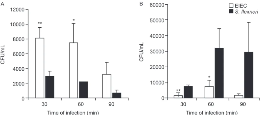
Bacterial survival inside the macrophages - Mac-rophages were infected with S. flexneri or EIEC for zero (cells harvested just after bacterial inoculation), 30, 60 and 90 min. The cells were then incubated with medium containing 50 µg/mL gentamicin for 50 min to kill non-phagocytosed bacteria and to prevent reinfection. The in-fected macrophages were washed threetimes with PBS. PBS containing 0.1% Triton X-100 was then addedto each well to lyse the macrophages. The recovered bacteria were resuspended in 0.9% NaCl, serial dilutions were plated on MacConkey agarand the CFU were counted following an overnight incubation of the plates at 37ºC.
Bacterial escape and macrophage death - To as-sess bacterial escape after phagocytosis and determine whether this process was accompanied by macrophage death, J774 cells were infected for an hour with EIEC or
S. flexneri and then the medium was replaced by fresh RPMI 1640 containing 10% FBS and 50 µg/mL of gen-tamicin. Following different periods of post-infection incubation (30, 60 or 90 min) in medium without anti-biotic, supernatant dilutions were plated to determine the CFU. The macrophages were then resuspended in a hypotonic fluorochrome solution (0.1% sodium citrate, 0.1% Triton X-100 and 50 µg/mL of propidium iodide) and analyzed on a FACScalibur flow cytometer (Becton Dickinson, Mountain View, CA, USA).
Detection of cytokines by enzyme-linked immunosor-bent assay (ELISA) - The J774 macrophages were infect-ed as describinfect-ed for the bacterial escape assay, however with an MOI of 1:10. After one hour of infection, the medium was replaced by fresh RPMI 1640 containing
10% FBS and 50 µg/mL of gentamicin. Supernatants of post-infected J774 cell cultures were collected after 6 h of incubation. Concentrations of IL-1, TNF and IL-10 were determined by ELISA according to the manufac-turer’s instructions (Peprotech, Mexico).
Expression of EIEC and S. flexneri virulence genes in infected macrophages - Total RNA from infected mac-rophages was extracted with a RNeasy Mini Kit (Qiagen, Hilden, Germany), as described by the manufacturer. Contaminating genomic DNA was removed using the RNase-free DNase Data Set (Qiagen, Hilden, Germany) and samples were frozen at -80°C until use. Total RNA was quantified by A260 measurements (NanoDrop® ND-1000 Spectrophotometer; Amersham Biosciences) and diluted to 10 ng/µL prior to cDNA synthesis. Bacterial mRNA was reverse transcribed into cDNA using the Su-perscript II First-Strand Synthesis System with random hexamers, as described by the manufacturer (Invitrogen, Carlsbad, CA, USA). The cDNA was sequenced to check the primer and probe adequacy. Reverse transcriptase polymerase chain reaction (RT-PCR) products were ob-tained by PCR amplification for 30 cycles as follows: 95°C for 45 s, 50-60ºC (Table I) for 45 s and 72ºC for 2 min, with an initial denaturation step of 95ºC for 5 min and final extension step of 72ºC for 7 min. The virulence genes studied here and their respective primers are listed in Table I.
Quantitative real-time PCR (qRT-PCR) analyses were carried out using a TaqMan assay on an ABI Prism 7000 Sequence Detection System version 1.6(Applied Biosystems). The parameters for PCR were as follows: 50ºC for 2 min, 95ºC for 10 min and 50 cycles of 95ºC for 15 s and 60ºC for 1 min. The primers and the fluorogenic probes (Assays-by-design, Applied Biosystems) used in this study are described in Table II. For each reaction, 16S rRNA was used as an endogenous housekeeping gene control. Data were normalized to control levels and analyzed by the standard curve method. The relative comparison method was used to evaluate the expression levels of the bacterial virulence genes.
Statistical analysis - The data were analyzed by a two way analysis of variance and the results were con-firmed through multiple comparisons by Tukey’s test. Differences between groups were considered significant when p < 0.05.
RESULTS
TABLE I
Virulence genes and respective primers used for quantitative reverse transcriptase polymerase chain reaction
Gene Primer sequence (5’-3’)
Fragment size (bp)
Hybridization temperature (ºC)
ipaB IpaB-F CCGGCAATTCCTTCATGGAAC
IpaB-R AGTTGAGAAGAAAAATTCTTG 310 50
ipaC IpaC-F GTCACACAAGTAGGTATAACG
IpaC-R TCTGGGTGTCAATTTTATCCT 301 50
icsA IcsA-F GAGTCAATCTACCCATAATC IcsA-R GTGTTCCATCATCTTGTTTAC
342
50
icsB IcsB-F TGCATCAAGTCTTTCGGCTGT
IcsB-R AACTCAATTCAACACTCTTTC 355 55
virF VirF-F CATTTCAACACTCCTATTC
VirF-R AACTAAGAGAAGAAGCTATCG 208 53
virB VirB-FCAGCAAAAGAGCATAGCATC
VirB-R AGAGATTCATTAGCCTTTTC 251 53
16S 16S-F CAGCAGCCGCGGTAATAC
16S-R ACCAGGGTATCTAATCCTGT 283 60
the primers were designed based on the known sequences of Shigella flexneri (Venkatesan et al. 2001).ics: intracellular move-ment;ipa: invasion plasmid antigen;vir: virulence factor production.
TABLE II
Primers and probes used for quantitative reverse transcriptase polymerase chain reaction
Gene Primer 5’ (5’-3’) Probe (5’FAM) Primer 3’ (5’-3’)
ipaB TTGGGCGTCGACTCGAAAA ATTGCCCCCAGAATAGA ACTGCTGCAACTAGGACAAGAG ipaC CCTCACCACAAACTAACTCTAGCA CTGGCGCCAGTTTAT
GAGAAGTTTTATGTTCAGTT-GACAGGGATA
icsA CTCTCTGTAATCAATAAGGGCACGTT CCGTAAGCAGCACCTCC CCACCGTAGCCATCATAACCATAA icsB CATTCCGCCGGGATACCA CTACAGGAACCAACTCATACCT GGCATAACCCATTTAGGGCATACTA virF TCTGAGGAGGAGGTTTCTATCGATT CCGAAAGGCATCTCTTTT GAAACAGCTGATAAAAGGCAAGCT virB TCTCGCGCGAAAGTCACT CCTTTCAGGCAGCAAGC GTTCTGACGCGATTGGAAATAGAGA 16S GCGGTTTGTTAAGTCAGATGTGAAA TCAACCTGGGAACTGC GACTCAAGCTTGCCAGTATCAGAT
ics: intracellular movement;ipa: invasion plasmid antigen;vir: virulence factor production.
12000
10000
8000
6000
4000
2000
0
CFU/mL
Time of infection (min) 30 60 90
** *
EIEC
S. flexneri
A 60000
50000
40000
30000
20000
10000
0
CFU/mL
Time of infection (min) 30 60 90
**
*
B
J774 cells death caused by bacterial infection - Mac-rophage death rates due to S. flexneri and EIEC infec-tions were analyzed throughout 8 h of infection (Fig. 2). We observed 50% cell death in cultures infected with S. flexneri at 2 h post-infection, whereas in EIEC infected cultures we observed only 20% cell death. This differ-ence was proportionally maintained until the 4th h of infection and convergence of cell death ratios was ob-served after 8 h of infection, indicating that EIEC is less efficient than S. flexneri at macrophage killing.
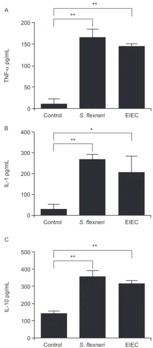
Cytokines expression after J774 infection with EIEC and S. flexneri - We determined the expression levels of IL-1, IL-10 and TNF-α following infection of J774 mac-rophages with EIEC or S. flexneri to evaluate if the cy-tokine expression profile differs based on the infecting bacterial species. We observed a similar increase in IL-1, IL-10 and TNF concentrations measured in the superna-tants of J774 cells infected with EIEC or S. flexneri com-pared to supernatants from uninfected cultures (Fig. 3).
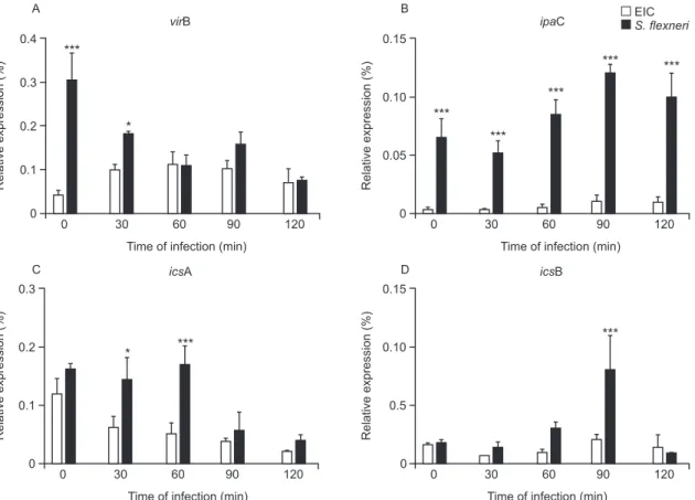
Differential expression of selected virulence genes in EIEC and S. flexneri during J774 cell infection - qRT-PCR was performed to measure virF, virB, ipaB, ipaC, icsA and icsB transcription rates every 30 min during the 1st 2 h of infection. We observed a very low ipaC tran-scription level in EIEC, but not in S. flexneri, throughout the course of the infection (Fig. 4). This was the only statistically significant difference between EIEC and S. flexneri virulence gene expression at all time intervals. Higher transcription levels were also observed for virB
and icsA in S. flexneri duringthe initial 30 min and dur-ing the 30-60 min interval, respectively. Transcription of
icsB was elevated in S. flexneri at 90 min after infection. No significant differences between EIEC and S. flexneri
were found in the transcription levels of virF and ipaB
(data not shown). The expression of EIEC or S. flexneri
virulence genes was also studied when bacteria were cultured in medium without contact with J774 cells and we observed no difference in gene expression between EIEC and S. flexneri (data not shown).
100
75
50
25
0
(%) Cells death
Time of infection (h) *** EIEC
S. flexneri *** ***
0.5 1 1.5 2 4 6 8
Fig. 2: J774 cell death ratios caused by enteroinvasive Escherichia coli (EIEC) (white bar) or Shigella flexneri (black bar) were determined on a FACScalibur flow cytometer. Statistical analysis was performed by two-way analysis of variance. p values of less than 0.05 were consid-ered as statistically significant. Asterisks mean p < 0.001.
200
150
100
50
0
TNF-
pg/mL
Control S. flexneri EIEC **
A **
400
300
200
100
0
IL-1 pg/mL
Control S. flexneri EIEC **
B *
500
400
300
200
100
0
IL-10 pg/mL
Control S. flexneri EIEC **
C
**
Fig. 3: production of TNF (A), IL-1 (B) and IL-10 (C) by J774
mac-rophages infected with enteroinvasive Escherichia coli (EIEC) or
Shigella flexneri at 6 h post-infection: data is presented as mean and standard deviation from quaduplicate infections in two independent experiments. Statistical analysis was performed by two-way analysis of variance. p values of less than 0.05 were considered as statistically significant. *: p < 0.05; **: p < 0.01.
DISCUSSION
Infection with S. flexneri causes rapid macrophage death (Clerc et al. 1987, Nonaka et al. 2003), which con-stitutes an evasion mechanism from the innate immune system (Kubota 2006, Schroeder & Hilbi 2008). There-fore, the first step in the present investigation consisted of determining whether EIEC and S. flexneri survive in and escape from macrophages in a similar way. EIEC was less efficient in macrophage killing during the initial 4 h of infection than S. flexneri (Figs 1, 2) which may indicate differences in the expression of virulence genes.
The virulence gene ipaC is involved in bacterial es-cape from the phagocytic vacuole and is responsible for lysis of the phagosome membrane (Harrington et al. 2006, Schroeder & Hilbi 2008). This gene is also responsible for human epithelial cell invasion by targeting the TTSS, which leads to the efficient injection of effector proteins that improve the invasion process (Jaumouillé et al. 2008). In the present study, we detected a very low expression of ipaC in EIEC throughout the infection compared to S. flexneri (Fig. 4). EIEC shares almost complete identity of the ipaC nucleotide sequence with S. flexneri (Gibotti et al. 2004), suggesting that an EIEC chromosomal factor(s) could modulate ipaC transcription inside macrophages. However, further studies are necessary to investigate this hypothesis. No differences in the expression of the pro-apoptotic gene ipaB were detected between EIEC and S.
0.4
0.3
0.2
0.1
0
Relative expression (%)
0 30 60 90 120 virB
***
*
Time of infection (min) A
0.3
0.2
0.1
0
Relative expression (%)
0 30 60 90 120 icsA
Time of infection (min)
* ***
C
EIC S. flexneri 0.15
0.10
0.05
0
Relative expression (%)
0 30 60 90 120 ipaC
***
Time of infection (min)
***
***
*** ***
B
0.5 0.15
0.10
0
Relative expression (%)
0 30 60 90 120 icsB
Time of infection (min)
***
D
Fig. 4: transcriptional profiles of virulence factor production (vir) B (A), invasion plasmid antigen (ipa) C (B), intracellular movement (ics) A (C) and icsB (D) genes of enteroinvasive Escherichia coli (EIEC) (white bar) or Shigella flexneri (black bar) obtained by quantitative real-time polymerase chain reaction during different periods of infection in J774 macrophages. Results are presented as mean and standard deviation from triplicate experiments. Statistical analysis was performed by two-way analysis of variance. p values of less than 0.05 were considered as statistically significant. *: p < 0.05; **: p < 0.01; ***: p < 0.001.
flexneri. This gene encodes for a protein that activates caspase-1, which initiates macrophage apoptosis and the cleavage of proinflammatory cytokines (Zychlinsky et al. 1994, Schroeder & Hilbi 2008).
Recently, we studied the expression of EIEC and S. flexneri VP genes in a different experimental model, fo-cusing on intestinal cell invasion and dissemination. We demonstrated that the disease triggered by EIEC may be restricted to a definite infection site, meaning that it is not capable of disseminating beyond a certain point to extend and worsen tissue injury as Shigella does. This phenomenon may be associated with the lower level of virulence factor expression in EIEC when compared to
Shigella (Moreno et al. 2009). However, the lower patho-genicity of EIEC could also be related to the host in-flammatory response against these bacteria.
In conclusion, EIEC demonstrates a delayed killing effect in J774 macrophage cultures when compared to S. flexneri. This in vitro behavior was found to be related to the diminished ipaC expression. Therefore, EIEC’s “slow invasion” of colonic epithelial cells in vivo could be relat-ed to the hypoexpression of ipaC, leading to a prolonged presence inside infected cells. All together, these data could explain why EIEC takes longer than Shigella spe-cies to cause diarrhea. We believe that more experiments with host cells should be conducted to better understand the inflammatory response generated by EIEC.
ACKNOWLEDGEMENTS
To Dr Patrícia Tobo, for help with real-time PCR analysis.
REFERENCES
Bryce J, Boschi-Pinto C, Shibuya K, Black RE, WHO Child Health Epidemiology Reference Group 2005. WHO estimates of the causes of death in children. Lancet 365: 1147-1152.
Clerc PL, Ryter A, Mounier J, Sansonetti PJ 1987. Plasmid-mediated early killing of eucaryotic cells by Shigella flexneri as studied by infection of J774 macrophages. Infect Immun 55: 521-527.
Dorman CJ, McKenna S, Beloin C2001. Regulation of virulence gene
expression in Shigella flexneri, a facultative intracellular patho-gen. Int J Med Microbiol 291: 89-96.
DuPont HL, Formal SB, Hornick RB, Snyder MJ, Libonati JP, Shea-han DG, LaBrec EH, Kalas JP 1971. Pathogenesis of Escherichia coli diarrhea. N Engl J Med 285: 1-9.
Gibotti A, Tanaka TL, Oliveira VR, Taddei CR, Martinez MB 2004.
Molecular characterization of enteroinvasive Escherichia coli
ipa genes by PCR-RFLP analysis. Braz J Microbiol 35: 74-80.
Hale TL1998. Bacillary dysentery. In WJ Hansler, M Shuman,
Top-ley and Wilson’s microbiology and microbial infections, Arnold, London, p. 479-493.
Harrington A, Darboe N, Kenjale R, Picking WL, Middaugh CR, Bir-ket S, Picking WD 2006. Characterization of the interaction of single tryptophan containing mutants of IpaC from Shigella flex-neri with phospholipid membranes. Biochemistry 45: 626-636.
Harris JR, Wachsmuth IK, Davis BR, Cohen ML1982.
High-molec-ular-weight plasmid correlates with Escherichia coli enteroinva-siveness. Infect Immun 37: 1295-1298.
Jaumouillé V, Francetic O, Sansonetti PJ, Tran Van Nhieu T 2008. Cytoplasmic targeting of IpaC to the bacterial pole directs polar type III secretion in Shigella. EMBO J 27: 447-457.
Kaper JB, Nataro JP, Mobley HL 2004. Pathogenic Escherichia coli. Nat Rev Microbiol 2: 123-140.
Kubota K 2006. A novel functional T cell hybridoma recognizes mac-rophage cell death induced by bacteria: a possible role for innate lymphocytes in bacterial infection. J Immunol 176: 7576-7588.
Ménard R, Dehio C, Sansonetti PJ 1996. Bacterial entry into epithe-lial cells: the paradigm of Shigella. Trends Microbiol 4: 220-226.
Moreno AC, Ferreira LG, Martinez MB 2009. Enteroinvasive Escher-ichia coli vs. Shigella flexneri: how different patterns of gene ex-pression affect virulence. FEMS Microbiol Lett 301: 156-163.
Moreno AC, Filho AF, Gomes T do A, Ramos ST, Montemor LP, Ta-vares VC, Filho L do S, Irino K, Martinez MB 2010. Etiology of childhood diarrhea in the northeast of Brazil: significant emer-gent diarrheal pathogens. Diagn Microbiol Infect Dis 66: 50-57.
Navarre WW, Zychlinsky A 2000. Pathogen-induced apoptosis of macrophages: a common end for different pathogenic strategies. Cell Microbiol 2: 265-273.
Nonaka T, Kuwabara T, Mimuro H, Kuwae A, Imajoh-Ohmi S 2003. Shigella-induced necrosis and apoptosis of U937 cells and J774 macrophages. Microbiology 149: 2513-2527.
Parsot C, Ageron E, Penno C, Mavris M, Jamoussi K, d’Hauteville H, Sansonetti P, Demers B 2005. A secreted anti-activator, OspD1, and its chaperone, Spa15, are involved in the control of transcrip-tion by the type III secretranscrip-tion apparatus activity in Shigella flex-neri. Mol Microbiol 56: 1627-1635.
Petri WA Jr, Miller M, Binder HJ, Levine MM, Dillingham R, Guer-rant RL 2008. Enteric infections, diarrhea and their impact on function and development. J Clin Invest 118: 1277-1290.
Sansonetti PJ 1991. Genetic and molecular basis of epithelial cell invasion by Shigella species. Rev Infect Dis 13 (Suppl. 4): 285-292.
Sansonetti PJ, Arondel J, Cantey JR, Prévost MC, Huerre M 1996. Infection of rabbit Peyer’s patches by Shigella flexneri: effect of adhesive or invasive bacterial phenotypes on follicle-associated epithelium. Infect Immun 64: 2752-2764.
Sansonetti PJ, Kopecko DJ, Formal SB 1982. Involvement of a
plas-mid in the invasive ability of Shigella flexneri. Infect Immun
35: 852-860.
Sansonetti PJ, Ryter A, Clerc P, Maurelli AT, Mounier J 1986.
Mul-tiplication of Shigella flexneri within HeLa cells: lysis of the
phagocytic vacuole and plasmid-mediated contact hemolysis. Infect Immun 51: 461-469.
Schroeder GN, Hilbi H 2008. Molecular pathogenesis of Shigella spp: controlling host cell signaling, invasion and death by type III se-cretion. Clin Microbiol Rev 21: 134-156.
Silva RM, Toledo MR, Trabulsi LR 1980. Biochemical and cultural
characteristics of invasive Escherichia coli. J Clin Microbiol
11:441-444.
Silva RM, Toledo MR, Trabulsi LR1982. Correlation of invasiveness
with plasmid in enteroinvasive strains of Escherichia coli. J In-fect Dis 146: 706.
Suzuki T, Franchi L, Toma C, Ashida H, Ogawa M, Yoshikawa Y, Mimuro H, Inohara N, Sasakawa C, Nuñez G 2007. Differential regulation of caspase-1 activation, pyroptosis and autophagy
via Ipaf and ASC in Shigella-infected macrophages. PLoS
Pathog 3: e111.
Trabulsi LR, Fernandes MR, Zuliani ME 1967. Novas bactérias patogênicas para o intestino do homem. Rev Inst Med Trop Sao Paulo 9: 31-39.
Venkatesan MM, Goldberg MB, Rose DJ, Grotbeck EJ, Burland V, Blattner FR 2001. Complete DNA sequence and analysis of the
large virulence plasmid of Shigella flexneri. Infect Immun 69:
3271-3285.
Zychlinsky A, Kenny B, Ménard R, Prévost MC, Holland IB, San-sonetti PJ 1994. IpaB mediates macrophage apoptosis induced by
Shigella flexneri. Mol Microbiol 11: 619-627.
Zychlinsky A, Prevost MC, Sansonetti PJ 1992. Shigella flexneri in-duces apoptosis in infected macrophages. Nature 358: 167-168.