A Theoretical Model of the Tridimensional Structure of
Bacillus
thuringiensis
subsp.
medellin
Cry 11Bb Toxin
Deduced by Homology Modelling
Pablo Gutierrez/
++Oscar Alzate*/
+++, Sergio Orduz/
+Unidad de Biotecnología y Control Biológico, Corporación para Investigaciones Biológicas, AA7378, Medellin, Colombia *Universidad Pontificia Bolivariana, Medellin, Colombia
Cry11Bb is an insecticidal crystal protein produced by Bacillus thuringiensis subsp. medellin during its stationary phase; this ∂-endotoxin is active against dipteran insects and has great potential for mos-quito borne disease control. Here, we report the first theoretical model of the tridimensional structure of a Cry11 toxin. The tridimensional structure of the Cry11Bb toxin was obtained by homology modelling on the structures of the Cry1Aa and Cry3Aa toxins. In this work we give a brief description of our model and hypothesize the residues of the Cry11Bb toxin that could be important in receptor recognition and pore formation. This model will serve as a starting point for the design of mutagenesis experiments aimed to the improvement of toxicity, and to provide a new tool for the elucidation of the mechanism of action of these mosquitocidal proteins.
Key words: homology modelling - ∂-endotoxin structure - Cry proteins - Bacillus thuringiensis
Bacillus thuringiensis is a Gram-positive
en-dospore forming bacterium characterized by the production of parasporal crystalline proteic inclu-sions (which contain ∂-endotoxins) during the sta-tionary phase. ∂-endotoxins are highly toxic and specific to insects of the Coleopteran, Lepidopteran and Dipteran orders (Schnepf et al. 1998), and once in the insect midgut, they are activated by gut pro-teases, followed by binding to specific receptors on the cells lining the larval midgut (Hofmann et al. 1988). This interaction promotes their insertion into the membrane, forming ion selective channels by oligomerization of toxin monomers (Gazit & Shai 1993, Aronson et al. 1999), and the insect dies by loss of osmotic pressure regulation (Knowles & Ellar 1987).
∂-endotoxins, also known as Cry proteins, are classified according to their degree of evolution-ary divergence into 22 groups (Crickmore et al.
This work received financial support from Colciencias, and Corporación para Investigaciones Biológicas, Medellin, Colombia.
+Corresponding author. Fax: +(574)-441-5514. E-mail:
++Current address: Department of Biochemistry, McGill
University, Montreal, Canada
+++Current address: Biochemistry Department, The Ohio
State University, 484 W 12th Ave., OH 43210, USA Received 15 May 2000
Accepted 25 August 2000
1998). The Cry11 family of ∂-endotoxins is com-prised of dipteran-active proteins where the Cry11Aa protein of B. thuringiensis subsp. israelensis has been the most intensively studied.
At present, there is interest in identifying new dipteran-active toxins for their importance in mos-quito and black fly control (Orduz et al. 1992, Ragni et al. 1996). Mosquitocidal activity has also been found in Cry1Ab, Cry1Ca, Cry2Aa, Cry11Ba, Cry11Aa, Cry16Aa, Cry19Ba, Cyt1Aa, Cyt1Ab, Cyt2Aa and some Cry-related proteins (Cry 17Aa, Cry18Aa and Cry 19Aa) produced by Clostridium bifermentans subsp. malaysia (Schnepf et al. 1998).
impor-tant for insect specifity and protein stability (Bosch et al. 1994, Masson et al. 1994, Lee et al. 1999).
B. thuringiensis subsp. medellin is a potentially
important strain for mosquito control (Orduz et al. 1992, Ragni et al. 1996, Thiéry et al. 1998). So-dium dodecyl sulfate polyacrylamide gel electro-phoresis (SDS-PAGE) analysis of the B. tharingiensis subsp. medellin parasporal inclusion
indicates that this strain produces a polypeptide of 94 kDa, multiple bands between 80 and 65 kDa, and two doublets at 40-41 and 28-30 kDa (Orduz et al. 1994). The 94 kDa protein (Cry11Bb) has been cloned into Escherichia coli and B. thuringiensis and
was shown to be responsible for most of the mosquitocidal activity. The mode of action of Cry11 ∂-endotoxins is still not clear, and though a series of studies have been made (Dai & Gill 1993, Feldmann et al. 1995, Orduz et al. 1998, Thiéry et al. 1998),
no structural information is still available.
In this work, we propose a model for the struc-ture of the Cry11Bb ∂-endotoxin based on the hy-potheses of structural similarity with Cry1Aa and Cry3Aa toxins. This model provides a starting point for the design of mutagenesis experiments aimed to elucidate the mechanism of action of the Cry11 family of toxins.
MATERIALS AND METHODS
Sequence alignment between Cry11Bb (Genbank accession AAC97162, Orduz et al. 1998), Cry1Aa and Cry3Aa (PDB entries 1CIY and 1DLC respectively), was generated using the struc-tural alignment tool of the program Swiss-PdbViewer (Guex & Peitsch 1997) and corrected manually until a satisfactory placement of con-served blocks and aminoacid identities was ob-tained. This alignment was submitted to Swiss-Model in the expasy server (http://www.expasy.ch/ spdbv/) and a preliminary model for Cry11Bb was retrieved. Loops and side chains conformations were recalculated with the OPLS force field with-out distance restrains of the Hyperchem program (Hypercube, Inc.) and the most severe steric over-laps removed. The model was validated with PROCHECK (Laskowski et al. 1993) and WHAT IF (Vriend 1990) programs by submitting the co-ordinates to the EMBL server (http://www.embl-heidelberg.de/). Sequence identities were calcu-lated with Needleman and Wunsch maximum matching algorithm of the MacDNASIS program (Hitachi, Software). Figures, electrostatic poten-tials, and Ca RMSD calculations were generated with SwissPdbViewer (Guex & Peitsch 1997).
RESULTS
Based on the structural alignment of the amionoacid sequence of the Cry11Bb toxin with
Cry1Aa and Cry3Aa toxins (Fig. 1) a theoretical model of the Cry11Bb toxin was obtained, and cor-responds to residues 15-620 of the primary struc-ture (Fig. 2). Alignment of domain I was straight-forward and the highly conserved nature of helix 5 in the Cry11Bb toxin made placement of the other residues in this domain reliable. According to ge-netic studies and the structural features observed in Cry1Aa and Cry3Aa toxins, large insertions and deletions seem unlikely to be present here. Align-ment of domain II was less reliable and had to be corrected manually. The limits of this domain could be determined because of the conserved nature of flanking domains I and III. Placements of large in-sertions or deletions were mostly done in regions corresponding to the loops connecting β sheets in-volved in receptor recognition, therefore the most variable region of the molecule, and their lengths were determined by selecting the combination that allowed the best conservative profile of the neigh-boring aminoacids. Domain III is quite well con-served on the N-terminal side, but on the C-termi-nal side, there was no clear correspondence of aminoacids (only one identity present at Ile114) making difficult to define the end of the molecule.
Structural comparison of the Cry1Aa, Cry3Aa toxins with the theoretical model of the Cry11Bb protein indicates correspondence to the general model for a Cry protein, and the few differences found were located in the loops of domains II and III (Fig. 3). The superimposed backbone traces of Cry1Aa and Cry3Aa displayed 0.66 and 0.83 Å RMS deviations for Cα (Fig. 4). The Rama-chandran plot (data not shown) indicated that most (95%) of residues have φ and ψ angles in the core and allowed regions, except for some proline and glycine residues and few residues located in the loop regions. Most bond lengths, bond angles, and torsion angles were in the range of values expected for a naturally folded protein (data not shown). The structural model shown in Fig. 2 indicates that it contains all the general features of the Cry toxins (an α+β structure with three domains). Four of the five Cys residues present in the primary structure were included in the Cry11Bb model (Cys157, Cys186, Cys225 and Cys436), but it seems that they are not involved in structure stabilization through disulfide bonds, since the shortest S-S dis-tance (Cys157-Cys225) between the six different possible bonds was 18.51 Å.
DISCUSSION
Domain I - Domain I is composed of residues
(Phe117-Pro138); α5 (Ser151-Ile168); α6 (Pro178-Arg209); α7a (Leu215-Phe230); α7b (Glu234-Tyr241); ß0 (Ala34-Ala36) and ß1a (Thr249-Leu252). All the helices in the Cry11Bb model were slightly shorter than those in Cry1Aa and Cry3Aa, and less amphiphilic (Table I). Accord-ing to the amphiphilicity calculated with the Hoops and Woods values, the most exposed helices are α1, α2a, α2b, α3 and α6, which corresponds well with the accessibility calculated with SwissPDB, except for α1, which is packed against domain II. It is possible that this helix will have some mobil-ity, if we take into consideration that one of the cutting sites by gut proteases is located between Ser56 and Ile58, close to the middle of this helix (Segura et al. 2000). The charge distribution pat-tern in the Cry11Bb theoretical model corresponds to a negatively charged patch along ß4 and ß13 of domains II and III respectively.
The Cry11Bb domain I model correlates well with data from Gazzit et al. (1998) who suggested that α4 and α5 insert into the membrane in an an-tiparallel manner as an helical hairpin. It is pos-sible that according to the surface electrostatic po-Cry11Bb 15 SSFPLFNSKI EPSIAPALIA VAPIAKYLAT ALAKWALKQG FAKLKSEIFP GNETATMEKV RLEVQTILNQ Cry3Aa 61 TTKDVIQKGI SVVGDLLGVV GFPFGGALVS FYTNFLNTIW -PSE-DPWKA FMEQVEALMD QKIADYAKNK Cry1Aa 33 YTPIDISL SLTQFLLSEF VPGAG--FVL GLVDI---IW GIFGPSQWDA FLVQIEQLIN QRIEEFARNQ . . . . . . . . . * Cry11Bb 85 TLQTDRVATL KAEYEGFIHL GKVFTDYVSQ STFTPATAKT HFLNMSNLLI QRLPQFEIAG YEGVSISLFT Cry3Aa 129 ALAELQGLQN NVEDYVSALS SWQKNPVSSR NPHSQGRIRE LFSQAESHFR NSMPSFAISG YEVLFLTTYA Cry1Aa 96 AISRLEGLSN LYQIYAESFR EWEAD--PTN P-ALREEMRI QFNDMNSALT TAIPLLAVQN YQVPLLSVYV .. . . . . * . . .* . . *. .. . Cry11Bb 155 QMCTLHLGLL KDGILAGSDW GFTPEDKDSL ICQFNRYVNE YNTRMMGLYS IEFGRLLAKN LNEALNFRNM Cry3Aa 199 QAANTHLFLL KDAQIYGEEW GYEKEDIAEF YKRQLKLTQE YTDHCVKWYN VGLDKLRGSS YESWVNFNRY Cry1Aa 164 QAANLHLSVL RDVSVFGQRW GFDAATINSR YNDLTRLIGN YTDYAVRWYN TGLERVWGPD SRDWVRYNQF * ** .* .* . * * *. . . * . *. . .. . . . Cry11Bb 225 CSLYVFPFSE AWYLLRYEGT KLENTLSLWN FVGEDIGGIL HNDWKGALYK LLMGATNQRL ANVRFNYSYF Cry3Aa 269 RREMTLTVLD LIALFPLYDV RLYPKEVKTE LTRDVLTDPI VG--VNNLRG YGTTFSNIEN YIRKPHLFDY Cry1Aa 234 RRELTLTVLD IVALFSNYDS RRYPIRTVSQ LTREIYT-NP VLENFDGSF- -RGMAQRIEQ NIRQPHLMDI . . *. . . . . . D2 Cry11Bb 295 SDTQGTIHRE NILGAHPTYN GEQTPTGWIG NGRLGRFSAP YSNELEITKV EQEITYNNKG DHSNSIVPAN Cry3Aa 337 LHRIQFHTRF QPG---YY GNDSFNYWSG NYVSTRPSIG -SND-IITSP FYGN--- ---KSSEPV-Cry1Aa 300 LNSITIYTDV HR--- ---GFNYWSG HQITASPVGF SGPE--FAFP LFGN--- ---AGNAAPP . * * . . . .. . . Cry11Bb 365 TRNEILTATV PITADPFFKT ADIN--- ---WR YFSQGLYYGW NIKFDDRVIL NSRVPGGIPS Cry3Aa 390 QNLEFN-GEK VYRAVANTNL AVWP--- SAVYSGVTKV EFSQYNDQTD EASTQTYDSK RNVGAVSWDS Cry1Aa 348 VLVSLT-GLG IFRTLSSPLY RRIILGSGPN NQELFVLDGT EFSFASLT-T NLPSTIYRQ- ----RGTVDS . . . . ** . . * Cry11Bb 420 NRLEYDGYYI RAVSACPRNV PLSYNHNYLT LTYNRLEYDA PTTQNIIVGF SPNNTKSFYA RNSHYLSATN Cry3Aa 453 IDQLP-PETT DEPLEKGYSH QLNYVMCFLM QGS---RGTI PVLTWTHKSV D----FFNMI DSKKITQLPL Cry1Aa 411 LDVIP-PQDN SVPPRAGFSH RLSHVTMLSQ AAGAVYTLRA PTFSWQHRSA E----FNNII PSSQITQIPL . *. . * . D3 .
Cry11Bb 491 DAYVIPALQF ATVSDRSFLE DTPDQATDGS IKFTETVLGN ---EAKYSIR LNTGFNTATR YRLVIRFKAT Cry3Aa 515 VKAYKLQSGA SVVAGPRFTG GDIIQCTENG SAATIYVTPD VSYSQKYRAR IHYASTSQIT FTLSLDGAPF Cry1Aa 476 TKSTNLGSGT SVVKGPGFTG GDILRRTSPG QISTLRVNIT APLSQRYRVR IRYASTTNLQ FHTSIDGRPI . * * . * . * * . * * .. . . . . . Cry11Bb 558 ARLAAGIRVR SQNSGNNRLL GGIPVEGNSG WVDYITDSFT FNDLGITTAS TNAFFSIDSD GVN Cry3Aa 585 NQYYFDKTIN K–GDTLTYNS FNLASFSTPF ELSG--NNLQ IGVTGLSAGD KVYIDKIEFI PVN Cry1Aa 546 NQGNFSATMS S–GSNLQSGS FRTVGFTTPF NFSNGSSVFT LSAHVFNSGN EVYIDRIEFV PAEVT . . . . . . . . .... . *. .
Fig. 1: sequence and structural alignment of Cry1Aa, Cry3Aa and Cry11Bb. * denotes identical residues, the point indicates conserved residues, D2 and D3, the initiation of domains 2 and 3 respectively.
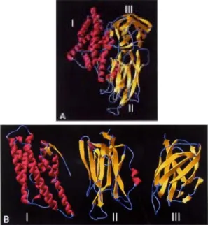
Fig. 2: homology model deduced for the tridimensional struc-ture of the Cry11Bb toxin. A: domain I is mostly alpha helical (fuchsia), specificity domain II is located in the middle and domain III on the right side; B: from left to right domains I, II and III
A
tential of helices 4 and 5 (Fig. 5), there was a neu-tral region in the middle of the helices which prob-ably indicates, if the umbrella model is correct, that both helices cross the membrane with their polar sides exposed to the solvent, as it has been sug-gested by the results of mutagenesis experiments done by Kumar and Aronson (1999) with the Cry1Ac toxin. This region is also the most con-served among the Cry toxins.
Kumar and Aronson (1999) demonstrated that mutations in the base of helix 3 and the loop be-tween α3 and α4 that cause alterations on the
bal-ance of negative charged residues can cause loss of toxicity by Cry1Ac. Mutations in helices α2, α6 and the surface residues of α3 have no impor-tant effect on toxicity; meanwhile, helices α4 and α5 seem to be very sensitive to mutations. Helix α1 probably does not play an important part in toxin activity after cleavage of the protoxin. It is pos-sible that mutations aimed to an increase in amphilicity in these helices will improve the pore forming activity of Cry11 type of toxins.
Domain II - This is the most variable domain
among Cry toxins, and it has been shown that it is
involved in receptor recognition and therefore con-sidered as the specificity determining region. As for other Cry toxins, domain II of the Cry11Bb toxin consists of three greek key beta sheets ar-ranged in a beta prism topology. It is comprised by residues 257-478, one helix (α8, Ala279-Ala285) and 11 ß-strands (ß2 Ser292-Asn305, ß3 Pro319-Ser332, ß4 Ile341-Lys343, ß5 Thr365-Iso369, ß6 Val374-Phe381, ß7 Trp389-Leu396, ß8 Asn401-Arg407, ß9 Ile418-420, ß10 Pro437-Thr450, ß11 Tyr458-Val468 and ß12 Phe470-Lys476). The main difference between the Cry11Bb theoretical model and Cry1Aa and Cry3Aa structures is the length of the two loops joining the apical ß-strands (ß2-ß3 and ß4-ß5) lo-cated between L307-Y313 and I348-N358. We propose that these residues are implicated in recep-tor binding, and consequently in specificity of the Cry11Bb toxin. The first insertion between strands ß2 and ß3, the N-terminal part of this loop (Ile306-Thr311) is mostly hydrophobic, while the C-termi-nal half (Thr312-Thr318) is polar and have one posi-tively charged residue (Glu316). This loop probably interacts with the receptor through both hydropho-bic and electrostatic interactions; Gly315 probably helps in receptor binding by providing more mobil-ity to Glu316 that may interact through salt bridges with the receptor. Loop ß4-ß5 is mostly hydrophilic, and the charged residues located at the tip of the loop (Lys353, Asp355 and His356) are probably im-portant determinants of insect specificity. As in loop ß2-ß3, a glycine residue (354) is also present before a negatively charged residue (Asp355) supporting the hypothesis that correct orientation of charged residues in the specificity loops could be important in receptor recognition. The third insertion (Ser471-Lys476) probably does not play an important role in insect recognition as it is located in the region joining domains II and III, but it probably gives more mobility to domain III modulating its function in receptor binding.
Mutations in defined regions of the Cry1Aa toxin have identified residues 365-371 (equivalent to residues in the Cry11 Bbß6-ß7 loop), as essen-tial for binding to the membrane of midgut cells of
Bombyx mori (Ge et al. 1989, Lu et al. 1994). In
the Cry11Bb model, this region is shorter than their counterparts in Cry1Aa and Cry3Aa. Loop ß2-ß3 seems also to be able to modulate the toxicity and specificity of Cry1C (Smith & Ellar 1994), in this region it was found a five residue insertion (Gly308-Thr312). The dual specificity of Cry2Aa for Lepidoptera and Diptera has been mapped to residues 307-382 that corresponds in the Cry11Bb theoretical model to sheet 1, strand ß6, and loop ß6-ß7, where most of the insertions/deletions and structural differences were located.
Fig. 4: superposition of the αC carbon traces of Cry1Aa (blue), Cry3Aa (green) and Cry11Bb (red) toxins of Bacillus thuringiensis. The main differences in the three toxins are lo-cated in loops of domain II, which is implilo-cated in receptor recognition and binding.
Domain III - This domain showed high
con-servation of residues and the only important modi-fication is a 3-residue deletion between ß16 and ß17. The ß-strands in this domain are ß13a (Tyr485-Asn490), ß3b(Ile495-Ala497), ß14 (Ala501-Val503), ß15 (Pro513-Ala516), ß16 (Ser520-Gly529), ß17 (Lys533-Asn543), ß18 (Thr546-Arg553), ß19 (Lys555-Ala562), ß20 (Gly579-Glu583), ß21 (Ile592-Leu601), ß22 (Thr608-Val619). We propose the sequence AKYSIRLNTGF as homolog to conserved block 4. In the case of conserved block 5 there was only one identity and two conserved residues (Ile614, Phe611 and Asp615, respectively). Several stud-ies indicate that site mutations in this block reduce toxicity and alter channel properties in Cry1Ac (Lee et al. 1999) and Cry1Aa (Chen et al. 1993, Schwartz et al. 1997), divergence in block 5 possi-bly reveals an alternative mechanism of membrane permeabilization.
In conclusion, evidences presented here, based on the identification of structural equivalent resi-dues of Cry1Aa and Cry3Aa in Cry11Bb toxin through homology modelling indicate that, despite the low aminoacid homology among these three toxins, they share a common tridimensional struc-ture. Cry1Aa, Cry3Aa, and Cry11Bb contain the most variable regions in the loops of domain II, which determine the specificity of these toxins. This is the first model of a Cry11 protein and its impor-tance can be perceived since members of this group of toxins are potentially important candidates for mosquito control programs. The coordinates of the Cry11Bb model can be obtained upon request to [email protected].
REFERENCES
Aronson A, Geng C, Wu L 1999. Aggregation of Bacil-lus thuringiensis Cry1A toxin upon binding to
tar-get insect larval midgut vesicles. Appl Environm Microbiol 65: 2503-2507.
Bosch D, Schipper B, Van der Kleij H, de Maagd RA, Stiekema WJ 1994. Recombinant Bacillus thu-ringiensis crystal proteins with new properties:
pos-sibilities for resistant management. Biotechnol-ogy 12: 915-919.
Chen XJ, Lee MK, Dean DH 1993. Site-directed muta-tions in a highly conserved region of Bacillus thurngiensis∂-endotoxin affect inhibition of short
circuit current across Bombyx mori midguts. Proc Natl Acad Sci USA 90: 9041-9045.
Crickmore N, Zeigler DR, Feitelson J, Schnepf E, Van Rie J, Lereclus D, Baum J, Dean DH 1998. Revi-sion of the nomenclature for the Bacillus thuringiensis pesticidal crystal proteins. Microbiol Mol Biol Rev 62: 807-813.
Dai S-M, Gill S 1993. In vitro and in vivo proteolysis of
the Bacillus thuringiensis subsp. israelensis CryIVD
protein by Culex quinquefasciatus larval midgut
proteases. Insect Biochem Molec Biol23: 273-283.
Feldmann F, Dullemans A, Waalwijk C 1995. Binding of the CryIVD toxin of Bacillus thuringiensis to
lar-val dipteran midgut proteins. Appl Environ Microbiol 61: 2601-2605.
Gazit E, Shai Y 1993. Structural and functional organi-zation of the α5 segment of Bacillusthuringiensis
endotoxins. Biochemistry 32: 3429-3436.
Gazit E, La Rocca P, Sansom MSP, Shai Y 1998. The structure and organization within the membrane of the helices composing the pore forming domain of
Bacillus thuringiensis∂-endotoxin are consistent
with an “umbrella-like” structure of the pore. Proc Natl Acad Sci USA 95: 12289-12294.
Ge AZ, Sivarova ND, Dean DH 1989. Location of the
Bombyx mori specificity domain of Bacillus thuringiensis ∂-endotoxin protein. Proc Natl Acad Sci USA 86: 4037-4041.
Gill SS, Cowless EA, Pietrantonio PV 1992. The mode of action of Bacillus thuringiensis endotoxins. Annu Rev Entomol 37: 615-636.
Grochulski P, Masson L, Borisova S, Pusztai-Carey M, Schwartz JL, Brousseau R, Cygler M 1995. Bacil-lus thuringiensis CryIA(a) insecticidal toxin:
crys-tal structure and channel formation. J Mol Biol 254:
447-464. TABLE
Length (Å) and hydrophobic moments of helices in domain I of Cry1Aa, Cry3Aa and Cry11Bb toxins of
Bacillus thuringiensis
Toxin α1 α2a α2b α3 α4 α5 α6 α7a
Cry11Bb 20.21a 20.69 17.40 43.10 33.64 26.27 46.75 22.71
(0.37)b (0.55) (0.20) (0.25) (0.04) (0.58) (0.48) (0.14)
Cry1Aa 18.23 12.22 14.99 40.08 34.53 19.75 46.45 15.32
(0.25) (0.62) (0.67) (0.63) (0.460 (0.51) (0.78) (0.51)
Cry3A 20.80 20.62 20.12 43.21 34.93 26.89 47.06 22.75
(0.96) (0.62) (1.26) (0.53) (0.58) (0.39) (1.05) (0.70)
Guex N, Peitsch MC 1997. SWISS-MODEL and the Swiss-PdbViewer: an environment for comparative protein modeling. Electrophoresis 18: 2714-2723.
Hofmann C, Vanderbruggen H, Hofte H, Van Mellaert H 1988. Specificity of Bacillus thuringiensis
delta-endotoxins is correlated with the presence of high affinity binding sites in the brushborder membrane of the insect membrane of target insect midguts. Proc Natl Acad Sci USA 85: 7844-7848.
Knowles BH 1994. Mechanism of action of Bacillus thuringiensis insecticidal ∂-endotoxins. Advan In-sect Physiol 24: 275-308.
Knowles BH, Ellar DJ 1987. Colloid osmotic lysis is a general feature of the mechanism of action of Bacil-lus thuringiensis delta-endotoxins with different
in-sect specificities. Biochim Biophys Acta 924: 509-518.
Kumar ASM, Aronson AI 1999. Analysis of mutations in the pore-forming region essential for insecticidal activity of a Bacillus thuringiensis∂-endotoxin. J Bacteriol 181: 6103-6107.
Laskowski RA, MacArthur MW, Moss DS, Thornton JM 1993. PROCHECK: a program to check the ste-reochemical quality of protein structures. J Appl Cryst 26: 283-291.
Lee MK, You TH, Gould FL, Dean DH 1999. Identifi-cation of residues in domain III of Bacillus thuringiensis Cry1Ac toxin that affect binding and
toxicity. Appl Environ Microbiol 65: 4513-4520.
Li J, Carroll J, Ellar DJ 1991. Crystal structure of insec-ticidal ∂-endotoxin from Bacillus thuringiensis at 2.5
A resolution. Nature 353: 815-821.
Lu H, Rajamohan F, Dean DH 1994. Identification of amino acid residues of Bacillus thuringiensis∂
-en-dotoxin CryIA(a) associated with membrane bind-ing and toxicity to Bombyx mori. J Bacteriol 176:
5554-5559.
Masson L, Mazza A, Gringorten JL, Baines D, Anelunias V, Brousseau R 1994. Specificity domain localiza-tion of Bacillus thuringiensis insecticidal CryIA
tox-ins is highly dependent on the bioassay system. Mol Microbiol 14: 851-860.
Orduz S, Diaz T, Thiéry I, Charles J-F, Rojas W 1994. Crystal proteins from Bacillus thuringiensis
subspe-cies medellin. Appl Microbiol Biotechnol 40:
794-799.
Orduz S, Realpe M, Arango R, Murillo LA, Delecluse A 1998. Sequence of the cry11Bb11 gene from Ba-cillus thuringiensis subsp. medellin and toxicity
analysis of its encoded protein. Biochim Biophys Acta 1388: 267-272.
Orduz S, Rojas W, Correa MM, Montoya AE, de Barjac H 1992. A new serotype of Bacillus thuringiensis
from Colombia toxic to mosquito larvae. J Invertebr Pathol 59: 99-103.
Ragni A, Thiery I, Delecluse A 1996. Characterization of six highly mosquitocidal Bacillus thuringiensis
strains that do not belong to H-14 serotype. Curr Microbiol 32: 48-54.
Schnepf E, Crickmore N, van Rie J, Lereclus D, Baum J, Feitelson J, Zeigler DR, Dean DH 1998. Bacillus thuringiensis and its pesticidal crystal proteins. Microbiol Mol Biol Rev 62: 772-806.
Schnepf E, Tomczak K, Ortega JP, Whiteley HR 1990. Specificity-determining regions of lepidopteran-spe-cific insecticidal proteins produced by Bacillus thuringiensis. J Biol Chem 265: 20923-20930.
Schwartz JL, Potvin L, Chen XJ, Brousseau R, Laprade R, Dean DH 1997. Single-site mutations in the con-served alternating-arginine region affect ion chan-nels formed by CryIAa, a Bacillus thuringiensis
toxin. Appl Environ Microbiol 63: 3978-3984.
Segura C, Guzman F, Patarroyo ME, Orduz S 2000. Activation pattern and toxicity of the Cry11Bb toxin of Bacillus thuringiensis subsp. medellin. J Invertebr Pathol 76: 56-62.
Smith GP, Ellar DJ 1994. Mutagenesis of two surface exposed loops of the Bacillus thuringiensis CryIC
∂-endotoxin affects insecticidal specificity. Biochem J302: 611-616.
Thiery I, Hamon S, Delecluse A, Orduz S 1998. The introduction into Bacillus sphaericus of the Bacil-lus thuringiensis subsp. medellin Cyt1Ab1 gene
re-sults in higher susceptibility of resistant mosquito larva population to B. sphaericus.Appl Environ Microbiol 64: 3910-3916.
Vriend G 1990. WHAT IF: a molecular modeling and drug design program. J Mol Graph 8: 52-56.
von Tersch MA, Slatin SL, Kuleszca CA, English LH 1994. Membrane-permeabilizing activities of Bacil-lus thuringiensis coleopteran-active toxin CryI-IIB2
and CryIIIB2 domain I peptide. Appl Environ Microbiol 60: 3711-3717.