Original article
in high-grade cervical intraepithelial neoplasia in women from
a densely populated Brazilian urban region
Classiicação ilogenética dos genótipos de papilomavírus humano em neoplasia intraepitelial
cervical de alto grau em mulheres de uma região urbana brasileira densamente povoada
Denise Rocha Pitta
1, Luis Otávio Sarian
2, Elisabete Aparecida Campos
1, Sílvia Helena Rabelo-Santos
3,
Kari Syrjänen
4, Sophie Françoise Derchain
5Department of Obstetrics and Gynecology, Universidade Estadual de Campinas (Unicamp), Campinas, São Paulo, Brazil
1MSc. Biologist, Department of Obstetrics and Gynecology, Universidade Estadual de Campinas (Unicamp), Campinas, São Paulo, Brazil. 2MD, PhD. Assistant professor, Department of Obstetrics and Gynecology, Universidade Estadual de Campinas (Unicamp), Campinas, São Paulo, Brazil. 3PhD. Pharmacist and assistant professor, School of Pharmacy, Universidade Federal de Goiás (UFG), Goiânia, Goiás, Brazil.
4MD, PhD, FIAC. Associate professor, Department of Oncology and Radiotherapy, Turku University Hospital, Turku, Finland
5MD, PhD. Associate professor, Department of Obstetrics and Gynecology, Universidade Estadual de Campinas (Unicamp), Campinas, São Paulo, Brazil.
ABSTRACT
CONTEXT AND OBJECTIVE: Differences in human papillomavirus (HPV) types may correlate with the biological potential and invasion risk of high-grade cervical intraepithelial neoplasia (CIN 2 and CIN 3). The objective of this study was to determine the relationship between different combinations of HPV types and CIN severity.
DESIGN AND SETTING: Cross-sectional study, at Universidade Estadual de Campinas (Unicamp).
METHODS: Cervical samples from 106 women treated due to CIN 2 (18) or CIN 3 (88) were examined for speciic HPV genotypes using Roche Linear Array® (LA-HPV). The proportions of CIN 2 and CIN 3 in groups of women infected with the HPV phylogenetic groups A7 and A9 were compared. Three groups were formed: women with single infections; multiple infections; and the whole sample.
RESULTS: Multiple infections were detected in 68 samples (64.7%). The most frequent high-risk genotypes detected (single/multiple) were HPV 16 (57.1%), HPV 58 (24.7%), HPV 33 (15.2%), HPV 52 (13.3%), HPV 31 (10.4%), HPV 51 (7.6%) and HPV 18 (6.6%). Women without infection with HPV species Alpha 9 were less likely to have CIN 3 than were their Alpha 9 HPV-infected counterparts. HPV 16 and/or HPV 18, with or without associations with other viral types, were more frequently found in women with CIN 3 than in those with CIN 2.
CONCLUSIONS: The severity of high-grade CIN may be aggravated by the presence of HPV types included in the Alpha 9 phylogenetic classiication and by infections including HPV 16 and 18, singly or in combination with other HPV genotypes.
RESUMO
CONTEXTO E OBJETIVO: Diferentes tipos de papilomavírus humano (human papillomavirus, HPV) podem ser correlacionados com a capacidade biológica e risco de invasão das neoplasias intra-epitelial de alto grau cervical (NIC 2 e NIC 3). O objetivo deste estudo foi determinar a relação de diferentes tipos de HPV com a gravidade da NIC.
TIPO DE ESTUDO E LOCAL: Estudo transversal na Universidade Estadual de Campinas (Unicamp).
MÉTODOS: Foram avaliados os genótipos especíicos de HPV da amostra cervical de 106 mulheres com NIC 2 (18) ou NIC 3 (88), utilizando Roche Linear Array® (LA) HPV genotyping assay. Foram comparadas as proporções de NIC 2 e NIC 3 em grupos de mulheres infectadas com tipos de HPV dos grupos ilogenéticos A7 e A9. Três grupos foram formados: mulheres com infecção simples; infecção múltipla; e infecção simples e múltipla.
RESULTADOS: Infecções múltiplas foram detectadas em 68 (64,7%) das amostras. Os genótipos de alto risco mais frequentemente detectados em infecção simples ou múltipla foram HPV 16 (57,1%), HPV 58 (24,7%), HPV 33 (15,2%), HPV 52 (13,3%), HPV 31 (10,4%), HPV 51 (7,6%) e HPV 18 (6,6%). A probabilidade de mulheres com NIC 3 serem infectadas com HPV que não da espécie Alfa 9 era menor do que com os tipos de HPV da espécie Alfa 9. HPV 16 e ou 18, associado ou não com outros tipos virais eram mais frequentemente encontrados nas mulheres com NIC 3 do que naquelas com NIC 2.
CONCLUSÃO: A gravidade da NIC de alto grau pode ser aumentada pela presença de tipos de HPV incluídos na classiicação ilogenética Alfa 9 e por infecções que incluem HPV 16 e 18 combinados ou não com outros genótipos de HPV.
KEY WORDS:
Human papillomavirus 11. Human papillomavirus 16. Human papillomavirus 18. Human papillomavirus 6. Genotype.
Cervical intraepithelial neoplasia. Cancer.
Polymerase chain reaction.
PALAVRAS-CHAVE: Papillomavirus 11 humano. Papillomavirus 16 humano. Papillomavirus 18 humano. Papillomavirus 6 humano. Genótipo.
Neoplasia intra-epitelial cervical. Câncer.
INTRODUCTION
Persistent infection with high-risk types of human papillomavirus (HPV) is known to be a unifying risk factor for the development of cervi-cal intraepithelial neoplasia (CIN) and invasive carcinoma.1-4 CIN is
clas-siied into three grades, based on progressive spreading of atypical cells from the proliferative layers to the full thickness of the epithelium.5
Al-though CIN 2 and CIN 3 represent high-grade lesions of the cervix, they are heterogeneous in their potential for progression to invasive cancer.6
Current evidence indicates that diferences in HPV types may cor-relate with the biological potential and invasion risk of CIN lesions.2,7,8
Recent data show that the HPV types found in CIN 2 are diferent from those observed in CIN 3, and CIN 2 frequently contains HPV types that are not commonly found as single types in invasive cancers.9
his could suggest that invasive cancer is unlikely to be the end point for such CIN 2 lesions.9,10
Genital HPV types are classiied as Alpha papillomavirus genera. he species within these genera are closely related in terms of phylo-genesis. Despite having distinct genomic sequences, they show identi-cal or very similar biologiidenti-cal or pathologiidenti-cal properties. Along with the type species HPV 16, species 9 also includes HPV types 31, 33, 35, 52, 58 and 67. Along with the type species HPV 18, species 7 also includes HPV types 39, 45, 59, 68, 70 and 85.11
Controversy exists regarding possible competition or synergy of in-dividual types in multiple infections. Some natural history studies have suggested that women who are already infected present greater risk of acquiring new HPV types than do those who are HPV-negative.12,13 An
alternative interpretation of these indings might be that more than one HPV type is transmitted simultaneously, and their sequential detection could be a consequence of replicated life cycles that are asynchronous and only occasionally overlap.14 hese life cycles might be
interdepen-dent.15 Multiple HPV types may be associated with a risk of
progres-sion that exceeds the risk due to single-type infections. In a recent study, there was evidence that this risk increased with the cumulative number of HPV types, in most combinations, and these associations seemed particularly strong over the short term.16
OBJECTIVE
In this report, our prime focus was to address the question of het-erogeneity of HPV types as a possible source of biological variation in CIN 2 and CIN 3. To add new information to the growing body of lit-erature in women with these lesions, we aimed to evaluate the distribu-tion of single and multiple infecdistribu-tions of diferent HPV types in women with high-grade cervical intraepithelial neoplasia (CIN 2 and CIN 3), and to compare the prevalence of diferent HPV types in CIN 2 and CIN 3, in view of the phylogenetic classiication of the virus.
METHODS
Type of study and setting
he patient sample for this cross-sectional study comprised 106 non-consecutive women who underwent large loop excision of the
transformation zone (LLETZ) to treat CIN 2 or 3, between February 2001 and April 2004.
he study was carried out at the colposcopy clinics of Universidade Estadual de Campinas (Unicamp), Brazil, a public health institution dedicated to comprehensive care for women.
Sample
he sample size for this study was estimated according to the re-lationship n = (p*q)/E2, where n is the sample size, p is the estimated
prevalence of the condition in afected women (in the present case, the prevalence of HPV in high-grade CIN is as high as 95%16), and q
is the prevalence of the condition in non-afected subjects. he overall prevalence of highly oncogenic types of HPV in an urban female pop-ulation (mean age: 33 years) without high-grade CIN has been esti-mated as 6.2%.16E is the standard error, set to 2.5% for the present
cal-culations. herefore, we estimated a sample size of roughly 94 women. he women in this study were selected after referral to Unicamp for specialized treatment for high-grade squamous intraepithelial lesions (HSIL). hey were invited to enter the study at the time of their pre-treatment visit (i.e. their irst visit after being accepted for pre-treatment). One of the researchers was always present at these visits and, after ex-plaining the study protocol and ethical concerns, made the invitation. All the patients who agreed to be enrolled in the study gave their writ-ten signed consent.
At the pretreatment visit, all of the women were interviewed to ob-tain clinical, social and demographic data. A complete gynecological ex-amination was performed, with collection of endocervical specimens for HPV testing, followed by colposcopic examination of the cervix. he de-cision to perform diathermal conization was based on the referral cytolo-gy and the clinical/colposcopic coniguration of the cervix. he study was approved by the local Ethics Committee (Protocol #CEP 309/2004).
Procedures
Histology
he histological samples consisted of 106 diathermal conization specimens. his material was ixed in 10% phosphate bufered formalin and then was embedded in parain and stained with hematoxylin and eosin. he samples were analyzed according to the World Health Orga-nization’s criteria.17 In the present series, only cases diagnosed as CIN 2
(n = 18) or CIN 3 (n = 88) were included. he patients’ mean age was 34.08 years (90% central range: 17.5 to 73.6 years). he ages of the women with CIN 2 (mean age: 32.01 years; 90% central range: 17.4 to 59.5 years) and CIN 3 (mean age: 34.05 years; 90% central range: 17.3 to 73.6 years) were similar (P = 0.32).
Main measurements
HPV detection
DNA extraction
HPV genotype
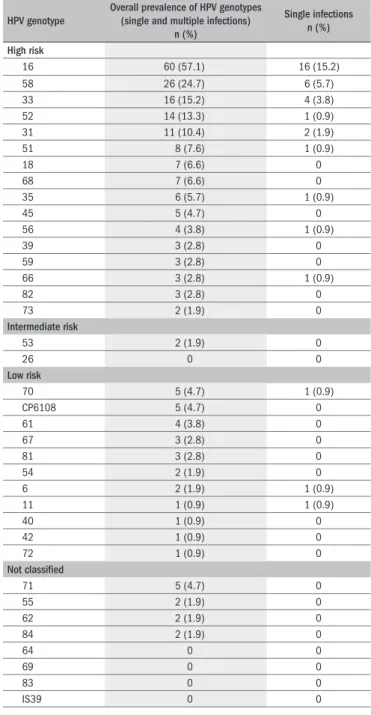
Overall prevalence of HPV genotypes (single and multiple infections)
n (%)
Single infections n (%)
High risk
16 60 (57.1) 16 (15.2)
58 26 (24.7) 6 (5.7)
33 16 (15.2) 4 (3.8)
52 14 (13.3) 1 (0.9)
31 11 (10.4) 2 (1.9)
51 8 (7.6) 1 (0.9)
18 7 (6.6) 0
68 7 (6.6) 0
35 6 (5.7) 1 (0.9)
45 5 (4.7) 0
56 4 (3.8) 1 (0.9)
39 3 (2.8) 0
59 3 (2.8) 0
66 3 (2.8) 1 (0.9)
82 3 (2.8) 0
73 2 (1.9) 0
Intermediate risk
53 2 (1.9) 0
26 0 0
Low risk
70 5 (4.7) 1 (0.9)
CP6108 5 (4.7) 0
61 4 (3.8) 0
67 3 (2.8) 0
81 3 (2.8) 0
54 2 (1.9) 0
6 2 (1.9) 1 (0.9)
11 1 (0.9) 1 (0.9)
40 1 (0.9) 0
42 1 (0.9) 0
72 1 (0.9) 0
Not classiied
71 5 (4.7) 0
55 2 (1.9) 0
62 2 (1.9) 0
84 2 (1.9) 0
64 0 0
69 0 0
83 0 0
IS39 0 0
Table 1. Distribution of human papillomavirus (HPV) genotypes in single
and multiple infections in Brazilian women
cells in the pellet were re-suspended in 200 μl of digestion solution (Tris 1 mM, 200 μg/ml of proteinase K and 0.5% sodium dodecyl sulfate, SDS). his suspension was shaken and incubated at 55 °C for two hours and at 95 °C for ive minutes. Next, 200 μl of solution phenol/chloroform/iso-amyl alcohol (25:24:1) was added and shaken vigorously before centrifu-gation at 5,000 g for 10 minutes. he aqueous phase was removed and transferred to a clean tube, and 1/10 (10%) of NaAc (sodium acetate) 3M pH 5.2 was added and mixed. Next, 2.5 volumes of 70% ice-cold etha-nol was added and shaken. he solution was centrifuged at 15,000 g for 15 minutes and the supernatant was removed. After the pellet of deoxy-ribonucleic acid (DNA) had dried, it was dissolved in 100 μl of Tris-eth-ylenediaminetetraacetic acid (TE-EDTA) solution (Tris 1 mM, EDTA 100 μM, pH 8.2). he nucleic acids were stored at -20 °C until use.
Roche Linear Array®
he Roche Linear Array® (LA) HPV genotyping assay (Roche Mo-lecular Systems, Alameda, California, United States) is based on poly-merase chain reaction (PCR) ampliication of the target DNA using HPV primers (PYGM09/11), hybridization of the ampliied product using oligonucleotide probes and then their detection using a colori-metric reaction. he master mix contains primers for ampliication of a 450-base pair (bp) fragment of the L1 region of more than 37 HPV genotypes18 and a 268-bp fragment of the human β-globin gene, as an
internal control.
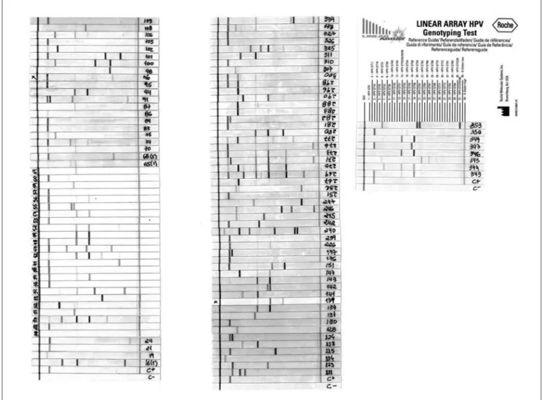
Detection and genotype determination were performed using the de-natured ampliied DNA and an array of oligonucleotide probes located in the polymorphic region of L1. his enabled independent identiica-tion of 37 individual HPV genotypes (Figure 1), as follows: 16 high-risk HPV types (16, 18, 31, 33, 35, 39, 45, 51, 52, 56, 58, 59, 66, 68, 73 and 82), 11 low-risk HPV types (6,11, 40, 42, 54, 61, 70, 72, 81, CP6108 and 67), two intermediate-risk HPV types (26 and 53) and eight HPV types of undetermined risk (55, 62, 64, 69, 71, 83, 84 and IS 39).
Statistical methods
he data were tabulated in OpenOice spreadsheets and analyzed in the R environment for statistical analyses. he proportions of CIN 2 and CIN 3 in groups of women infected with various A7 and A9 phylo-genetic HPV groups were compared using Fisher’s exact test. hree situ-ations were considered: women with single infections; multiple infec-tions; and the whole sample. A linear regression model was then itted to assess the proportions of CIN 2 and CIN 3 in groups of women with diferent single or multiple combinations of infecting HPV types.
RESULTS
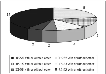
he distribution of HPV types as single and multiple infections is shown in Table 1. Of the 37 HPV types included in LA-HPV, 32 were detected in the samples analyzed. HPV 16 was the most prevalent gen-otype (present in 57.1% of the samples), either as a single infection or co-infecting the cervix together with other HPV types. It was the only infecting type in 15.2% of the samples. Other frequent genotypes, in ei-ther multiple or single infections, were HPV 58 (26/105; 24.7%), HPV 33 (16/105; 15.2%), HPV 52 (14/105; 13.3%), HPV 31 (11/105; 10.4%) and HPV 51 (8/105; 7.6%), which are all high-risk HPV types. HPV 18 appeared in only 6.6% of the lesions, and it was not a single agent in any of them. Only 3/105 (2.87%) of the women had infections composed exclusively of low and intermediate-risk HPV types (as either single or multiple types). he precise composition of the various mul-tiple HPV infections is shown in Figure 1.
Figure 1. Linear array strips for all cases, aligned with the reference card.
16/52 with or without other types (n = 8), HPV 16/18 with or without other types (n = 5), HPV 16/33 with or without other types (n = 4) and HPV 33/58 and HPV 35/52 with or without other types (n = 2, each).
he distribution of HPV types in accordance with their phyloge-netic species (genus Alpha, species 7: HPV 18, 39, 45, 59, 68, 70 and 85; and genus Alpha, species 9: HPV 16, 31, 33, 35, 52, 58, and 67) in CIN 2 and CIN 3 lesions is shown in Table 2. he majority of the women had an HPV type of species Alpha 9 and others, while very few had Alpha 7 and others (5/105). here were 22 women (22/105) with both species (Alpha 9 and Alpha 7). Proportionally, women not infect-ed with HPV species Alpha 9 were less likely to have CIN 3, comparinfect-ed with their Alpha 9 HPV-infected counterparts (P = 0.01). his tendency was also found when analyzing the subset of women with multiple in-fections only (P = 0.001). In women infected with a single HPV type, the proportion of CIN 3/CIN 2 was the same, regardless of the HPV species present in their cervix (P = 0.20).
he overall detection of HPV by LA was 105/106 women (99%); for CIN 2 it was 100% and for CIN 3 it was 99%. he HPV 16 and 18 single infections, HPV 16/18 co-infections and HPV single or mul-tiple infections without HPV 16 and 18 are summarized in Table 3.
Infections including HPV 16 and 18 singly or in association with oth-er HPV genotypes woth-ere signiicantly more common in CIN 3 than in CIN 2 (P = 0.01).
DISCUSSION
In this study using the Roche LA-HPV genotyping assay, HPV was detected in 99% of CIN 2 and CIN 3 samples. Similar results were re-ported by Castle et al.19 and Gargiulo et al.20 LA-HPV is a commercial
HPV test, manufactured following good manufacturing practices with standardized reagents.19 In this assay, the same amplicon can be directly
used for both detection of β globin and 37 diferent HPV genotypes in a single reaction. In the present series, this method was easy to use and highly accurate for detecting the majority of clinically signiicant HPV genotypes.
Unlike DNA sequencing, LA-HPV is capable of identifying multi-ple infections.21,22 In the present series, 64.7% of the samples had
mul-tiple HPV genotypes. his high prevalence of mulmul-tiple infections is evi-dent in most studies using LA-HPV, ranging from 49.7% to 79.0%.20-22
Species in sample
Single n (%) Multiple n (%) Total n (%) CIN 2 CIN 3 CIN 2 CIN 3 CIN 2 CIN 3
A9 and others (except A7)
1 (20) 29 (88) 8 (61) 34 (62) 9 (50) 63 (72)
A7 and others (except A9)
2 (40) 0 1 (8) 2 (4) 3 (17) 2 (2)
A9 and A7 0 0 3 (23) 19 (34) 3 (17) 19 (22) Only others 2 (40) 4* (12) 1 (8) 0 3 (17) 4 (4)
Total 5 (100) 33 (100) 13 (100) 55 (100) 18 (100) 88 (100)
P = 0.20 P = 0.001 P = 0.01
Table 2. Distribution of human papillomavirus (HPV) genus A, species 7
and 9 genotypes in cervical intraepithelial neoplasia (CIN 2 and CIN 3) among Brazilian women
A9 including 16, 31, 33, 35, 52, 58 and 67; A7 including 18, 39, 45, 59, 68, 70 and 85 *One CIN 3 HPV was HPV negative according to the Linear Array®
Comparing CIN 2 and CIN 3 and infection including A7 or A9; P = 0.01
HPV Infection
Histological grade CIN 2
n (%)
CIN 3 n (%)
Multiple infections including HPV 16 and/or 18 6 (33) 40 (45) Single infections including HPV 16 and/or 18 0 16 (19) Multiple infections including neither HPV 16 nor 18 7 (39) 15 (17)
Single infections including neither HPV 16 nor 18 5 (27) 16 (19)
Total 18 (100) 87 (100)
One CIN 3 HPV was HPV negative according to the Linear Array® (not included in the table)
Table 3. Occurrence of human papillomavirus (HPV 16 and HPV 18) in
cervical intraepithelial neoplasia (CIN 2 and CIN 3) in Brazilian women
Figure 2. Most prevalent associations of human papillomavirus (HPV)
types in multiple infections.
8
5
4 2
2 14
16-58 with or without other
16-18 with or without other
33-58 with or without other
16-52 with or without other
16-33 with or without other
35-52 with or without other
containing multiple genotypes, because of competition for reagents dur-ing ampliication and discrimination of the types ampliied by PCR. he correlation between the numbers of additional types detected with LA-HPV suggests that less competition during ampliication was en-countered with LA-HPV.23
One clear advantage of full HPV genotyping is the ability to increase the performance of screening programs using HPV, while identifying the HPV-positive women who have persistent oncogenic HPV. here is some recent evidence that the risk of developing the lesion increases with the cumulative number of HPV types, and these associations seemed particu-larly strong during the irst year of follow-up.16 hus, detection of
short-term HPV persistence may increase the speciicity of the screening based on HPV. If full genotyping is introduced into the screening, the question of how often women who repeatedly test positive for oncogenic HPV (with or without HPV 16 and HPV 18) present persistent oncogenic HPV infection can be resolved.24 However, it should be emphasized that
current guidelines on cervical screening suggest that HPV testing should be used at intervals no shorter than three years, which ofsets the applica-bility of HPV genotyping, in the form described above.
In this series of Brazilian women, the overall prevalence of HPV 16 was 57.1%, although HPV 16 as a single type was present only in 15.2% of the cases. he associations most frequently found were of HPV 16 to-gether with HPV 58, 52, 18 and 33. In a study very similar to the pres-ent one, in terms of design, Prétet et al.25 examined a sample of CIN 2
and 3 and found that HPV 16 was the most frequent type. However, in contrast to the present indings, that type was the only agent in 40.4% of the samples. In a recent meta-analysis, Smith et al.8 found that the
prevalence of HPV 16 (both single and co-infections) in high-grade le-sions collected from all around the world was 45.3%.
In our study, the HPV 18 genotype appears in seventh position in terms of prevalence, occurring in 6.6% of the samples. Invariably, HPV 18 was associated with other HPV genotypes. his is very similar to the report by Prétet et al.,25 with HPV 18 prevalence of 4.3% (6.6% of 121
CIN 2 lesions and 3.5% of 372 CIN 3 lesions) and only three CIN 3 le-sions presented HPV 18 as the single type of infection. In the report by Smith et al.,8 HPV 18 appears in ifth position with an overall prevalence
of 6.9%. here are some minor diferences between this meta-analysis8
and our results, regarding the order of prevalence of the most common HPV genotypes: 16, 31, 33, 58, 18, 52, 35 and 51 in the meta-analysis; and 16, 58, 33, 52, 31, 51, 18 and 68 in the present series. However, these authors stated that there was an important lack of South American data in their meta-analysis, and our present study adds new information concerning the HPV type distribution in high-grade lesions among Bra-zilian women.
Comparing CIN 2 and CIN 3, HPV 16 and 18 were signiicantly more prevalent in CIN 3 lesions. his is consistent with the data of Pré-tet et al.,25 who analyzed 121 CIN 2 and 372 CIN 3 lesions in France,
and also found higher prevalence of HPV 16 in CIN 3 (64.2%) than in CIN 2 lesions (56.2%). his diference was even more marked in a study by Zuna et al.,9 who reported HPV 16 infection in 65.6% of CIN
3 and only 19.0% in CIN 2 lesions. Despite the fact that in all report-ed series, CIN 2 lesions representreport-ed a minority of cases, these data sug-gest that CIN 2 and 3 might difer in their biological potential, and that HPV genotypes might interfere with the risk of progression in these two categories of CIN lesions.
the Alpha 9 species, possibly has a greater oncogenic potential than HPV 18 and 45.7 In another epidemiological study, Wheeler et al.26 observed
a higher risk of CIN 3 among women with HPV 31 (Alpha 9) and rela-tively low oncogenic potential for HPV 56 and 59 (Alpha 7).
he present study provides new information on the distribution of individual HPV genotypes and multiple infections, obtained using the Roche LA-HPV assay. hese data suggest that a prophylactic vaccine against HPV 16/18 has the potential to prevent approximately half of the high-grade lesions. For one of the two candidate vaccines, Harper et al.27 stated that this proportion might even be increased, if
cross-pro-tection were achieved against some other high-risk HPV types, e.g. total protection against HPV 45, partial protection against HPV 31 and no protection against HPV 33, 52 and 58. However, the full implications of this observation are diicult to deine, because HPV 45 was under-represented in our cohort, in the same way as in the recent meta-analy-sis.8 hese data on HPV genotype distribution among diferent
popula-tions are of crucial importance for designing second-generation prophy-lactic HPV vaccines in the near future. Also of relevance, future thera-peutic HPV vaccines would have greater potential beneit if their design for treating HPV infections were not restricted to HPV 16 and 18. he best hypothetical scenario is one in which vaccine constituents are cho-sen taking into consideration the epidemiological distribution of HPV types in the speciic geographical regions where they are to be used.
CONCLUSIONS
he present series irmly corroborates the assumption that, in most CIN 2 and CIN 3 cases, multiple high-oncogenic HPV types may be found. In our series, the prevalence of HPV 58 and HPV 33 was unex-pectedly high. CIN 3 was typically associated with HPV genus Alpha, species 9, most often consisting of types 16 and 58, alone or in combi-nation with other HPV types.
REFERENCES
1. Walboomers JM, Jacobs MV, Manos MM, et al. Human papillomavirus is a necessary cause of invasive cervical cancer worldwide. J Pathol. 1999;189(1):12-9.
2. Clifford GM, Smith JS, Aguado T, Franceschi S. Comparison of HPV type distribution in hi-gh-grade cervical lesions and cervical cancer: a meta-analysis. Br J Cancer. 2003;89(1): 101-5.
3. Muñoz N, Bosch FX, de Sanjosé S, et al. Epidemiologic classiication of human papillomavi-rus types associated with cervical cancer. N Engl J Med. 2003;348(6):518-27. 4. Moscicki AB, Schiffman M, Kjaer S, Villa LL. Chapter 5: Updating the natural history of HPV
and anogenital cancer. Vaccine. 2006;24 Suppl 3:S3/S42-51.
5. Baak JP, Kruse AJ, Robboy SJ, Janssen EA, van Diermen B, Skaland I. Dynamic behavioural interpretation of cervical intraepithelial neoplasia with molecular biomarkers. J Clin Pathol. 2006;59(10):1017-28.
6. Syrjänen KJ, Syrjänen SM. Papillomavirus infections in human pathology. England: John Wiley & Sons Ltd; 2000.
7. Snijders PJ, Steenbergen RD, Heideman DA, Meijer CJ. HPV-mediated cervical carcinogene-sis: concepts and clinical implications. J Pathol. 2006;208(2):152-64.
8. Smith JS, Lindsay L, Hoots B, et al. Human papillomavirus type distribution in invasive cervical cancer and high-grade cervical lesions: a meta-analysis update. Int J Cancer. 2007;121(3):621-32.
9. Zuna RE, Allen RA, Moore WE, Lu Y, Mattu R, Dunn ST. Distribution of HPV genotypes in 282 women with cervical lesions: evidence for three categories of intraepithelial lesions based on morphology and HPV type. Mod Pathol. 2007;20(2):167-74.
10. Zuna RE, Allen RA, Moore WE, Mattu R, Dunn ST. Comparison of human papillomavirus genotypes in high-grade squamous intraepithelial lesions and invasive cervical
carci-noma: evidence or differences in biologic potential of precursor lesions. Mod Pathol. 2004;17(11):1314-22.
11. de Villiers EM, Fauquet C, Broker TR, Bernard HU, zur Hausen H. Classiication of papilloma-viruses. Virology. 2004;324(1):17-27.
12. Liaw KL, Hildesheim A, Burk RD, et al. A prospective study of human papillomavirus (HPV) type 16 DNA detection by polymerase chain reaction and its association with acquisition and persistence of other HPV types. J Infec Dis. 2001;183(1):8-15.
13. Rousseau MC, Pereira JS, Prado JC, Villa LL, Rohan TE, Franco EL. Cervical coinfection with human papillomavirus (HPV) types as a predictor of acquisition and persistence of HPV infection. J Infect Dis. 2001;184(12):1508-17.
14. Weissenborn SJ, Funke AM, Hellmich M, et al. Oncogenic human papillomavirus DNA loa-ds in human immunodeiciency virus-positive women with high-grade cervical lesions are strongly elevated. J Clin Microbiol. 2003;41(6):2763-7.
15. Woodman CB, Collins SI, Young LS. The natural history of cervical HPV infection: unresolved issues. Nat Rev Cancer. 2007;7(1):11-22.
16. Trottier H, Mahmud S, Costa MC, et al. Human papillomavirus infections with multiple types and risk of cervical neoplasia. Cancer Epidemiol Biomarkers Prev. 2006;15(7):1274-80. 17. Scully RE, Boniglio TA, Kurman RJ, Silverberg SG, Wilkinson EJ. Histological typing of
fema-le genital tract tumors. (International histological classiication of tumors). 2nd ed. Berlin:
Springer-Verlag; 1994.
18. Gravitt PE, Peyton CL, Alessi TQ, et al. Improved ampliication of genital human papillomavi-ruses. J Clin Microbiol. 2000;38(1):357-61.
19. Castle PE, Sadorra M, Garcia F, Holladay EB, Kornegay J. Pilot study of a commercialized human papillomavirus (HPV) genotyping assay: comparison of HPV risk group to cytology and histology. J Clin Microbiol. 2006;44(11):3915-7.
20. Gargiulo F, De Francesco MA, Schreiber C, et al. Prevalence and distribution of single and multiple HPV infections in cytologically abnormal cervical samples from Italian women. Virus Res. 2007;125(2):176-82.
21. Giuliani L, Coletti A, Syrjänen K, Favalli C, Ciotti M. Comparison of DNA sequencing and Roche Linear array in human papillomavirus (HPV) genotyping. Anticancer Res. 2006;26(5B):3939-41.
22. Woo YL, Damay I, Stanley M, Crawford R, Sterling J. The use of HPV Linear Array Assay for multiple HPV typing on archival frozen tissue and DNA specimens. J Virol Methods. 2007;142(1-2):226-30.
23. Coutlée F, Rouleau D, Petignat P, et al. Enhanced detection and typing of human papillo-mavirus (HPV) DNA in anogenital samples with PGMY primers and the Linear array HPV genotyping test. J Clin Microbiol. 2006;44(6):1998-2006.
24. Meijer CJ, Snijders PJ, Castle PE. Clinical utility of HPV genotyping. Gynecol Oncol. 2006;103(1):12-7.
25. Prétet JL, Jacquard AC, Carcopino X, et al. Human papillomavirus genotype distribution in high grade cervical lesions (CIN 2/3) in France: EDITH study. Int J Cancer. 2008;122(2):424-7. 26. Wheeler CM, Hunt WC, Schiffman M, Castle PE; Atypical Squamous Cells of Undetermined
Signiicance/Low-Grade Squamous Intraepithelial Lesions Triage Study Group. Human pa-pillomavirus genotypes and the cumulative 2-year risk of cervical precancer. J Infect Dis. 2006;194(9):1291-9.
27. Harper DM, Franco EL, Wheeler CM, et al. Sustained eficacy up to 4.5 years of a bivalent L1 virus-like particle vaccine against human papillomavirus types 16 and 18: follow-up from a randomised control trial. Lancet. 2006;367(9518):1247-55.
Acknowledgements: The authors acknowledge the Research Support Foundation of the State of São Paulo (Fundação de Amparo à Pesquisa do Estado de São Paulo, Fapesp) for Grant 04/09309-5. S.D is a researcher accredited by the National Council for Scientiic and Technological Development (Conselho Nacional de Desenvolvimento Cientíico e Tecnológico, CNPq)
Sources of funding: Fundação de Amparo à Pesquisa do Estado de São Paulo (Fapesp), grant number 04/09309-5
Conlict of interest: Not declared
Date of irst submission: February 22, 2008
Last received: June 23, 2009
Accepted: June 24, 2009
Address for correspondence:
Sophie Françoise Derchain Rua Dr. Antonio Hossri, 629 Campinas (SP) — Brasil CEP 13083-370