INTRODUCTION
Article/Artigo
1. Programa de Pós-graduação em Doenças Infecciosas e Medicina Tropical, Universidade Federal de Minas Gerais, Belo Horizonte, MG. 2. Hospital das Clínicas, Universidade Federal de Minas Gerais, Belo Horizonte, MG. 3. ECOCENTER, Hospital Geral Socor, Belo Horizonte, MG.
Address to: Dra. Marselha Marques Barral. Avenida Barão do Rio Branco 3865/1401, Bom Pastor, 36100-000 Juiz de Fora, MG, Brasil.
Phone: 55 32 3232-6103 e-mail: [email protected] Received in 06/02/2011 Accepted in 09/08/2011
Echocardiographic parameters associated with pulmonary congestion in
outpatients with Chagas’ cardiomyopathy and non-chagasic cardiomyopathy
Parâmetros ecocardiográicos associados com a congestão pulmonar nas miocardiopatias
chagásica e não-chagásica
Marselha Marques Barral
1, Maria do Carmo Pereira Nunes
1,2,3, Marcia Melo Barbosa
3, Cid Sérgio Ferreira
2,
Wilson Campos Tavares Júnior
2and Manoel Otávio da Costa Rocha
1,2ABSTaCT
Introduction: Despite signiicant let ventricular (LV) systolic dysfunction and cardiomegaly, pulmonary congestion does not seem to be a major inding in Chagas’ cardiomyopathy (CC). his study sought to identify echocardiographic parameters associated with pulmonary congestion in CC and in dilated cardiomyopathy of other etiologies, such as non-CC (NCC), and to compare pulmonary venous hypertension between the two entities. Methods: A total of 130 consecutive patients with CC and NCC, with similar echocardiographic characteristics, were assessed using Doppler echocardiography and chest radiography. Pulmonary venous vessel abnormalities were graded using a previously described pulmonary congestion score, and this score was compared with Doppler echocardiographic parameters. Results: NCC patients were older than CC patients (62.4 ± 13.5 × 47.8 ± 11.2, p = 0.00), and there were more male subjects in the CC group (66.2% × 58.5%, p = 0.4). Pulmonary venous hypertension was present in 41 patients in the CC group (63.1%) and in 63 (96.9%) in the NCC group (p = 0.0), the mean lung congestion score being 3.2 ± 2.3 and 5.9 ± 2.6 (p = 0.0), respectively. On linear regression multivariate analysis, the E/e’ ratio (β = 0.13; p = 0.0), LV diastolic diameter (β = 0.06; p = 0.06), let atrial diameter (β = 0.51; p = 0.08), and right ventricular (RV) end-diastolic diameter (β = 0.02; p = 0.48) were the variables that correlated with pulmonary congestion in both groups. Conclusions: Pulmonary congestion was less signiicant in patients with CC. he degree of LV of systolic and diastolic dysfunction and the RV diameter correlated with pulmonary congestion in both groups. he E/e’ ratio was the hallmark of pulmonary congestion in both groups.
Keywords: Chagas’ cardiomyopathy. Pulmonary congestion. Dilated cardiomyopathy. Echocardiography.
RESUMO
Introdução: Na miocardiopatia chagásica, ocorre uma discrepância entre os achados de disfunção ventricular e uma menor magnitude de congestão pulmonar em relação a outras miocardiopatias. Foram associados parâmetros morfofuncionais ecocardiográicos com achados de congestão pulmonar à radiograia do tórax em pacientes portadores de miocardiopatia chagásica e não chagásica, sendo a intensidade dos achados radiológicos comparada nos dois grupos. Métodos: Foram recrutados 130 pacientes portadores de miocardiopatia chagásica e não chagásica, tendo os dois grupos parâmetros ecocardiográicos semelhantes. Todos realizaram o estudo radiológico do tórax, sendo atribuída uma pontuação aos achados sugestivos de congestão pulmonar, conforme escore já previamente estabelecido, sendo este comparado com os achados ecocardiográicos de disfunção ventricular. Resultados: Os pacientes não chagásicos eram mais idosos (62,4±13,5 x 47,8±11,2, p=0,0), havendo um predomínio do sexo feminino nos chagásicos (66,2% x 58,5%, p=0,4). A hipertensão venocapilar pulmonar esteve presente em 41 chagásicos (63,1%) e 63 (96,9%) não-chagásicos (p=0,0), com escore da congestão pulmonar de 3,2±2,3 e 5,9±2,6 (p=0,0) respectivamente. Na análise de regressão linear, a relação E/e’ (β=0,13; p=0,0), o diâmetro diastólico do ventrículo esquerdo (β=0,06; p=0,06), o diâmetro do átrio esquerdo (β=0,51; p=0,08) e o diâmetro diastólico inal do ventrículo direito (β=0,02; p=0,48) foram as variáveis que mais se associaram com a congestão pulmonar nos dois grupos. Conclusões: Os pacientes chagásicos apresentaram um menor grau de congestão pulmonar. Os parâmetros de disfunção sistólica e diastólica associaram com a intensidade da congestão pulmonar, sendo a relação E/e’ a variável que mais determinou a congestão pulmonar nos dois grupos.
Palavras-chaves: Miocardiopatia chagásica. Congestão pulmonar. Miocardiopatia dilatada. Ecocardiograia.
A century ater its description by the Brazilian Carlos Chagas, Chagas’ disease continuesto be a serious health and economic problem in several Latin American countries1-3. Recent estimates
from the World Health Organizationindicate
that 12 million persons are chronically infected with Trypanosoma cruzi, with 100,000 new cases occurring each year4.
Chest radiography is an important and inexpensive tool for the evaluation of patients with Chagas’ dilated cardiomyopathy (CC). he inding of an enlarged heart by this method indicates a poor prognosis5. However, the intensity of pulmonary
congestion is usually less than expected for the degree of cardiomegaly and of let ventricular (LV) systolic dysfunction6. he mechanism underlying
this discrepancy remains unknown7.
Doppler echocardiography represents one of the most important methods for the evaluation of CC8-9. In a previous study, we compared the degree
of pulmonary congestion with echocardiographic parameters in CC. In that study, the degree of pulmonary congestion correlated with Doppler echocardiographic left and right ventricular dysfunction parameters10. Thus, the objective of
the present study was to verify if CC presents with less pulmonary congestion when compared with non-CC (NCC) with the same echocardiographic characteristics.
METHODS
Sixty-ive consecutive patients with CC and 65 patients with NCC were prospectively enrolled.
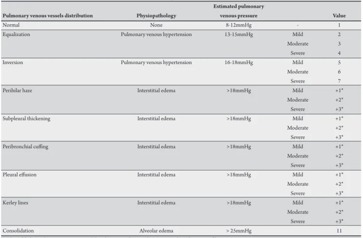
TABLE 1 -Pulmonary venous vessels on chest X-ray of patients with Chagas and non-chagasic dilated cardiomyopathy. Estimated pulmonary
Pulmonary venous vessels distribution Physiopathology venous pressure Value
Normal None 8-12mmHg - 1
Equalization Pulmonary venous hypertension 13-15mmHg Mild 2
Moderate 3
Severe 4
Inversion Pulmonary venous hypertension 16-18mmHg Mild 5
Moderate 6
Severe 7
Perihilar haze Interstitial edema >18mmHg Mild +1*
Moderate +2*
Severe +3*
Subpleural thickening Interstitial edema >18mmHg Mild +1*
Moderate +2*
Severe +3*
Peribronchial cuing Interstitial edema >18mmHg Mild +1*
Moderate +2*
Severe +3*
Pleural efusion Interstitial edema >18mmHg Mild +1*
Moderate +2*
Severe +3*
Kerley lines Interstitial edema >18mmHg Mild +1*
Moderate +2*
Severe +3*
Consolidation Alveolar edema > 25mmHg 11
*Value added to pulmonary venous vessel distribution grading on the corresponding chest X-ray ilm.
(ischemic, hypertensive, idiopathic, and peripartum) and who were negative for Chagas’ disease. Dilated cardiomyopathy found in both Chagas and non-Chagas’ disease is characterized by the echocardiographic inding of a dilated let ventricle with impaired ventricular systolic function.
Patients were selected if they had a let ventricular diastolic diameter/body surface area ≥31mm and a let ventricular ejection fraction <55%11. All patients underwent clinical examination,
and their New York Heart Association (NYHA) functional class was established. Medical therapy was individually adjusted, according to a standardized treatment regimen, and all patients received optimized treatment for heart failure. Patients who had associated heart diseases or systemic arterial hypertension were not included in the Chagas’ group. Patients with renal failure, low serum albumin levels, hypothyroidism, pregnancy, alcoholism, anemia, or other conditions leading to pulmonary congestion were excluded from both study groups. Chest radiographs were obtained at the same time Doppler echocardiograms were performed in all patients. Radiographs were obtained in the posteroanterior and lateral positions. All ilms were acquired using the conventional technique of the Radiology Department of the University Teaching Hospital12. All radiological parameters were graded, the intensity
of changes in the pulmonary venous vessels on chest radiographs being established (Table 1) with the use of a previously described pulmonary congestion score (PCS)13-14. Two independent observers
simultaneously evaluated all chest radiographs, any discordance being solved by consensus. he interobserver variability was analyzed.
Echocardiography
Images were acquired using a Sonos 5,500 echocardiographer (Hewlet-Packard Corporation, Palo Alto, CA), with 2.5-MHz to 3.5-MHz transducers. All recordings were performed by one investigator blind to the clinical evaluation of the patients. he echocardiographic techniques and calculations of cardiac chamber dimensions and volumes were performed according to the recommendations of the American Society of Echocardiography. Let ventricular ejection fraction was calculated according to the modiied Simpson’s rule11.
Right ventricular (RV) morphology and function were evaluated qualitatively on multiple echocardiographic views11. Quantitative
evaluation of global RV function was performed with the use of the Doppler-derived index of myocardial performance (Tei index), as previously described15.
he presence and degree of mitral and tricuspid regurgitation were evaluated using pulsed and continuous wave Doppler, guided by color low mapping16. Maximal velocity of the tricuspid regurgitation
low was obtained, and systolic pulmonary pressure was calculated17.
Diastolic function was assessed using pulsed-wave Doppler examination of mitral, pulmonary venous inflow and Doppler tissue imaging (S, e’, and A’ waves)18-19. he ratio between peak
early diastolic transmitral low velocity (E) and e’ was calculated. Color Doppler M-mode also was used to assess ventricular diastolic function19. According to these Doppler parameters,
RESULTS
TABLE 2 - Clinical characteristics and pharmacologic therapy in CG and NCG.
Characteristics CG NCG p
Age 47.8 ± 11.2 62.43 ± 13.5 0.0
Body surface (m²) 1.7 ± 0.1 1.8 ± 0.2 0.7
Systolic blood pressure (mmHg) 117.6 ± 12.8 127.8 ± 19.6 0.1 Diastolic blood pressure (mmHg) 74.1 ± 9.4 77.8 ± 13.3 0.1 Heart rate (bpm) 62.7 ± 11.0 78.0 ± 11.7 0.0
ACE inhibitors (%) 67.7 83.1 0.0
Digital 13.8 26.2 0.1
Amiodarone 50.8 6.2 0.0
Anticoagulants 4.6 47.7 0.0
Spironolactone 41.5 18.5 0.0
Nitrate 0 56.9 0.0
Carvedilol 4.6 18.5 0.0
Furosemide 32.3 66.2 0.0
NYHA Class I 49.2 67.7
NYHA Class II 43.1 23.1
NYHA Class III e IV 7.7 9.2
Gender male 66.2 58.5
Gender female 33.8 48.5
CG: Chagas’ cardiomyopathy; NCG: non-Chagas’ dilated cardiomyopathy; ACE: angiotensin-converting enzyme; NYHA: New York Heart Association.
TABLE 3 -Pulmonary congestion score.
CG NCG
Characteristics absolute relative absolute relative PCS
Normal 24 36.9 2 3.1 1
Mild equalization 16 24.6 17 26.2 2
Moderate equalization 10 15.4 13 20.0 3
Severe equalization 3 4.6 6 9.2 4
Mild inversion 6 9.2 18 27.7 5
Moderate inversion 6 9.2 8 12.6 6
Severe inversion 0 0.0 1 1.5 7
Perihilar haze 1 1.5 36 5.4 1
Mild subpleural thickening 7 7.7 9 13.8 1 Moderate subpleural thickening 5 4.0 1 1.5 2
Pleural efusion 1 1.5 16 24.6 1
CG: Chagas’ cardiomyopathy; NCG: non-Chagas’ dilated cardiomyopathy, PCS: pulmonary congestion score.
Statistical analysis
Data were expressed as the mean value ± standard deviation for continuous variables and absolute or relative frequencies for categorical variables. The interobserver variability on chest radiograph interpretation was assessed with the Kappa coeicient, a value close to 1 being considered satisfactory.
Correlations between PCS and Doppler echocardiographic variables were assessed using linear regression analysis.
Multiple linear regression analysis was performed to obtain the best regression model, with the PCS as the dependent variable and the echocardiographic parameters as independent variables. he permanence of all variables in the inal model was established when the signiicance level was <0.05. he 13th version of the SPSS
(SPSS Inc., Chicago, Illinois) was used for all analyses.
Ethical considerations
The research protocol was approved by the research ethics commitee of both institutions, and a writen informed consent was obtained from all patients.
A total of 65 patients with CC and 65 patients with NCC were enrolled. Among the NCC patients, 35 (53.8%) had ischemic cardiomyopathy, 20 (30.8%) were hypertensive, 8 (12.3%) were hypertensive and had ischemic cardiomyopathy, 1 (1.5%) had peripartum cardiomyopathy, and 1 (1.5%) had idiopathic cardiomyopathy.
heir clinical data are shown on Table 2. Gender and functional class did not difer between the two groups (p = 0.4 and p = 0.6, respectively) (Table 2). Age and heart rate difered between the two groups, but the diference did not inluence the degree of pulmonary congestion (p = 0.3 for age and p = 0.1 for heart rate). Age diferences
between the two groups probably occurred because CC usually manifests at a younger age, compared with other cardiomyopathies.
here was a signiicant association between the NYHA functional class and the degree of pulmonary congestion in CC patients (p = 0.0) and in NCC patients (p = 0.0). As for treatment of heart failure, results are shown on Table 2. Diferences between the drugs used did not inluence the degree of pulmonary congestion in both groups (p = 0.1 for spironolactone, p = 0.1 for amiodarone, p = 0.1 for furosemide, p = 0.6 for nitrate, p = 0.6 for anticoagulants, p = 0.7 for carvedilol, and p = 0.1 to ACE (angiotensin-converting enzyme) inhibitors.
he radiological indings of pulmonary congestion are shown on
Table 3. he mean value of lung congestion score was 3.23 ± 2.32 in CC patients and 5.91 ± 2.6 in the control group (p = 0.0). In the CC group, the kappa coeicient was 0.58 for perihilar haze; 0.86 for the presence of subpleural thickening; 0.78 for peribronchial cuing; and 1.0 for equalization, inversion, consolidation, and pleural efusion. In the NCC group, the kappa coeicient was 0.7 for perilar haze, 0.85 for subpleural thickening, 0.87 for peribronchial cuing, 0.95 for pulmonary venous low distribution, and 0.89 for pleural efusion.
Doppler echocardiographic parameters are shown on Table 4. NCC patients had a larger right ventricle (18.4 ± 75mm × 13.5 ± 4.6mm; p = 0.0), a larger let ventricle (65.6 ± 7.2mm × 62.8 ± 6.3mm; p = 0.0), and a larger let atrial diastolic diameter (47.5 ± 6.8mm × 42.6 ± 9.1mm; p = 0.0). Let ventricular ejection, calculated by the area-length method, was 37.8 ± 10.7 in CC patients and 40.8 ± 9.2 in the NCC group (p = 0.1). he LV ejection fraction correlated with the degree of pulmonary congestion in CC (r = -0.5; p = 0.0) and NCC patients (r = -0.3; p = 0.0), as shown in Figure 1.
DISCUSSION
TABLE 4 -Doppler echocardiographic parameters associated with the pulmonary congestion score.
Variables R value CG P value CG R value NCG P value NCG P value groups
RVd (mm) 0.2 0.0 0.2 0.0 0.0
LVd (mm) 0.3 0.0 0.2 0.0 0.0
LVs (mm) 0.4 0.0 0.2 0.0 0.3
LAd (mm) 0.2 0.0 0.2 0.0 0.0
FS (%) -0.4 0.0 -0.2 0.0 0.1
EF (%) -0.4 0.0 -0.2 0.0 0.1
LA vol 0.28 0.03 0.22 0.04 0.27
E wave (cm/s) 0.5 0.0 0.3 0.0 0.0
A wave (cm/s) -0.3 0.0 -0.2 0.0 0.2
E/A -0.1 0.5 -0.3 0.0 0.0
DT (ms) 0.5 0.0 0.0 0.7 0.3
IVRT (ms) -0.4 0.0 -0.3 0.0 0.4
E´/E 0.3 0.0 0.3 0.0 0.0
E’/A’ 0.5 0.0 0.5 0.0 0.1
SPAP (mmHg) 0.6 0.0 0.4 0.0 0.1
CG: Chagas’ cardiomyopathy; NCG: non-Chagas’ dilated cardiomyopathy; RVd: right ventricular diastolic diameter; LVd: let ventricular diastolic diameter; LVs: let ventricular systolic diameter; LAd: let atrium diastolic diameter; FS: fractional shortening; EF: ejection fraction; LA vol: let atrium volume; E wave: early diastolic transmitral low velocity; A wave: late transmitral low velocity; E/A: ratio of early to late transmitral flow velocity; DT: deceleration time of the E wave; IVRT: isovolumic relaxation time; E/E’: ratio of the early diastolic transmitral low velocity to early diastolic mitral annular velocity; E’/A’: ratio of the early diastolic mitral annular velocity to late diastolic mitral annular velocity; SPAP: systolic pulmonary artery pressure.
had grade III (p = 0.094 between the two groups). he degree of diastolic dysfunction correlated with the PCS (r = 0.6; p = 0.0 in CC patients, and r = 0.3; p = 0.0 in NCC patients).
Among the CC patients, mitral regurgitation was mild in 47 (72.3%), moderate in 13 (20%), and severe in 5 (7.7%). Among NCC patients, it was mild in 58 (89.2%), moderate in 3 (4.6%), severe in 1 (1.5%), and absent in 3 (4.6%). he degree of mitral regurgitation also was associated with PCS (in CC patients: r = 0.4, p = 0.0; and in NCC patients: r = 0.3, p = 0.0; p = 0.3 between the two groups).
On multivariate analysis, the echocardiographic variables that were found to correlate with the PCS were E/e´ (β = 0.133), let ventricular end-diastolic diameter (β = 0.065), let atrium diastolic diameter (β = 0.051), and right ventricular diastolic diameter (β = 0.024, r = 0.494).
he present study showed that in CC patients, pulmonary congestion is less significant than in patients with the same degree of ventricular involvement but with other etiologies for their dilated cardiomyopathy. Pulmonary congestion
score correlated with LV function and right ventricular diameter; E/e’ was found to be a hallmark of PCS in both groups. PCS was used by Milne et al.,13 who analyzed 216 chest radiographic studies
to establish the diferences among several etiologies of pulmonary congestion (cardiac, renal, or increased capillary permeability), with an accuracy varying from 86% to 89%.
It has been previously reported that in CC, the lungs usually do not show indings of severe pulmonary congestion6. his is a peculiar
aspect of this cardiomyopathy, which shows more congestion in the systemic circulation than in the pulmonary territory. In fact, in the present study, the degree of pulmonary congestion was lower in the CC than in the NCC group. In LV dysfunction, an increase in let atrial pressure and pulmonary venous congestion leads to low across the pulmonary microvasculature20.
In our study, left ventricular and atrial diastolic diameters remained important factors associated with pulmonary congestion.
10
R Nh=-0,286 (p=0.0) R Ch=-0,497 (p=0.0) 8
6
4
2
Chagas disease Linear regression
Pulmona
ry cong
es
�
n
sc
or
e
non-Chagas` dilated cardiomyopathy Chagas cardiomyopathy
Ejec�on-Frac�on
10 20 30 40 50
he authors declare that there is no conlict of interest.
CONFLICT OF INTEREST
REFERENCES
Right ventricular diastolic diameter also was associated with the degree of pulmonary congestion in the two groups, and it was included in the inal model. Right ventricular function is more associated with aterload, which is established by the pulmonary vascular resistance, than with RV intrinsic contractility or with preload. his may explain the fact that the intensity of pulmonary congestion, associated with elevated pulmonary vascular resistance, promotes RV enlargement21. In CC, the right ventricular diastolic
diameter was larger than that in NCC, probably relecting the fact that CC shows more involvement of the RV than other etiologies of dilated cardiomyopathy7.
Let atrial volume relects the duration and severity of increased let atrial pressure, and it is determined by the same factors that inluence diastolic illing of the let ventricle22. Accordingly, in the
present study, there was a correlation between the let atrial volume and the PCS in both groups.
he current study demonstrates that, in both groups, an elevated E/e´ ratio correlated with pulmonary congestion. A previous study has shown that an E/e’ >10 was the optimal cutof point for the prediction of a pulmonary capillary wedge pressure higher than 15mmHg in patients with depressed ejection fraction, with 97% sensitivity and 78% speciicity23,24. Based on the present study, the
E/e’ ratio relected higher pulmonary wedge pressure in both CC and NCC patients. Pulmonary congestion was more signiicant, and E/e’ was higher in NCC patients.
Study limitations
Our patients were NYHA functional class I or II; thus, these results cannot be extrapolated to the whole population of patients with cardiomyopathy. his is due to the fact that our subjects were selected from an outpatient referral center, most of them being clinically stable. Further studies enrolling class III or IV patients are required to assess pulmonary congestion in this group.
Conclusion
Pulmonary congestion was less marked in CC patients than in patients with dilated cardiomyopathy of other etiologies. he degree of LV systolic and diastolic dysfunction, as well as the size of the RV and the let atrium, correlated with pulmonary congestion, both in CC and NCC patients.
he E/E’ ratio was the hallmark of pulmonary congestion in both groups.
1. Cubillos-Garzón LA, Casas JP, Morillo CA, Bautista LE. Congestive heart failure in Latin America: the next epidemic. Am Heart J 2004; 147:412-417. 2. Mendez GF, Cowie MR. The epidemiological features of heart failure in
developing countries. Int J Cardiol 2001; 80:213-219.
3. Prata A. Clinical and epidemiological aspects of Chagas disease. Lancet Infect Dis 2001; 1:92-100.
4. Dias JC, Prata A, Correia D. Problems and perspectives for Chagas disease control: in search of a realistic analysis. Rev Soc Bras Med Trop 2008; 41:193-196. 5. Perez AA, Ribeiro AL, Barros MV, Sousa MR, Bitencourt RJ, Machado FSR,
et al. Value of the radiological study of the thorax for diagnosing let ventricular dysfunction in Chagas’ disease. Arq Bras Cardiol 2003; 80:208-213.
6. Ferreira CS. Aspectos radiológicos. In: Cançado JR, Chuster M, editors. Cardiopatia Chagásica. Belo Horizonte: Fundação Carlos Chagas; 1985. p. 181-188.
7. Marin-Neto JA, Andrade Z. Por que é usualmente predominante a insuiciência cardíaca direita na doença de Chagas? Arq Bras Cardiol 1991; 57:181-183. 8. Acquatella H, Schiller NB, Puigbó JJ, Giordano H, Suárez JA, Casal H, et al.
M mode and two- dimensional echocardiography in chronic Chagas’ heart disease. Circulation 1980; 62:787-799.
9. Barros MV, Rocha MO, Ribeiro AL, Machado FS. Doppler tissue imaging to evaluate early myocardium damage in patients with undetermined form of Chagas’ disease and normal echocardiogram. Echocardiography 2001; 18:131-136. 10. Barral MM, Nunes MC, Barbosa MM, Ferreira CS, Tavares Júnior WC,
Rocha MO. Echocardiographic parameters associated with pulmonary congestion in Chagas cardiomyopathy. Rev Soc Bras Med Trop 2010; 43:244-248. 11. Lang RM, Bierig M, Devereux RB, Flachskampf FA, Foster E, Pellikka PA,
et al. Recommendations for chamber quantiication: a report from the American Society of Echocardiography’s guidelines and Standards commitee and the chamber quantification writing group, developed in conjunction with the European Association of Echocardiography, a Branch of the European Society of Cardiology. J Am Soc Echocardiogr 2005; 18:1440-1463.
12. Bontragr KL. Tratado de técnica radiológica e bases anatômicas. Rio de Janeiro: Guanabara Koogan; 2003.
13. Milne EN, Pistolesi M, Miniati M, Giuntini C. he radiologic distinction of cardiogenic and non cardiogenic edema. Am J Roentgenol 1985; 144:879-894. 14. Chinard FP. Estimation of extravascular lung water by indicator dilution
techniques. Circ Res 1975; 37:137-145.
15. Tei C, Dujardin KS, Hodge DO, Bailey KR, McGoon MD, Tajik AJ, et al. Doppler echocardiographic index for assessment of global right ventricular function. J Am Soc Echocardiogr 1996; 9:838-847.
16. Helmcke F, Nanda NC, Hsiung MC, Soto-Badey CK, Goyal RG, Gatewood Jr RP. Color Doppler assessment of mitral regurgitation with orthogonal planes. Circulation 1987; 75:175-183.
17. Currie PJ, Seward JB, Chan KL, Fyfe DA, Hagler DJ, Mair DD, et al. Continuous wave Doppler determination of right ventricular pressure: a simultaneous Doppler catheterization study in 127 patients. J Am Coll Cardiol 1985; 6:750-756. 18. Nagueh SF, Middleton KJ, Kopelen HA, Zoghbi WA, Quiñones MA.
Doppler tissue imaging: a non-invasive technique for evaluation of left ventricular relaxation and estimation of illing pressures. J Am Coll Cardiol 1997; 30:1527-1533.
19. Garcia MJ, homas JD, Klein AL. New Doppler echocardiographic applications for the study of diastolic function. J Am Coll Cardiol 1998; 32:865-875. 20. Ravi K, Kappagoda CT. Let ventricular dysfunction and extravascular luid
in the lung: physiological basis for symptoms. Indian J Chest Dis Allied Sci 2008; 50:7-18.
21. Baker BJ, Wilen MM, Boyd CM. Relation of right ventricular ejection fraction to exercise capacity in chronic left ventricular failure. Am J Cardiol 1984; 54:596-599.
22. Abhayaratna WP, Seward JB, Appleton CP, Douglas PS, Oh JK, Tajik AJ, et al. Let atrial size: physiologic determinants and clinical applications. J Am Coll Cardiol 2006; 47: 2357-2363.
23. Nagueh SF, Middleton KJ, Kopelen HA, Zoghbi WA, Quinones MA. Doppler tissue imaging: A noninvasive technique for evaluation of left ventricular relaxation and estimation of illing pressures. J Am Coll Cardiol 1997; 30:1527-1533.