Takeshi Kato Segundo(a) Giovanna Ribeiro Souto(b) Ricardo Alves Mesquita(b) Fernando Oliveira Costa(a)
(a) Department of Periodontology, Dental School, Federal University of Minas Gerais, Belo Horizonte, MG, Brazil.
(b) Department of Oral Surgery and Pathology, Dental School, Federal University of Minas Gerais, Belo Horizonte, MG, Brazil.
Corresponding author: Takeshi Kato Segundo Av. Brasil, 283/1108 CEP: 30140-001
Belo Horizonte - MG - Brazil E-mail [email protected]
Received for publication on Dec 02, 2010 Accepted for publication on Feb 15, 2011
Langerhans cells in periodontal disease
of HIV
−
and HIV+ patients undergoing
highly active antiretroviral therapy
Abstract: The aim of this study was to assess and compare quantitatively the presence of S100+ Langerhans cells (LC) by immunochemistry tech-niques in HIV+ and HIV− gingivitis and periodontitis subjects. Addition-ally, it aimed to evaluate the correlation among densities of these cells with CD4+ and CD8+ T cells, and viral load levels in HIV+ subjects, all using Highly Active Antiretroviral Therapy (HAART). The samples were allocated into four groups: 1) 15 subjects with moderate chronic peri-odontitis (MCP), HIV+; 2) 15 subjects with MCP, HIV−; 3) 10 subjects with gingivitis (G), HIV+; and 4) 10 subjects with G, HIV−. The S100+ cells were assessed in the pocket epithelium, gingival epithelium, and lamina propria. A statistically signiicant increase of total S100+ cells in HIV+ periodontitis subjects was observed in relation to HIV− periodonti-tis subjects. No increase of S100+ cells with increased inlammation was observed. No statistically signiicant correlation among S100+ cells and blood levels of CD4, CD8, and viral load was observed. In conclusion, the use of HAART can aid in achieving viral loads, and it is suggested that it may prevent the destruction of the LC.
Descriptors: Langerhans Cells; Gingivitis; Periodontitis; HIV.
Introduction
Langerhans cells (LC) are the irst cells of the immune system capable of uptaking, processing, and presenting foreign antigens to T-lympho-cytes.1 Experimental studies have shown differences in the number of LC among patients with gingivitis, periodontitis, and clinically healthy gin-giva.2,3 Although a larger number of cells was observed with increased inlammation,4,5 a low quantitative difference was observed between gin-givitis and periodontitis.6
HIV infection is a modiier factor for periodontal disease, responsi-ble for the depletion of CD4+ T lymphocytes, macrophages and LC.7,8 As a result of this infection, in HIV+ patients with periodontitis, LC were decreased as compared with HIV− subjects9; moreover, the former showed a signiicant decrease in major histocompatibility complex class II (MHC-II) expression.10
highly active anti-retroviral therapy (HAART).8 Although a large part of LC immunologic activ-ity in HIV− patients has been described,1 we have little knowledge about the distribution of these cells in inlamed periodontal tissue in HIV+ subjects us-ing HAART. Strong evidence has shown that the in-troduction of HAART for the medical management of HIV+ subjects has resulted in a marked decrease in the severity of periodontal diseases in this popu-lation.8 However, the impact of this therapy on LC needs to be determined.
Thus, to gain a better understanding of the im-mune response in HIV+ subjects, we proposed this study: to assess and compare quantitatively S100+ LC in plaque-induced human gingivitis and peri-odontitis among both HIV− subjects and HIV+ sub-jects using HAART; also, to evaluate the correlation among densities of these cells with CD4+ and CD8+ T cells, and viral load levels in HIV+ subjects, all us-ing HAART.
Methodology
This study was approved by the Committee of Bioethics in Research from the Federal University of Minas Gerais (COEP number 514/07).
LC are identiied by their immune-reactivity against the S100 protein and the CD1a antigen; in the present study, the antibody anti-S100 protein was used to verify their presence in both the epithe-lium and lamina propria regions.13
The sample comprised 25 HIV+ and 25 HIV− patients. The two groups presented both chronic moderate periodontitis (CMP) (15 HIV− with CMP and 15 HIV+ with CMP) and gingivitis (G) (10 HIV− with G and 10 HIV+ with G). The patients ranged from 30 to 60 years of age and represented both genders. The periodontal conditions were de-ined according to the following criteria:
• Gingivitis: probing depth (PD) < 4 mm, bleeding on probing > 25% sites present.14
• Chronic moderate periodontitis: considered to-gether with the following criteria, case deinition: presence of proximal attachment loss ≥ 3 mm in ≥ 2 non-adjacent teeth.15
• Severity deinition: moderate form, sites (PD) ≥ 4 mm ≤ 6 mm.16
HIV+ patients were recruited from the Orestes Diniz Center between 2007 and 2008. HIV− pa-tients were recruited from the Periodontology Clinic of the School of Dentistry at the Federal University of Minas Gerais (Belo Horizonte, Brazil). All sub-jects from the HIV− group were sent to the Anony-mous Testing Center to conirm negativity for HIV. Those subjects with systemic disease that contrain-dicated periodontal treatment at that time were ex-cluded.
Only those HIV+ and HIV− subjects with CMP were provided oral hygiene instruction and scal-ing and root planscal-ing prior to surgery. After 45 to 60 days, in cases where patients presented both PD > 5 mm and also bleeding on probing, a modi-ied Widman surgery was indicated. In the gingivitis group, gingival tissue was removed during extrac-tion, indicated for orthodontic or prosthetic rea-sons. Later, those subjects with gingivitis received oral hygiene instruction and scaling and root plan-ing, if necessary.
HIV+ data for the sample were obtained from medical records to identify CD4+ and CD8+ T cells and viral load levels (determined no more than 2 months prior to performing the biopsy). The amount of time HAART was used by the patients ranged from 5 to 13 years. No oral disease was observed in any of the cases included.
Immunohistochemistry
in Permount (Fisher Scientiic, Fair Lawn, USA). Appropriate positive and negative controls were used.
Quantitative and statistical analysis
Assessment and quantiication of immunostain were carried out by a trained investigator (Kap-pa > 0.90). Counting was carried out with a micro-scope (Axioskop 2 Plus, Zeiss, Gottingen, Germany) (x400) using a meshwork eyepiece (0.1024 mm²). Counting of positive cells was irst carried out by ield, thereafter by slice, and then the density/LC per mm² was found.
Additionally, to observe the inlammation inten-sity, the iniltrate of mononuclear plasma cells and lymphocytes was counted. The iniltrate was classi-ied as mild (G ≤ 150 cells; CMP ≤ 500 cells), mod-erate (G > 151 ≤ 300 cells; CMP > 501 ≤ 1000 cells), and intense (G > 301 cells; CMP > 1001 cells).
The results were expressed as the average ± stan-dard deviation (s.d.) of the LC number per mm². Normal distribution was tested using the Shapiro-Wilk procedure. Statistical analysis was performed using two-tailed Student’s t-test. Correlations were determined by calculating Pearson’s correlation co-eficients (r). A P value of less than 0.05 was consid-ered statistically signiicant. The statistical package used was SPSS for Windows 15.0 (IBM Corpora-tion, Chicago, USA).
Results
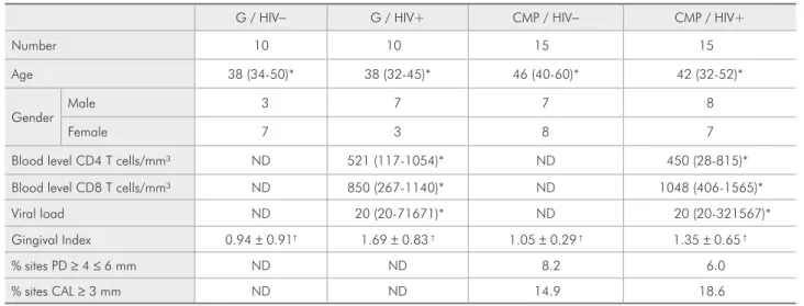
Clinically, the groups were homogenous in re-lation to age and periodontitis severity. Low viral load levels were observed in the HIV+ sample (Ta-ble1).
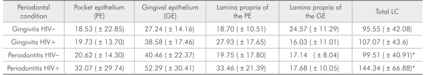
Quantitative analysis of the S100+ cells The LC were assessed in four distinct regions and throughout the histological section (Table2). When we compared the regions assessed in HIV+ and HIV− subjects with gingivitis, no statistically signiicant differences were observed. The same oc-curred in the comparison of subjects with periodon-titis.
No statistically signiicant difference was ob-served in the total S100+ LC between HIV+ subjects and HIV− subjects with gingivitis. However, a sta-tistically signiicant increase in HIV+ (p = 0.035) was observed when compared to HIV− subjects with periodontitis.
The samples were also compared according to in-iltrate intensity, and no statistically signiicant dif-ference was observed between HIV+ and HIV− sub-jects with the same iniltrate intensity. Additionally, no statistically signiicant difference was observed when a comparison between samples with mild and intense iniltrate intensity was made. This analysis was carried out to observe possible changes corre-lated with an increase in inlammation (Table3).
Table 1 - Status samples and periodontal condition.
G / HIV− G / HIV+ CMP / HIV− CMP / HIV+
Number 10 10 15 15
Age 38 (34-50)* 38 (32-45)* 46 (40-60)* 42 (32-52)*
Gender
Male 3 7 7 8
Female 7 3 8 7
Blood level CD4 T cells/mm³ ND 521 (117-1054)* ND 450 (28-815)*
Blood level CD8 T cells/mm³ ND 850 (267-1140)* ND 1048 (406-1565)*
Viral load ND 20 (20-71671)* ND 20 (20-321567)*
Gingival Index 0.94 ± 0.91† 1.69 ± 0.83 † 1.05 ± 0.29 † 1.35 ± 0.65 †
% sites PD ≥ 4 ≤ 6 mm ND ND 8.2 6.0
% sites CAL ≥ 3 mm ND ND 14.9 18.6
CD4 and CD8 T cells, viral load levels and S100+ LC correlations
To observe the correlation among S100+ LC den-sity with CD4 and CD8 T-cells, and viral load levels in peripheral blood, the Pearson correlation coef-icients were calculated. No statistically signiicant correlations were observed (Data not shown).
Discussion
In the present study, the quantitative analysis of total LC revealed a higher count for MCP in HIV+ subjects when compared to HIV− subjects. In the gingivitis samples, no statistically signiicant differ-ence was observed between HIV+ and HIV− sub-jects. These results are not in accordance with a pre-vious study9 that reported a decrease of LC in AIDS (HIV+) subjects. However, all HIV+ individuals in
the present study were using HAART, with a low viral load, and, consequently, showed less direct ag-gression by LC. Moreover, both MCP samples were deined by two different criteria, including case dei-nition and severity,18 which resulted in homogenous disease severity in HIV+ and HIV− subjects. Thus, this increased LC count in MCP among HIV+ sub-jects could be associated with the presence of spe-ciic pathogens.19-21
When we compared samples according to inil-trate intensity, no statistically signiicant difference was observed between HIV+ and HIV− subjects with the same iniltrate intensity for both gingivi-tis and periodontigingivi-tis. Additionally, no stagingivi-tistically signiicant difference was observed in the LC count with an increase in inlammation, also between HIV+ and HIV− subjects. It is dificult to compare these data to those of previous studies because of different analysis methods, tissue removal tech-niques, and the antibodies that were used, which might cause divergent results.1,22 Moreover, a large number of previous studies compared gingivitis and periodontitis. 2,4,6 In our understanding, this com-parison is not correct because they are distinct peri-odontal diseases, with different inlammatory cells, cytokines, and pathogens. Moreover, in the major-ity of previous studies 4,23, scaling and root planing and oral hygiene instructions were given only to the periodontitis patients, before tissue samples were re-moved, which would change the number of inlam-matory cells. In this study, all inlaminlam-matory mono-nuclear plasma cells and lymphocytes were counted; thus, we can afirm that no changes in LC density were observed to be correlated with increased in-lammation, which is in accordance with previous studies.2,6 One other study13 observed that calculus Table 3 - Total S100+ LC distribution according to
peri-odontal condition and inflammatory infiltrate intensity.
Periodontal condition Number of
subjects Total S100+ LC
HIV− Mild gingivitis 2 59.02 ± 54.60 HIV+ Mild gingivitis 2 138.40 ± 16.51 HIV− Moderate gingivitis 4 104.26 ± 29.34 HIV+ Moderate gingivitis 3 104.51 ± 39.26 HIV− Severe gingivitis 4 105.09 ± 48.43 HIV+ Severe gingivitis 5 96.06 ± 52.91 HIV− Mild periodontitis 2 59.39 ± 26.46 HIV+ Mild periodontitis 2 123.00 ± 42.77 HIV− Moderate periodontitis 4 83.94 ± 22.86 HIV+ Moderate periodontitis 7 163.51 ± 81.07 HIV− Severe periodontitis 9 115.34 ± 42.88 HIV+ Severe periodontitis 6 129.08 ± 57.40
(Student’s t, p > 0.05)
Table 2 - Distribution of S100+ LC density according to tissue localization. Periodontal
condition
Pocket epithelium (PE)
Gingival epithelium (GE)
Lamina propria of the PE
Lamina propria of
the GE Total LC
Gingivitis HIV− 18.53 ( ± 22.85) 27.24 ( ± 14.16) 18.70 ( ± 10.51) 24.57 ( ± 11.29) 95.55 ( ± 42.08) Gingivitis HIV+ 19.73 ( ± 13.70) 38.58 ( ± 17.46) 27.93 ( ± 17.65) 16.03 ( ± 11.01) 107.07 ( ± 43.6) Periodontitis HIV− 20.62 ( ± 14.30) 40.46 ( ± 22.37) 19.75 ( ± 17.80) 17.14 ( ± 8.04) 99.51 ( ± 40.91)* Periodontitis HIV+ 32.07 ( ± 29.74) 52.29 ( ± 30.41) 33.46 ( ± 21.39) 17.68 ( ± 10.05) 144.34 ( ± 66.88)*
removal, and subsequent decreased inlammation, was responsible for an LC count decrease even in the epithelium and gingival connective tissue. It is important to point out that only this study used the same antibody.
A comparison among the four regions assessed showed no statistically signiicant differences be-tween HIV+ and HIV− subjects. Another study 23 observed an increase of LC in the lamina propria and gingival epithelium, comparing gingivitis with periodontitis, in HIV− subjects, thus indicating dif-ferent characteristics of the immune response be-tween the two diseases. It is also interesting to note that in the pocket epithelium and lamina propria, no changes in LC count were observed. This fact may be related to junctional epithelium permeabil-ity, the presence of a local defense through gingival luid rich in IgG, and the presence of phagocyte cells in the lamina propria iniltrate.
No statistically signiicant correlation was
ob-served between S100+ LC, CD4 cells, CD8 T-cells, or viral load. This may also be related to the proile of HIV+ samples, in which all subjects were using HAART with a low viral load and levels of CD4 and CD8 T-cells within the accepted stan-dards.
Conclusions
The use of HAART can aid in achieving a low viral load and may prevent the destruction of LC in the gingiva of subjects with periodontitis. However, more studies are needed to evaluate changes in the functions of LC in HIV+ subjects with MCP.
Acknowledgments
This work was supported by grants from the Minas Gerais State Research Foundation (FAPE-MIG) and the National Council for Scientiic and Technological Research (CNPQ, # 301490/2007-4).
References
1. Cutler CW, Jotwani R. Antigen-presentation and the role of dendritic cells in periodontitis. Periodontol 2000. 2004 Jun;35(1):135-57.
2. Lins RD, Figueiredo CR, Queiroz LM, Silveira EJ, Freitas RA. Immunohistochemical evaluation of the inflammatory re-sponse in periodontal disease. Braz Dent J. 2008 Jan;19(1):9-14.
3. DiFranco CF, Toto PD, Rowden G, Gargiulo AW, Keene JJ, Connelly E. Identification of Langerhans cells in human gin-gival epithelium. J Periodontol. 1985 Jan;56(1):48-54. 4. Newcomb GM, Seymour GJ, Powell RN. Association between
plaque accumulation and Langerhans cell numbers in the oral epithelium of attached gingival. J Clin Periodontol. 1982 Jul; 9(4):297-304.
5. Jotwani R, Cutler CW. Multiple Dendritic cell (DC) subpopu-lations in human gingival and association of mature DCs with CD4+ T-cells in situ. J Dent Res. 2003 Set;82(9):736-41. 6. Jotwani R, Palucka AK, Al-Quotub M, Nouri-Shirazi M, Kim
J, Bell D et al. Mature dendritc Cells infiltrate the T cell-rich region of oral mucosa in chronic periodontitis: in situ, in vivo and in vitro studies. J Immunol. 2001 Oct;167(8):4693-700. 7. Wood GS, Warner NL, Warnke RA. Leu3/T4 antibodies react
with cells of monocyte/macrophage and Langerhans lineage. J Immunol. 1983 Jul;131(1):212-6.
8. Yin MT, Dobkin JF, Grbic JT. Epidemiology, pathogenesis and management of human immunodeficiency virus
infec-tion in patients with periodontal disease. Periodontol 2000. 2007 Jan; 44(1):55-81.
9. Myint M, Yuan ZN, Schenck K. Reduced numbers of Langer-hans cells and increased HLA-DR expression in Keratinocytes in the oral gingival epithelium of HIV-infected patients with periodontitis. J Clin Periodontol. 2000 Jul; 27(7):513-519. 10. Pimpinelli N, Riccardi R, Piluso S, Mori M, Ficarra G,
Ro-magnoli P. Immune cell infiltration in periodontal lesions of HIV infected subjects. Antigenic and ultra-structural features. Eur J Dermatol. 1995 May; 5(7):607-13.
11. Hofer D, Hämmerle CH, Grassi M Lang NP. Long-term results of supportive periodontal therapy in HIV-seropos-itive and HIV-seronegative patients.J Clin Periodontol. 2002 Jul;29(7):630-37.
12. Gonçalves LS, Ferreira SMS, Silva Jr. A, Villoria GE, Costinha LH, Colombo AP. Association of T CD4+ limphocyte levels and chronic periodontitis in HIV-infected Brazilian patients undergoing highly active anti-retroviral therapy: Clinical re-sults. J Periodontol. 2005 Jun;76(6):915-22.
13. Dereka XE, Tosios KI, Chrysomali E. Factor XIIIa+ dendritic cells and S-100 protein+ Langerhans Cells in adult periodon-titis. J Periodontol Res. 2004 Dec;39(6):447-52.
15. Tonetti MS, Claffey N. Advances in the progression of peri-odontitis and proposal of definitions of a periperi-odontitis case and disease progression for use in risk factor research. Group C consensus report of the 5th European Workshop in Peri-odontology. J Clin Periodontol. 2005 Jun;(6 suppl):210-3. 16. American Academy of Periodontology. Parameter on chronic
periodontitis with slight to moderate loss of periodontal sup-port. J Periodontol. 2000 May;71(5 suppl):856-8.
17. Miller KT, Kubier PH, Reynolds BH. Blocking on endogenous avidin-biotinactivity in immunohistochemistry: The use of skim milk as an economical and effective substitute for commercial biotin solutions. Appl Immunohistochem Mol Morphol. 1999 Jan;7(1):63-5.
18. Costa FO, Guimarães AN, Segundo TK, Cota LOM, Pa-taro AL, Cortelli SC, et al. Impact of different periodon-titis case definitions on periodontal research. J Oral Sci. 2009 Jun;51(2):199-206.
19. Odden K., Schenck K., Hurlen B. High numbers of T cells in gingival from patients with human immunodeficiency virus (HIV) infection. J Oral Pathol Med. 1995 Oct;24(9):413-9.
20. Myint M, Odden K, Schreurs O, Halstensen TS, Schenck K. The gingival plasma cell infiltrate in HIV-positive pa-tients with periodontitis is disorganized. J Clin Periodontol. 1999 Jun;26(6):358-365.
21. Gonçalves LS, Ferreira SMS, Souza CO,Souto R, Colombo AP. Clinical and microviological profiles of human immunodefi-ciency vírus (HIV)-seropositive brazilians undergoing highly active antiretroviral therapy and HIV-seronegative brazilians with chronic periodontitis. J Periodontol. 2007Jan;78(1):87-96.
22. Saglie FR, Pertuiset JH, Smith CT, Nestor MG, Carranza FA, Newman MG, et al. The presence of bacteria in oral epithe-lium in periodontal disease. III. Correlation with Langerhans cells. J Periodontol. 1987 Jun;58(6):417-22.