Braz Dent J 22(3) 2011
Bond strength of brackets to ceramic 245
INTRODUCTION
In addition to dental enamel, restorative materials may sometimes serve as substrates for bonding of orthodontic brackets under clinical conditions. Bonding to ceramic is usually achieved after etching using hydrofluoric acid. The literature presents, however, controversial findings regarding the etching time required for optimal bonding to ceramic. While some authors (1-3) found lower bond strengths associated with increasing etching times, others (4,5) observed that longer etching times may increase the bond strength to ceramic.
Following the acid treatment, the irregularities created on ceramic need to be infiltrated by an adhesive material for bonding. Photo-activated resin agents
Effect of Etching Time and Light Source on the
Bond Strength of Metallic Brackets to Ceramic
Paulo Roberto Amaral GONÇALVES1
Rafael Ratto de MORAES2
Ana Rosa COSTA3
Américo Bortolazzo CORRER3
Paulo Roberto Aranha NOUER1
Mário Alexandre Coelho SINHORETI3
Lourenço CORRER-SOBRINHO3
1Department of Orthodontics, Dental School, São Leopoldo Mandic Research Center, Campinas, SP, Brazil 2Department of Restorative Dentistry, Dental School, UFPel - Federal University of Pelotas, Pelotas, RS, Brazil
3Department of Restorative Dentistry, Dental Materials Division, Piracicaba Dental School,
UNICAMP - University of Campinas, Piracicaba, SP, Brazil
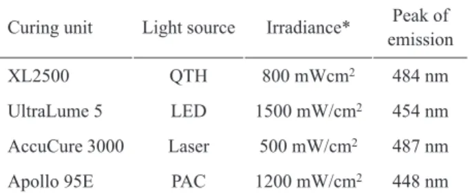
This study evaluated the bond strength of brackets to ceramic testing different etching times and light sources for photo-activation of the bonding agent. Cylinders of feldspathic ceramic were etched with 10% hydrofluoric acid for 20 or 60 s. After application of silane on the ceramic surface, metallic brackets were bonded to the cylinders using Transbond XT (3M Unitek). The specimens for each etching time were assigned to 4 groups (n=15), according to the light source: XL2500 halogen light, UltraLume 5 LED, AccuCure 3000 argon laser, and Apollo 95E plasma arc. Light-activation was carried out with total exposure times of 40, 40, 20 and 12 s, respectively. Shear strength testing was carried out after 24 h. The adhesive remnant index (ARI) was evaluated under magnification.
Data were subjected to two-way ANOVA and Tukey’s test (α=0.05). Specimens etched for 20 s presented significantly lower bond
strength (p<0.05) compared with those etched for 60 s. No significant differences (p>0.05) were detected among the light sources. The ARI showed a predominance of scores 0 in all groups, with an increase in scores 1, 2 and 3 for the 60 s time. In conclusion, only the etching time had significant influence on the bond strength of brackets to ceramic.
Key Words: bonding, ceramic, etching time, light source, shear bond strength.
are usually used for this purpose (6,7). Several light-curing units (LCUs) presenting varied light sources are available for photo-activation (8-14). In addition to the traditional quartz-tungsten-halogen (QTH) LCUs and the increasingly popular light-emitting diodes (LEDs), plasma arc (PAC) and laser units have been introduced to reduce the curing time. However, literature is still not conclusive regarding the effectiveness of different LCUs for bracket bonding, especially when the bonding substrate is a dental ceramic.
The aim of this study was to investigate the bond strength of metallic orthodontic brackets to feldspathic ceramic mediated by an adhesive resin, testing different times for ceramic etching (20 or 60 s) and different light sources for photo-activation (QTH, LED, PAC or laser). The null-hypotheses tested were that (i) no significant
Correspondence: Prof. Dr. Lourenço Correr-Sobrinho, Faculdade de Odontologia de Piracicaba, UNICAMP, Área Materiais Dentários, Avenida Limeira, 901, 13414-903 Piracicaba, SP, Brasil. Tel: +55-19-2106-5348. Fax: +55-19-2106-218. e-mail: [email protected]
Braz Dent J 22(3) 2011
246 P.R.A. Gonçalves et al.
differences would be detected between the etching times, and (ii) no significant differences would be detected among the light sources.
MATERIAL AND METHODS
Preparation of Specimens
Four feldspathic ceramic cylinders (Certec Advanced Ceramics, Barueri, SP, Brazil) measuring 15 mm in diameter and 20 mm in height were used. The surface of all cylinders was cleaned with pumice/ water slurry before testing. Two cylinders were etched for 20 and two for 60 s with 10% hydrofluoric acid gel (Dentsply Caulk, Milford, DE, USA). After etching, the surfaces were rinsed with air-water spray for 20 s and dried with air for 20 s. Two layers of a silane coupling agent (RelyX Ceramic Primer; 3M ESPE, St. Paul, MN, USA) were applied and dried for 60 s. Stainless steel, standard edgewise incisor brackets (slot 0.022”; Dental Morelli, Sorocaba, SP, Brazil) were bonded to the specimens using Transbond XT light-cured bonding resin (3M Unitek, Monrovia, CA, USA), following the manufacturer instructions.
Light-Curing Procedures
The brackets were seated and positioned firmly in the ceramic surface. Excess resin was removed using a microbrush and light-activation was carried out using one of the four LCUs tested, as shown in Table 1. The irradiance and light emission spectrum of each LCU were assessed with a power meter (Ophir Optronics Inc., Danvers, MA, USA) and a computer-controlled spectrometer (USB2000; Ocean Optics, Dunedin, FL, USA). Light-activation was carried out with 4 exposures on each side of the bracket. Total exposure times were 40, 40, 20 and 12 s for XL 2500 (3M ESPE), UltraLume 5 (Ultradent, South Jordan, UT, USA), AccuCure 3000 (LaserMed, West Jordan, UT, USA) and Apollo 95E (DMD, Westlake, CA, USA), respectively. In total, 30 brackets were bonded to each ceramic cylinder (n=15 for each etching time-light source combination). As several brackets were bonded to the same ceramic cylinder, a punch-holed strip of black adhesive tape was used to avoid light exposure to adjacent brackets, restricting the polymerization light solely to the specimen being bonded. The specimens were stored at 37oC and 100%
relative humidity.
Bond Strength Testing and Failure Analysis
The shear bond test was conducted in a mechanical testing machine after 24 h. A mounting jig was used to align the bracket-ceramic interface parallel to the testing device. The shear load was applied using a knife-edged rod at a crosshead speed of 1 mm/min until failure. Bond strength values were calculated in MPa. Data were submitted to two-way ANOVA and Tukey’s test (p<0.05).
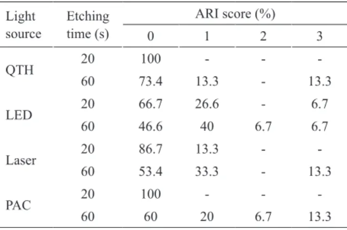
A stereomicroscope was used to analyze the debonded ceramic and bracket surfaces (40× magnification). The adhesive remnant index (ARI) was used to classify the failure modes as follows: 0 = no bonding resin left on the tooth, 1 = less than half of the bonding resin left on the tooth, 2 = more than half of the bonding resin left on the tooth, and 3 = all bonding resin left on the tooth, with distinct impression of the bracket mesh.
RESULTS
Results for bond strength are shown in Table 2. The statistical analysis showed the factor ‘etching time’ was significant (p<0.001), with specimens etched for 20 s showing significantly lower bond strengths compared with specimens etched for 60 s. The factor ‘light source’ was not significant (p=0.698), neither was the interaction between the two factors studied (p=0.919). No significant differences in bond strength were detected among the LCUs, irrespective of the etching time.
Results for ARI are shown in Table 3. A predominance of score 0 was observed for specimens etched for 20 s, irrespective of the LCU. On the other hand, despite the large number of scores 0 observed for specimens etched for 60 s, there was an increase in the number of scores 1, 2 and 3 for the longer etching time.
Table 1. Characteristics of the light-curing units.
Curing unit Light source Irradiance* Peak of emission
XL2500 QTH 800 mWcm2 484 nm
UltraLume 5 LED 1500 mW/cm2 454 nm
AccuCure 3000 Laser 500 mW/cm2 487 nm
Apollo 95E PAC 1200 mW/cm2 448 nm
*The radiant exposure was 32, 60, 10 and 14.4 J/cm2 for the QTH,
Braz Dent J 22(3) 2011
Bond strength of brackets to ceramic 247
Regarding the LCUs, no appreciable differences were detected with respect to the failure analysis. Failure scores were independent of the light source.
DISCUSSION
The first null hypothesis was rejected, as higher bond strengths were detected for specimens etched for 60 s compared with 20 s, irrespective of the LCU. Effective bonding to ceramic is dependent on creating sufficient retention for micromechanical interlocking with the bonding resin. According to Yen et al. (15), hydrofluoric acid starts etching the ceramic by reacting preferentially with the silica phase, creating retentive microchannels. With time, the acid reacts with the glassy matrix, partially dissolving it, increasing the formation of retentive channels. Therefore, longer etching times may create more irregularities for bonding, explaining the results of the present study.
According to Reynolds (16), bond strength values in the range of 6 to 8 MPa are necessary for bonded brackets to withstand the mechanical forces taking place in the oral environment. In the present study, bond strength values lower than 6 MPa were detected only for some groups in which the ceramic surfaces were etched for 20 s. This indicates that a 20 s etching time with 10% hydrofluoric acid might be insufficient for creating retention for proper bonding of orthodontic brackets to feldspathic ceramic.
With regard to the light-curing procedures, no significant differences among the LCU were detected, confirming the second null hypothesis. Dall’Igna et al. (8) also found no significant differences among the LCU units. However, Cekic-Nagas et al. (10) showed significant difference between LED and QTH and PAC units. Photo-polymerization is dependent on the
radiant exposure (17). Radiant exposure is the product of irradiance and exposure time (17). LED and QTH supplied the highest radiant exposure during photo-activation (60 and 32 J/cm2). Laser (10J/cm2) and PAC
(14.4 J/cm2) presented radiant exposure much lower
than the others light sources. However, as the emission spectrum of the laser is concentrated on the absorption peak of camphorquinone (at 468 nm), it promoted effective photoactivation. The absence of differences for PAC might be explained by the fact that a very thin resin layer is necessary for bracket bonding, and thus the differences in energy dose were probably not great enough to influence the bond strengths.
The increase in the number of ARI scores 1, 2 and 3 for the 60 s etching time is probably a result of increased retention created on the surface, enhancing the mechanical interlocking of the bonding resin to ceramic and, thus, generating failures involving the bonding material, not only adhesive failures. This finding is in line with those from Olsen et al. (18), who showed decreased etching times might affect not only the bond strengths of orthodontic brackets, but also the mode of failure to the bonding substrate.
The present results indicate the etching time is a decisive factor for the bond strength of brackets to ceramic. Although clinicians should follow the manufacturers’ recommendations for bonding procedures, a 60 s etching time for bonding to feldspathic ceramic might be recommended. However, care should be taken when selecting the etching time for each substrate, as previous studies have shown negative effects of over-etching ceramics, such as difficult penetration of the bonding agent into the irregularities and possible cohesive failure of the substrate (1-4,19,20). On the other hand, although the light source presented
Table 3. Results for the failure analysis (ARI scores).
Light source
Etching time (s)
ARI score (%)
0 1 2 3
QTH 20 100 - -
-60 73.4 13.3 - 13.3
LED 20 66.7 26.6 - 6.7 60 46.6 40 6.7 6.7
Laser 20 86.7 13.3 - -60 53.4 33.3 - 13.3
PAC 20 100 - -
-60 60 20 6.7 13.3 Table 2. Means (SD) for shear bond strength (MPa).
Light source
Etching time
20 s 60 s
QTH 6.26 (1.2) a, B 8.63 (2.0) a, A
LED 6.45 (1.0) a, B 8.38 (1.3) a, A
Laser 6.44 (1.0) a, B 8.21 (1.1) a, A
PAC 5.95 (1.4) a, B 8.00 (1.1) a, A
Braz Dent J 22(3) 2011
248 P.R.A. Gonçalves et al.
no significant influence, the use of high-intensity units is still recommended to ensure effectiveness of polymerization of the bonding resin. Finally, clinicians should be aware of the harmful effects of hydrofluoric acid and take care during its clinical application.
RESUMO
Este estudo avaliou a resistência de união de bráquetes à cerâmica testando diferentes tempos de condicionamento e fontes de luz para fotoativação do agente de união. Cilindros de cerâmica feldspática foram condicionados com ácido fluorídrico 10% por 20 ou 60 s. Após aplicação de silano na superfície da cerâmica, bráquetes metálicos foram colados aos cilindros utilizando Transbond XT (3M Unitek). Os espécimes de cada tempo de condicionamento foram divididos em 4 grupos (n=15): fonte halógena XL2500, LED UltraLume 5, laser de argônio AccuCure 3000 e arco de plasma Apollo 95E. A fotoativação foi realizada com tempo total de 40, 40, 20 e 12 s, respectivamente. O teste de cisalhamento foi realizado após 24 h. O índice de adesivo remanescente (IAR) foi avaliado sob aumento. Os dados foram submetidos a ANOVA de duas vias e teste de Tukey (p<0,05). Espécimes condicionados por 20 s apresentaram resistência de união significativamente menor que espécimes condicionados por 60 s. Não foram detectadas diferenças significativas entre as fontes de luz. O IAR mostrou predominância de escores 0 para todos os grupos, com aumento nos escores 1, 2 e 3 para o tempo 60 s. Em conclusão, apenas o tempo de condicionamento teve influência significativa na resistência de união de bráquetes à cerâmica.
REFERENCES
1. Barghi N, Fischer DE, Vatani L. Effects of porcelain leucite content, types of etchants, and etching time on porcelain-composite bond. J Esthet Restor Dent 2006;18:47-52.
2. Nagayassu MP, Shintome LK, Uemura ES, Araújo JE. Effect of surface treatment on the shear bond strength of a resin-based cement to porcelain. Braz Dent J 2006;17:290-295.
3. Shimada Y, Yamaguchi S, Tagami J. Micro-shear bond strength of dual-cured resin cement to glass ceramics. Dent Mater 2002;18:380-388.
4. Chen JH, Matsumura H, Atsuta M. Effect of different etching periods on the bond strength of a composite resin to a machinable porcelain. J Dent 1998;26:53-58.
5. Guler AU, Yilmaz F, Yenisey M, Guler E, Ural C. Effect of acid etching time and a self-etching adhesive on the shear bond strength
of composite resin to porcelain. J Adhes Dent 2006;8:21-25. 6. Kasuya K, Miyazaki Y, Ogawa N, Maki K, Manabe A, Itoh
K, et al.. Efficacy of experimental dual-cure resin cement for orthodontic direct bond system. Orthod Waves 2006;65:107-111. 7. Meguro D, Hayakawa T, Kasai K. Efficacy of using orthodontic
adhesive resin in bonding and debonding characteristics of a calcium phosphate ceramic bracket. Orthod Waves 2006;65:148-154.
8. Dall’Igna CM, Marchioro EM, Spohr AM, Mota EG. Effect of curing time on the bond strength of a bracket-bonding system cured with a light-emitting diode or plasma arc light. Eur J Orthod 2011;33:55-59.
9. Correr AB, Sinhoreti MA, Sobrinho LC, Tango RN, Schneider LF, Consani S. Effect of the increase of energy density on Knoop hardness of dental composites light-cured by conventional QTH, LED and xenon plasma arc. Braz Dent J 2005;16:218-224. 10. Cekic-Nagas I, Egilmez F, Ergun G. The effect of irradiation
distance on microhardness of resin composites cured with different light curing units. Eur J Dent 2010;4:440-446.
11. Thind BS, Stirrups DR, Lloyd CH. A comparison of tungsten-quartz-halogen, plasma arc and light-emitting diode light sources for the polymerization of an orthodontic adhesive. Eur J Orthod 2006;28:78-82.
12. Filipov IA, Vladimirov SB. Residual monomer in a composite resin after light-curing with different sources, light intensities and spectra of radiation. Braz Dent J 2006;17:34-38.
13. Park JK, Hur B, Ko CC, García-Godoy F, Kim HI, Kwon YH. Effect of light-curing units on the thermal expansion of resin nanocomposites. Am J Dent 2010;23:331-334.
14. Price RB, Labrie D, Rueggeberg FA, Felix CM. Irraciance differences in the violet (405 nm) and blue (460 nm) spectral ranges among dental light-curing units. J Esthet Restor Dent 2010;22:363-377.
15. Yen TW, Blackman RB, Baez RJ. Effect of acid etching on the flexural strength of a feldspathic porcelain and a castable glass ceramic. J Prosthet Dent 1993;70:224-233.
16. Reynolds IR. Composite filling materials as adhesives in orthodontics. Br Dent J 1975;138:83.
17. Rueggeberg F. Contemporary issues in photocuring. Compend Contin Educ Dent Suppl 1999;4-15.
18. Olsen ME, Bishara SE, Boyer DB, Jakobsen JR. Effect of varying etching times on the bond strength of ceramic brackets. Am J Orthod Dentofacial Orthop 1996;109:403-409.
19. Sant’Anna EF, Monnerat ME, Chevitarese O, Stuani MB. Bonding brackets to porcelain - in vitro study. Braz Dent J 2002;3:191-196. 20. Canay S, Hersek N, Ertan A. Effect of different acid treatments on
a porcelain surface. J Oral Rehabil 2001;28:95-101.