Supramolecular architectures in crystals of melamine and aromatic carboxylic acids
Genivaldo Julio Perpétuo
a, Jan Janczak
b,*aDepartamento de Física, Instituto de Ciências Exatas e Biológicas, Universidade Federal de Ouro Preto, 35400-000 Ouro Preto, MG, Brazil bInstitute of Low Temperature and Structure Research, Polish Academy of Sciences, PO Box 1410, 50950 Wroclaw, Poland
a r t i c l e
i n f o
Article history:
Received 24 January 2008
Received in revised form 9 April 2008 Accepted 12 April 2008
Available online 25 April 2008 Keywords:
Co-crystals Melamine
Supramolecular architecture Hydrogen bond
Carboxylic acid Crystal engineering
a b s t r a c t
Two crystals containing melamine (M), melaminium (MH+), and aromatic carboxylic acids are synthe-sized and characterized by single-crystal X-ray diffraction analysis: melamine–melaminium 3,4,5-trihy-droxybenzoate (THB ) dihydrate (1), and melamine–melaminium 2-acetylbenzoate (AB ) dihydrate (2). In the crystal structures protonated (MH+) and non-protonated melamine (M) molecules are intercon-nected via NAHN hydrogen bonds into a planar dimer in (1) and one-dimensional polymer in (2). The NAHO hydrogen bonding interaction between the protonated nitrogen atom of MH+and the car-boxylate oxygen atom of the corresponding anion is observed in both structures. However, only in (1) it results in an almost planar ionic unit (THB MH+) where additionalp–pinteractions between the anions appear, that are missing in (2). The water molecules act as donor and as acceptor of hydrogen bonds link-ing the substructures observed in the (1) and (2) crystals into 3D supramolecular networks.
Ó2008 Elsevier B.V. All rights reserved.
1. Introduction
A productive strategy in the crystal engineering is to build supramolecular structures from molecules that are programmed to engage in multiple interactions with the neighbours [1–3]. Hydrogen bonding system is the main key for organisation of mol-ecules containing complementary arrays of the hydrogen bonding sites[4–8]. The NAHN, NAHO, and OAHO hydrogen bonds
are widely used for organisation of the components in the design of a large number and different types of supramolecular architec-tures as ribbons, rosettes, layers, tubes, rods, spheres and sheets [9–14]. Some supramolecular motifs of hydrogen bonding patterns of symmetric and translational repetitive occurrence are important in the crystal and chemical engineering and technology[15]. The usually weak CAHN and CAHO hydrogen bonds play also a
sig-nificant role in the architectures, especially in the biological sys-tems[16–18]. A non-covalent association of proteins, formation of a phospholipid bilayer, interaction of a transcription factor with DNA, folding of a tRNA into its three-dimensional conformation are examples of processes that depend on the non-covalent interac-tions and organisainterac-tions[18].
Melamine is an example of a compound containing comple-mentary arrays of hydrogen bonding sites that forms in the solid state a two dimensional network (Scheme 1a). Protonation of its triazine ring decreases the number of the active sites resulting in decreasing of the dimensionality of the arrangement; for example
singly protonated melaminium cations form one-dimensional hydrogen bonded chains (Scheme 1b), while double protonated melaminium cations form only a dimeric structure (Scheme 1c). Our interest in melamine and its complexes or salts arises from the possibility to obtain a new material for non-linear optics. Crys-tals of melaminium selenate with non-centrosymmetric space group are optically active and generate the SHG (second harmonic generation) with c.a. 40% efficiency of KDP (KDP = KH2PO4) [19]. High efficiency of SHG is observed in the crystals of double proton-ated melaminium bis(trichloroacetate) dihydrate (about 3-times greater than that of KDP)[20]. Another non centrosymmetric crys-tal of melaminium phosphate may also be used as a material for non-linear optics [21]. One of the most interesting hydrogen bonded structure formed by melamine is with cyanuric and thioc-yanuric acids[22]. In both co-crystals of the 1:1 hydrogen bonded adducts of melamine (M) and cyanuric acid (CA) or thiocyanuric acid (TCA) form pseudohexagonal rosette arrangement with chan-nels along the shortest crystallographic axis[22].
Compounds containing partially protonated melaminium cat-ions (MH+, MH
22+) combined with different organic and inorganic counterions are widely reported in the literature[23–24]. In stud-ies of the crystalline melamine and its organic or inorganic salts, the most crystals contain only one melamine form, i.e. the neutral molecule, singly or doubly protonated melaminium cations [25], and only two works reporting the crystalline structure of neutral and protonated melminie (M and MH+) [26,27]. In the present work we describe two co-crystals containing protonated (MH+) and non-protonated melamine: melamine–melaminium 3,4,5-tri-hydroxybenzoate dihydrate (1) and melamine–melaninium
0022-2860/$ - see front matterÓ2008 Elsevier B.V. All rights reserved. doi:10.1016/j.molstruc.2008.04.032
* Corresponding author. Fax: +48 71 344 1029. E-mail address:[email protected](J. Janczak).
Contents lists available atScienceDirect
Journal of Molecular Structure
2-acetylbenzoate dihydrate (2). Both crystals investigated here are the examples containing neutral melamine M and singly proton-ated melaminium MH+cation in the same crystal.
Another feature observed in several structures containing the melaminium cation concerns its tendance to dimerization; gener-ally pairs of MH+ units are linked via two almost linear N
amine
AHNazahydrogen bonds to form a planar dimer[25]. The ques-tion if these dimers will provide formaques-tion of especially long ordered arrangement in the crystals like 1D polymeric chains or tapes, or even 2D layers, depends not only on the surrounding counterions and water molecules, but also on the site of the pro-tonation within the triazine ring relative to the dimer as a building unit. In the present study we attempt to answer the crucial questions concerning the competitive forces toward the stabilization of the crystal structures, such as thep–pinteractions,
the attractive and repulsive Coulomb forces and the participation of aza and amine nitrogen sites. The role of water molecules in formation of short and long ordered molecular arrangements is also discussed. For completeness, in each case the building units in the asymmetric unit of 1 and 2 are optimized and theirs geometries are analysed in relation to those in the crystal.
2. Experimental
All materials were commercially available and used as received. The Fourier transform infrared spectra were recorded from nujol mulls between 400 and 4000 cm 1on a Bruker IFS 113 V FTIR
spec-trometer at room temperature. Elemental analyses were carried out with a Perkin-Elmer 240 elemental analyzer. Thermogravimet-ric analyses (TGA) were performed under dinitrogen atmosphere using a Perkin-Elmer 7 thermogravimetric analyzer with a heating rate of 5°C min. The co-crystals were prepared as follows.
2.1. Preparation of (1)
Melamine and 3,4,5-trihydroxybenzoic acid (Aldrich, purity: 99% and 98%) were solved in hot water in 1:1 molar ratio. The hot solution was cooled slowly and kept at room temperature. After several days transparent yellowish crystals were formed. Anal. calculated for C13H22N12O7: C, 34.06; H, 4.84; N, 36.67; O, 24.43%. Found: C, 34.28; H, 4.76; N, 36.43. IR (cm 1): 3468, 3418, 3329, 3128, 1653, 1551, 1377, 1210, 1027, 813.
2.2. Preparation of (2)
Melamine and 2-acetylbenzoic acid (Aldrich, both purity of 99%) were solved in hot water in 1:1 molar ratio. The hot solution was cooled slowly and kept at room temperature. After several days transparent yellowish crystals were formed. Anal. calculated for C15H24N12O5: C, 40.02; H, 5.01; N, 36.98; O, 17.98%. Found: C, 40.12; H, 4.93; N, 36.81. IR (cm-1): 3466, 3415, 3327, 3129, 1695, 1613, 1580, 1493, 1450, 1372, 1203, 1030, 814.
2.3. X-ray single crystal analysis
X-ray intensity data for both crystals were collected using graphite monochromatic MoKaradiation on a four-circlej
-geom-etry KUMA KM-4 diffractometer with a two-dimensional area + + + + N N N N N H H H N H H N N N N N N H H H H H H H N N N N N N H H H H H H H N N N N N N H H H H H H H H H + +N N N N N H H H H H N H H N N N N N N H H H H H H H H H + + N N N N N N H H H H H H N N N N N N H H H H H N N N N N N H H H H H H N N N N N N H H H H H H N N N N N N H H H H H H H
Scheme 1.Possible dimensionality of melamine arrangements by increasing
deg-ree of protonation: 2D-layer of non-protonated (a), polymeric 1D-chain (b), and discrete dimeric form (c).
Table 1
Crystallographic data and structure refinement parameters
Crystal data 1 2
Formula C7H5O5C3H7N6C3H6N62(H2O) C9H7O3C3H7N6C3H6N62(H2O)
MW (g mol1) 458.43 452.46
Crystal size 0.450.240.18 mm 0.350.280.24 mm Crystal system Orthorhombic Monoclinic
Space group Pcnn C2/c
a(Å) 32.878(5) 12.286(3)
b(Å) 9.392(2) 18.934(4)
c(Å) 12.785(2) 18.515(4)
b(°) 95.26(3)
V(Å3) 3947.9(12) 4288.9(17)
Z 8 8
l(mm 1) 0.127 0.109
qobs;qcalc
(g cm 3) 1.54; 1.543 1.40; 1.401 Data collection
Radiation,k(Å) Mo Ka(0.71073) Mo Ka(0.71073)
hmax(°) 28.49 28.40
Absorpt. correct. (Tmin;Tmax)
0.9451; 0.9775 0.9603; 0.9715
Rint 0.0222 0.0176
No. collected reflections 45212 24078 No. unique reflections 4992 5350 No. observed reflections 3997 3571 Refinement R[F2> 2
r(F2)] 0.0456 0.0518
wR(F2)a,b 0.1086 0.1489
GooF 1.005 1.006
Dqmin;Dqmax (e Å3)
0.426; 0.642 0.211; 0.231
a
w= 1/[r2(Fo2) + (0.0466P)2+ 1.600P] where
P= (Fo2+ 2Fc2)/3. b
CCD detector. Thex-scan technique withDx= 0.75°for each
im-age was used for data collection. The 960 imim-ages for six different runs covering over 95% of the Ewald sphere were performed. Ini-tially the lattice parameters were refined on 150 reflections ob-tained from 40 images for eight runs with different orientation in the reciprocal space. Finally the lattice parameters were refined by least-squares methods based on all the reflections with I>2r(F2). One image was used as a standard after every 40 images for monitoring of the crystal’s stability as well as for mon-itoring the data collection, and no correction on the relative intensity variation was necessary. Integration of the intensities, correction for Lorenz and polarization effects was performed using a KUMA KM-4 CCD program[28]. The face-indexed analyt-ical absorption was calculated using theSHELXTL program[29]. The structures were solved by direct methods usingSHELXS of the SHELXL97 program [30]. The structures were refined with the anisotropic thermal parameters for all non-hydrogen atoms. Difference Fourier maps gave electron density concentrations approximately located for all hydrogen atoms positions; these positions were idealised (HFIX 43 for all H atoms of the phenyl rings with isotropic thermal parameters of 1.2Ueq of the carbon atoms joined directly to the hydrogen atoms). Final difference Fourier maps showed no peaks of chemically significance. Details of the data collection parameters and final agreement factors are collected inTable 1. Selected bond lengths and angles and torsion angles are listed inTable 2.
2.4. Quantum calculations
Ab-initiomolecular orbital calculations full geometry optimisa-tion were performed with the Gaussian98 program package[31].
Table 2
Selected geometric parameters (bond lengths in Å, bond angles in degrees)
(MH)+ X-ray Melamine X-ray Theoretical
1 2 1 2
N11AC12 1.344(2) 1.348(2) N21AC22 1.349(2) 1.344(2) 1.325
N11AC16 1.332(2) 1.326(2) N21AC26 1.351(2) 1.345(2) 1.326
N13AC12 1.352(2) 1.353(2) N23AC22 1.339(2) 1.348(2) 1.325
N13AC14 1.331(2) 1.332(2) N23AC24 1.345(2) 1.344(2) 1.326
N15AC14 1.362(2) 1.359(2) N25AC24 1.346(2) 1.343(2) 1.325
N15AC16 1.358(2) 1.361(2) N25AC26 1.335(2) 1.348(2) 1.326
C12AN17 1.337(2) 1.328(2) C22AN27 1.340(2) 1.337(2) 1.344
C14AN18 1.324(2) 1.320(2) C24AN28 1.333(2) 1.348(2) 1.344
C16AN19 1.319(2) 1.324(2) C26AN29 1.337(2) 1.333(2) 1.344
C12AN11AC16 115.4(2) 115.8(2) C22AN21AC26 114.2(2) 114.8(2) 114.03
N11AC12AN13 126.7(2) 126.0(2) N21AC22AN23 125.2(2) 125.3(2) 125.97
C12AN13AC14 114.8(2) 115.9(2) C22AN23AC24 115.3(2) 114.3(2) 114.03
N13AC14AN15 122.1(2) 121.1(2) N23AC24AN25 124.5(2) 125.9(2) 125.97
C14AN15AC16 118.9(2) 119.8(2) C24AN25AC26 115.2(2) 114.3(2) 114.03
N11AC16AN15 121.8(2) 121.5(2) N21AC26AN21 125.6(2) 125.3(2) 125.97
(THB) (1) X-ray Theoretical (AB) (2) X-ray Theoretical
C1AC2 1.397(2) 1.402 C1AC2 1.405(2) 1.405
C2AC3 1.386(2) 1.390 C2AC3 1.399(2) 1.403
C3AC4 1.391(2) 1.394 C3AC4 1.389(2) 1.398
C4AC5 1.387(2) 1.397 C4AC5 1.373(2) 1.400
C5AC6 1.388(2) 1.394 C5AC6 1.388(2) 1.397
C6AC1 1.389(2) 1.401 C6AC1 1.388(2) 1.400
C1AC7 1.504(2) 1.554 C1AC7 1.510(2) 1.538
C7AO1 1.257(2) 1.258 C7AO1 1.255(2) 1.266
C7AO2 1.273(2) 1.261 C7AO2 1.248(2) 1.254
C3AO3 1.370(2) 1.397 C2AC8 1.487(2) 1.513
C4AO4 1.362(2) 1.391 C8AC9 1.499(2) 1.522
C5AO5 1.377(2) 1.392 C8AO3 1.224(2) 1.228
C1AC7AO1 119.75(12) 115.6 C1AC7AO1 116.01(13) 113.6
C1AC7AO2 117.57(13) 115.1 C1AC7AO2 118.79(13) 117.2
O1AC7AO2 122.67(13) 129.3 O1AC7AO2 125.11(14) 129.2
C2AC8AC9 119.46(15) 118.0
C2AC8AO3 120.94(14) 120.0
C9AC8AO3 119.61(16) 120.3
N N N
N
N N
H
H
H H
H
N
N
N+ N
N
N H
H
H H
H
H H
H
N N N
N
N N
H
H
H H
H H
H O
O
O
O
O H
H
H
O CH3
O
O
N
N
N N
N
N H
H
H
H H
H H
Scheme 2.Hydrogen bonding motifs in the crystal structures of1(a and b) and2(a
All calculations were performed by the density functional three-parameters hybrid (B3LYP) methods[32,33] with the 6-31G(d,p) basis set starting from the X-ray geometry. As convergence criteri-ons the threshold limits of 0.00025 and 0.0012 a.u. were applied for the maximum force and the displacement, respectively.
3. Results and discussion
The crystallizations of melamine in hot water solution of 3,4,5-trihydroxybenzoic acid as well as in 2-acetylbenzoic acid were car-ried out in a 1:1 molar ratio, but the formed crystals contained 2:1 ratios of melamine to carboxylic acid. The crystallizations were also performed in a molar proportion of 2:1, but the formed crys-tals were not suitable for the X-ray analysis. In both co-cryscrys-tals, the carboxylic proton is transferred to the melamine molecule leading to singly protonated (MH+) unit, which interacts with non-protonated melamine (M) to form a dimeric unit (MH+M) (Scheme 2a); the latter interacts with anionic part of the crystals by a pair of NAHO hydrogen bonds with a graph set of R22(8) in 1 or of R22(11) in 2 as illustrated in Scheme 2b and c. The first graph (R22(8)) is one of the 24 most frequently observed bimolec-ular cyclic hydrogen-bonded synthons in organic crystal structures [34,35].
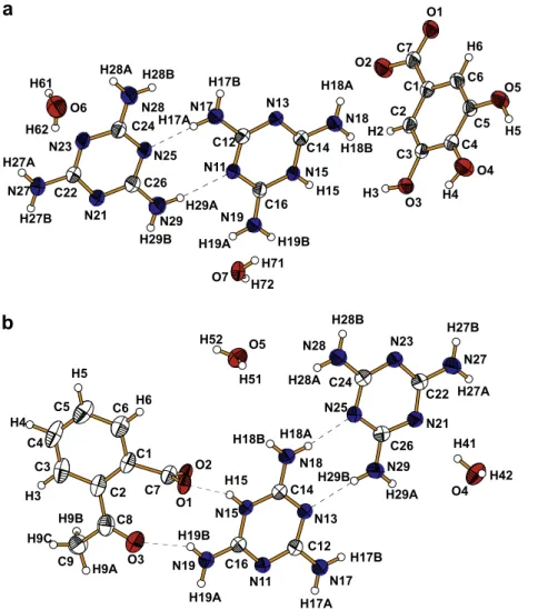
The asymmetric unit of 1 consists of a melamine molecule (M), a protonated melaminium (MH+) cation, a 3,4,5-trihydroxybenzo-ate (THB-) anion, and two water molecules (O6 and O7), (Fig. 1a),
and the asymmetric unit of 2 consists of the melamine molecule, a protonated melaminium cation, 2-acetyl-benzoate (AB ) anion, and two water molecules (O4 and O5), (Fig. 1b). A search in the Cambridge Structural Data Base for structures containing mela-minium residues yield 26 structures of single-protonated, four structures of double-protonated melamine residues and only three structure containing neutral melamine and singly protonated mel-amine[25–27]. Thus the structures described here are rare exam-ples of both neutral (M) and protonated (MH+) units co-existing in the same environment. In nearly all cases, as in the present work, the triazine rings of M+and M are almost planar, but the bond lengths and especially the internal angles within the rings deviate significantly from the ideal hexagonal form. A correlation between the NringAC bond lengths and their position in relation to the
pro-tonated nitrogen atom in the ring is observed (seeTable 2). For example, generally the non-protonated NAC bond values are
with-in the expected range for CarAN, 1.328(2) to 1.348(2) Å[36], while
the protonated NringAC bonds are slightly above of this range. The
internal CANAC bond angles at the protonated nitrogen atom is
about four degrees larger than at the non-protonated nitrogen atoms. The differences between the internal CANAC angles within
the protonated melaminium ring are in agreement with the va-lence-shell electron-pair repulsion model, VSEPR [37], according to which the lone pair on non-protonated aza nitrogen atoms af-ford a wider region than the covalent bond NprotAH, causing the
internal angle of the last to be greater than on the nonprotonated
H15
H19B
H18B
N19
N15
H19A
N18
C16
H18A
C14 N11
N13
C12
H29B H29A N29
N17 H17A
H17B
C26
N21
N25
H27B C22 N27
C24
N23
H28B
H27A
N28 H28A
H72 H61
O7 H62
O2
H71 O6
O1
C7
H2 C1
C2
H3
H6
C6
C3
O3
C5
C4 O5
O4
H4
H5
H27A
H17B
H17A
H29A
N27
N17
H27B
N21
N29 C22
H41
H29B
C26
O4 O5
H52
C12
N23
N13
N11
N25
H42 C24
H51
H9B
C14
C16
H18A
H19A
N18 N28
N15
H9A N19
H28B
C9
H28A
H18B
H15
H19B H9C
O1 C8
H3
O3 C3
C2 H4
C4
C7 C1
C5 C6
H5
H6
O2
Fig. 1.Asymmetric unit of1(a) and2(b) with the atom labelling scheme. Displacement ellipsoids are show at the 50% probability level. Dashed lines represent the hydrogen
bonds.
Nringatoms. As a result of the protonation of the melamine ring at one of three N atoms, the internal NACAN angle involving
non-protonated N atoms is significantly greater than the remaining two NACAN angles involving protonated and non-protonated N
atoms. The ab-initio gas-phase geometry calculated for isolated melamine molecule and it’s singly protonated cation shows similar correlation between the internal CANAC and CANAC angles
with-in the rwith-ings as found with-in these crystals (Table 2). Thus the ring dis-tortions of MH+ in comparison to M result mainly from the protonation, and to a lesser degree, from the hydrogen-bonding system and crystal packing forces.
The whole THB anion in the crystal of 1 is almost planar. The greatest deviations from the mean plane defined by the benzene ring are 0.061(2) Å for C7 and 0.194(2) Å for O2 atoms. All of the CC bond distances within the ring are in the range from 1.375(2) to 1.391(2) Å, and the internal angles are slightly different from 120°, and they agree well with the average geometry
param-eters observed in aromatic organic compounds[34]. Furthermore, the hydroxyl groups are bonded to Caratoms through the typical single-bond value with the average of 1.370 Å. A weak intramolec-ular O4AH4O3 hydrogen bond is observed (O4AH4O3, with a O4O3 distance of 2.701(2) Å). The values of the two CO bond lengths of carboxylate group are intermediate between the single Csp2AO (1.308–1.320 Å) and double Csp2@O bonds (1.214–
1.224 Å)[36]indicating delocalization of the charge on both oxy-gen atoms of the COO group. However, the small difference be-tween the two CAO distances results from the hydrogen bonds,
in which the O atoms act as acceptors H. The oxygen of the longer CAO bond is involved in two stronger hydrogen bonds, while the
second oxygen is also involved in two hydrogen bonds, but they are weaker (seeTable 3). Comparison of the X-ray experimental and gas-phase molecular orbital geometries of the TBA- anion shows that the greatest differences are observed in the OACAO
an-gle of the COO group and in the CarACOO bond length (Table 2).
The shortening of the CarACOO bond and decreasing of the
OACAO angle in the crystal geometry in relation to MO geometry
is probably due to the interactions with the neighbourhood by al-most linear NAHO and OAHO hydrogen bonds that diminish
the steric effect of the lone-pairs of electron on both O atoms of the COO group in the crystals.
The benzene ring (mean plane P1: C1–C6) of the AB anion in the crystal of 2 is planar. The acetyl C8 and carboxyl C7 atoms are bonded to the benzene ring on C2 and C1, respectively, with the Car–C8 and Car–C7 bond lengths of 1.487(2) and 1.510(2) Å, typical values for CarAC single bonds [36]. Due to the repulsive
interaction between acetyl and carboxyl groups, C8 and C7 atoms deviate from the mean plane of benzene ring by 0.101(2) and 0.050(2) Å respectively, while the C1AC2 bond (1.405(2) Å) is
long-er than the remaining CAC bonds within the ring (average value of
1.387 Å). The charge of deprotonated carboxyl group is delocalized over both CAO bonds. However, the slight difference in the CAO
bond lengths of the COO-group results from the different involve-ment in the NAHO and OAHO hydrogen bonds. The C@O bond
length in the acetyl group shows a typical value (1.224(2) Å) ob-served for the carbonyl groups[36]. The X-ray values of CAO bonds
compare well with the values obtained by molecular orbital calcu-lations. When the conformations in the gas-phase and in the crys-tal are compared, different orientations of the acetyl (plane P2: C8, O3, C9) and carboxyl (plane P3: C7, O1, O2) groups around single bonds of C2–C8 and C1–C7, respectively, appear. The X-ray inter-planar angles between planes P1, P2, and P3 are: P1P2 = 19.1(2)°,
P1P3 = 76.5(2)°, and P2P3 = 71.5(2)°, while in the gas-phase
con-formation the respective angles are: P1P2 = 112.5°, P1P3 = 6.4°,
and P2P3 = 110.5°. Thus in the gas phase conformation, the
carbox-ylate group is almost coplanar with the benzene ring, while the carbonyl is oriented to enable interaction between one H methyl
hydrogen and one carboxyl oxygen. In the crystal, the carboxylate group adopts rather an nearly orthogonal conformation in relation to the benzene ring, while the carbonyl double bond of acetyl group is almost eclipsed to the CringACOO single bond. Despite this
unusual internal conformation in the crystal, the oxygen atoms are involved in up to six hydrogen bonds toward melamine dimers and water molecules, which probably compensates the carbonyl–car-boxyl steric and repulsion interactions.
The crystal packing of compounds 1 and 2 show rich set of the NAHN, NAHO and OAHO hydrogen-bonding systems (Table 3) and p–p interactions between the aromatic rings. The best
way to realize such complex 3D crystal architectures is to analyse the substructures generated by basic synthons, as shown in Scheme 2. The interactions between these basic units lead to for-mation of stacks, chains, layers, tapes and channel patterns, devel-oping interesting supramolecular 3D architectures.
The synthon MH+M (Scheme 2a) is observed in the crystal struc-ture of 1 as shown inFig. 1a. They form a cationic layer nearly paral-lel to the crystallographic plane of (100) atx=1=4,3=4(Fig. 2a). The protonated and non-protonated melamine units are interconnected by two almost linear NAHN hydrogen bonds, N17AH17AN25
and N29AH29AN11 (Fig. 1a). An offsetp–pinteraction between
two MH+M dimers related to each other by a twofold axis along thec-axis is observed (Fig. 2b); the mean interplanar distance of 3.45(2) Å indicates strongp–pinteraction[38]. Thec-glide plane perpendicular to the a-axis provides that mutually orthogonal stacks interact with each other via hydrogen bonds through
Table 3
Geometric parameters of the hydrogen bonds in the structure of1and2
DAHA DAH (Å) HA (Å) DA (Å) <DHA(°)
Compound1
N15AH15O2i 0.86 1.78 2.633(2) 170
N17AH17AN25 0.86 2.11 2.950(2) 165
N17AH17BN21ii 0.86 2.32 3.014(2) 139
N18AH18AO5iii 0.86 2.13 2.911(2) 151
N18AH18BO1i 0.86 1.96 2.819(2) 179
N19AH19AN13i 0.86 2.27 3.089(2) 160
N19AH19BO3iv 0.86 2.15 2.928(2) 150
O3AH3O1v 0.823(5) 1.81(1) 2.630(2) 172(2) O4AH4O7vi 0.822(5) 1.96(1) 2.715(2) 153(2)
O4AH4O3 0.822(5) 2.30(2) 2.701(2) 110(2)
O5AH5O6vii 0.822(5) 2.03(1) 2.828(2) 165(2) N27AH27AO7viii 0.86 2.15 2.946(2) 153
N27AH27BO6v 0.86 2.35 3.163(2) 158
N28AH28BN21ii 0.86 2.27 3.103(2) 163
N29AH29AN11 0.86 2.24 3.082(2) 166
N29AH29BN13i 0.86 2.49 3.097(2) 128
O6AH62N23 0.820(1) 2.30(3) 2.835(2) 123(3) O7AH71O2v 0.820(1) 1.91(1) 2.729(2) 175(2) Compound2
N15AH15O1 0.86 1.81 2.640(2) 162
N17AH17AO5i 0.86 2.15 2.985(2) 165
N17AH17BO4iv 0.86 2.10 2.945(2) 166
N18AH18AN25 0.86 2.11 2.965(2) 170
N18AH18BO2iii 0.86 2.08 2.800(2) 141
N19AH19AN23ii 0.86 2.13 2.980(2) 172
N19AH19BO3 0.86 2.17 2.934(2) 148
N27AH27AO5v 0.86 2.34 2.970(2) 131
N27AH27BN11vi 0.86 2.14 2.999(2) 175
N28AH28AO2iii 0.86 2.37 3.219(2) 172
N29AH29AO4iv 0.86 2.30 2.970(2) 135
N29AH29BN13 0.86 2.12 2.979(2) 175
O4AH41N21 0.84(3) 2.07(3) 2.906(2) 176(2) O4AH42O1vii 0.89(2) 1.94(3) 2.792(2) 159(2) O5AH51O2iii 0.91(3) 1.83(3) 2.735(2) 170(2) Codes for symmetry operations(i)x, 3/2 y, 1/2+z;(ii)1/2 x,y, 1/2+z;(iii)1 x, 1 y,
2 z;(iv)1 x, 1 y, 1 z;(v)x, 1/2 y, -1/2+z;(vi)1 x, y, 1 z;(vii)1/2+x, y, 3/2 z; (viii)1/2 x, 1/2 y,z.
aza-nitrogen atoms, N13 and N21, which act as proton-acceptors from the amine nitrogen atoms of adjacent neighbouring stacks. In this way, several NAHN bonds are summed up to the dimeric
p–pinteraction, once involving two amine nitrogen as donors H of
MH+and M units toward a single aza nitrogen acceptor on M, v.z., N17ii
AH17BiiN2 and N28iiAH28BiiN21; and also from two amine
nitrogen H-donors of MH+and M units toward a single aza nitrogen
acceptor on MH+, v.z., N19
AH19AN13iand N29AH29BN13i. The MH+MMH+Mcluster of mutually orthogonal stacking dimers forming a polymeric substructure along thec-axis (Fig. 2b). More-over, the protonated side of dimer MH+M, i.e., the ring nitrogen atom N15 and its amine nitrogen atoms (N18) in MH+, interacts with the THB anion while the other side interacts with water molecule (O6). The stacking unit of two parallel THB anions, related by the
x=3/4
x=0
x=1/4 (see Fig. 2b) x=1/2 (see Fig. 2c)
a
b
c
See Fig
.2d
N19 H19A N13i N28ii
N29 N28Bii
H29B N21
N21
3.45(2) Ao 3.45(2) Ao
a
b
c
O7v H71v O1v
O7vi
O2 H3
C7 H4
O3
C2
O1 C1 C3 C6
C4 O4
C5 O5
H5 O6vii a
b c
3.52(1)A
O
3.52(1)A
O
O3i O4i
N29 N23
H29A O2i
N25
N11 H17A H15
C7i
N15 N17
O5i
H62 O6 O1i
H18B
N18 b a
c
Fig. 2.View of the crystal structure of1along thec-axis (a); rectangular polymer parallel to thec-axis formed by alternating single stacks of interacting MH+M dimers
orthogonal to each other (b); offsetp–pinteractions of the THB anions forming stacks interconnected by OAHO hydrogen bonds; angle between stacking direction and c-axis is90°(c) and hydrogen bonded THB and MH+M units into 1D polymer via water molecules (d). Symmetry code seeTable 3.
inversion symmetry and with an estimated interplanar distance of 3.52(1) Å, characterizes a strong offset ofp–pinteraction between the benzoate anions (Fig. 2c). There are two stacking directions orthogonal toc-axis. Furthermore, single THB stacks and water O7 form an anionic layer parallel to (100) crystallographic plane at x=0, ½, 1. Within these layers, THB anions of distinct stacks are linked strictly via OAHO hydrogen bonds: once directly from
hydroxyl to carboxylate oxygen atoms onto O3AH3O1v, and sec-ondly by means of water O7, which bridges the anions as acceptor from the hydroxyl proton in O4AH4O7viiand as donor to the
car-boxylate oxygen in O7AH71O2v. Additionally, the water O6 mole-cule is bridging THB MH+and MH+M units via O
AHO bonds, being
proton acceptor of hydroxyl oxygen in O5AH5O6, and proton
do-nor to the aromatic nitrogen N23 of M. An interesting feature of this
3D arrangement is the continuous THB MH+MO6
THB MH+MO6polymeric chains (Fig. 2d), which runs approxi-mately along the [1,3,0] directions throughout the two substruc-tures described above.
The observed synthon MH+M in the crystal structure of 2 is shown inFig. 1b. Each protonated and non-protonated melamine participates in the N18AH18AN25 and N29AH29BN13 hydro-gen bonds to form the planar dimer MH+M. An infinite chain MH+MMH+M is generated by translation symmetry along the a-axis, enabling two additional N19AH19AN23ii and N27ii
AH27iiN15 hydrogen bonds between two adjacent dimers,
to form a tape-like 1D polymer (Fig. 3a). Dimeric pairs of neigh-bouring polymeric tapes, related by inversion center, are parallel to each other and separated by an interplanar distance of 3.55(1) Å (between the triazine rings), characterising strong off-setp–pinteractions. Distinct non-interacting pairs of melamine–
melaminium tapes, related to each other by thec-glide planes, ful-fil the limited region close toy=0, ½, 1crystallographic planes
perpendicular tob-axis, defining positive 2D layers (Fig. 3b). Fur-thermore, these layers interact directly with the AB anions within the intermediate layers aty=1=4,3=4by means of NAHO
hydro-gen bonds, or via bridging water molecules. No noticeable intermo-lecular interaction between the AB anions takes place within an anionic layer in which the angle between two adjacent bezene rings is 13.6(1)°. However, the active lone electron pairs on
car-bonyl and carboxylate oxygen atoms (Scheme 2c) are envolved in NAHO hydrogen bonds to neighbouring MH+M dimers,
N15AH15O1, N19AH19BO3, N28AH28AO2iii, and N17AH17BO2iii. Additionally, each water molecule acts as
H-do-nor to O atoms of COO group, to form OAHO bonds,
O4AH42O1vii and O5AH51O2iii. Furthermore, the water O4 and O5 molecules act twicely as acceptor in two hydrogen bonds: N29AH29AO4iiand N17AH17BO4iihydrogen bonds within one
dimer and N27AH27AO5vand N1AH17AO5ihydrogen bonds of
distinct dimers, which can be seen as bridges between low-dimen-sional arrangements in a 3D network.
Although the water molecules are hydrogen bonded to the or-ganic framework in both crystals, the TGA study (Fig. 4) shows a weight loss of about 8% in the temperature range of 90–110oC, which is close to the theoretical value of 7.85% and 7.96% in 1 and 2, respectively, for the loss of water molecules. Both TGA curves imply that the host framework remains stable below 200°C, upon removing the water molecules. Further heating leads
to sublimation of the compounds, what illustrates the quick weight loss of the samples.
4. Conclusions
Upon self-recognition two new melamine–melaminium aro-matic carboxylate dihydrate co-crystals are formed. X-ray single crystal results have been compared with those obtained by theo-retical calculations. The quantum-mechanical predicted geome-tries resulted in a conformation of the AB anion quite different from that observed in the crystal. These co-crystals are the exam-ples where protonated and non-protonated melamine molecules coexist in the crystalline state. They have important structural properties: besides the strong tendency to dimerize, the proton-ation site enables interactions with the neighbouring anions, at the same time preserving strongp–pinteractions between the
tri-azine rings.
5. Supplementary material
The X-ray crystallographic data for the structures reported in this paper have been deposited at the CCDC as supplementary data, H17A
N29ii
H29Bii H17B N17
N27ii
N13ii C26ii
H29A
H27Bii C12 H27A
N29
N25ii N11
H29B N21
N23ii N27
N13 C26
H17Aii
N17ii C22
H27B
H19A C16
N19 N25
C14
N28ii N15
N23
H19B C24
H18A
N18
H15 H18B N28 H28A H28B
a b
C8 N28iii
O3
C2 C1
O2 C7 O1
O4 O5
N28
a b
c
Fig. 3.View of the crystal structure of2showing polymeric MH+M chain running
along thea-axis (a) and the alternating positive and negative charged layers parallel to (010) plane (b). Symmetry code seeTable 3.
50 100 150 200 250 300
0 20 40 60 80
100 ~ 8%
Weight loss [%]
T [ºC]
CCDC Nos. 667507 and 667506 for (1) and (2), respectively. Copies of the data can be obtained on application to CCDC, 12 Union Road, Cambridge CB2 1EZ, UK. E-mail: [email protected].
Acknowledgment
This work was financially supported by the Ministry of Science and Information Society Technologies (Project No. 3 T09A 121 28).
References
[1] J.D. Wuest, Chem. Commun. (2005) 5830. [2] G.R. Desiraju, J. Mol. Struct. 374 (1996) 191. [3] G.R. Desiraju, J. Mol. Struct. 656 (2003) 5.
[4] J.C. MacDonald, G.M. Whitesides, Chem. Rev. 94 (1994) 2382.
[5] G.M. Whitesides, E.E. Siemanek, J.P. Mathias, C.T. Seto, D.N. Chin, M. Mammen, D.M. Gordon, Acc. Chem. Res. 28 (1995) 37.
[6] G.R. Desiraju, Angew. Chem. Int. Ed. Engl. 34 (1995) 2311. [7] T. Steiner, Angew. Chem. Int. Ed. Engl. 41 (2002) 48.
[8] J.A. Zerkowski, C.T. Seto, D.A. Wierda, G.M. Whitesides, J. Am. Chem. Soc. 112 (1990) 9025.
[9] G.R. Desiraju (Ed.), Perespectives in Supramolecular Chemistry: The Crystal as a Supramolecular Entity, vol. 2, Wiley, Chichester, 1996.
[10] M.J. Krische, J.M. Lehn, Struct. Bonding 96 (2000) 3.
[11] J.A. Zerkowski, G.M. Whitesides, J. Am. Chem. Soc. 116 (1994) 4298. [12] T.G.N. Row, Coord. Chem. Rev. 183 (1999) 81.
[13] J.A. Zerkowski, J.C. MacDonald, C.T. Seto, D.A. Wierda, G.M. Whitesides, J. Am. Chem. Soc. 116 (1994) 2382.
[14] J.A. Zerkowski, J.C. MacDonald, G.M. Whitesides, Chem. Mater. 6 (1994) 1250. [15] G.R. Desiraju, Crystal Engineering. The Design of Organic Solids, Elsevier
Science Publisher B.V., Amsterdam, 1989. [16] G.R. Desiraju, Acc. Chem. Res. 29 (1996) 441.
[17] M. Muthuraman, Y. Le-Fur, M. Bagien-Beucher, R. Masse, J.F. Nicoud, S. George, A. Nangia, G.R. Desiraju, J. Solid State Chem. 152 (2000) 211.
[18] G.R. Desiraju, T. Steiner, The Weak Hydrogen Bonds in Structural Chemistry and Biology, Oxford University Press, Oxford, 1999.
[19] M. Marchewka, J. Janczak, S. Debrus, J. Baran, H. Ratajczak, Solid State Sci. 5 (2003) 643.
[20] G.J. Perpétuo, J. Janczak, Acta Crystallogr. C62 (2006) o372. [21] J. Janczak, G.J. Perpétuo, Acta Crystallogr. C58 (2006) o455.
[22] A. Ranganathan, V.R. Pedireddi, C.N.R. Rao, J. Am. Chem. Soc. 121 (1999) 1752. [23] (a) J. Janczak, G.J. Perpétuo, Acta Crystallogr. C57 (2001) 123;
(b) J. Janczak, G.J. Perpétuo, Acta Crystallogr. C57 (2001) 873; (c) J. Janczak, G.J. Perpétuo, Acta Crystallogr. C57 (2001) 1120; (d) J. Janczak, G.J. Perpétuo, Acta Crystallogr. C57 (2001) 1431;
(e) G.J. Perpétuo, J. Janczak, Acta Crystallogr. C58 (2002) o112; (f) J. Janczak, G.J. Perpétuo, Acta Crystallogr. C58 (2002) o339; (g) J. Janczak, G.J. Perpétuo, Acta Crystallogr. C59 (2003) o349; (h) G.J. Perpétuo, J. Janczak, Polish J. Chem. 77 (2003) 1323; (i) J. Janczak, G.J. Perpétuo, Acta Crystallogr. C60 (2004) o211;
(j) G.J. Perpétuo, M.A. Ribero, J. Janczak, Acta Crystallogr. E61 (2005) o287. [24] (a) R. Tanbug, K. Kirschbaum, A.A. Pinkerton, J. Chem. Crystallogr. 29 (1999)
45;
(b) A. Roy, A. Choudhury, C.N.R. Rao, J. Mol. Struct. 613 (2002) 61; (c) L.E. Gordon, W.T.A. Harrison, Acta Crystallogr. E59 (2003) o195; (d) M.K. Marchewka, J. Baran, A. Pietraszko, A. Haznar, S. Debrus, H. Ratajczak, Solid State Sci. 5 (2003) 509;
(e) J. Zhang, Y. Kang, Y.H. Wen, Z.J. Li, Y.Y. Qin, Y. Yao, Acta Crystallogr. E60 (2004) o462;
(f) C.S. Choi, R. Venkatraman, E.H. Kim, H.S. Hwang, S.K. Kang, Acta Crystallogr. C60 (2004) o295;
(g) X.M. Li, L.P. Lu, S.S. Feng, H.M. Znang, S.D. Qin, M.L. Zhu, Acta Crystallogr. E61 (2005) o811;
(h) X.L. Zhang, X.M. Chen, Cryst. Growth Des. 5 (2005) 617; (i) X.L. Zhang, X.M. Chen, S.W. Ng, Acta Crystallogr. E61 (2005) o156; (j) K.U. Lakshmi, S. Thamotharan, K. Ramamurthi, B. Varghese, Acta Crystallogr. E62 (2006) o455.
[25] F.A. Allen, Acta Crystallogr. B58 (2002) 380.
[26] X.L. Zhang, B.H. Ye, X.M. Chen, Cryst. Growth Des. 5 (2005) 1609. [27] J. Janczak, G.J. Perpétuo, Acta Crystallogr. C64 (2008) o91.
[28] KUMA DIFFRACTION, KUMA KM-4 CCD Software, Ver. 171.3, Wrocław, Poland, 2004.
[29] G.M. Sheldrick, Acta Crystallogr. A32 (1990) 751.
[30] G.M. Sheldrick, SHELXS97, SHELXL97, Programs for Crystal Structures Solution and Refinement, University of Göttingen, Göttingen, Germany, 1997. [31] J.M. Frisch, G.W. Trucks, H.B. Schlegel, P.M.W. Gill, B.G. Johnson, M.A. Robb, J.
Cheeseman, T. Keith, G.A. Petersson, J.A. Montgomery, K. Raghavachari, M.A. Al-Laham, V.G. Zakrzewski, J.V. Ortiz, J.B. Foresman, J. Cislowski, B.B. Stefanov, A. Nanayakkara, M. Challacombe, C.Y. Peng, P.Y. Ayala, W. Chen, W.M. Wong, J.L. Andres, E.S. Replogle, R. Gomperts, R.L. Martin, D.J. Fox, J.S. Binkley, D.J. Defrees, J. Baker, B.B. Stewart, M. Head-Gordon, C. Gonzales, J.A. Pople, Gaussian98 Revision A.3, Gaussian, Inc., Pittsburgh PA, 1998.
[32] A.D. Becke, J. Chem. Phys. 104 (1996) 1040–1046. [33] C. Lee, W. Yang, R.G. Parr, Phys. Rev. B37 (1988) 785.
[34] N. Stanley, V. Sethraman, P.T. Muthiah, P. Luger, M. Weber, Cryst. Growth Des. 2 (2002) 631.
[35] S.B. Raj, N. Stanley, P.T. Muthiah, G. Bocelli, R. Ollá, A. Cantoni, Cryst. Growth Des. 3 (2003) 567.
[36] F.H. Allen, O. Kennard, D.G. Watson, L. Brammer, A.G. Orpen, J. Chem. Soc. Perkin Trans. 2 (1987) S1–S19.
[37] R.J. Gillespie, Chem. Soc. Rev. 21 (1992) 59.