Musculoskeletal alterations
associated factors physical
and environmental in dental
students
Alteraciones osteomusculares
asociadas a factores físicos y
ambientales en estudiantes de
odontología
Juntzo Fals Martínez
Farith González Martínez
Jennifer Orozco Páez
Sandra Patricia Correal Castillo
Cindy Vanessa Pernett Gómez
Facultad de Odontología de la Universidad de Cartagena, en Colombia.
Correspondence address: Farith González Martínez. Facultad de Odontología, Universidad de Cartagena campus de la salud, barrio Zaragocilla, Cartagena de indias. Tel. 6698184 ext. 110 fax ext. 124 correo [email protected]
Abstract
Objective: To describe the musculoskeletal disorders and association with physical and environmental in students of Dentistry.
Methods: Cross sectional study. Simple random sampling was conducted obtai-ning a proportional sample of 182 students per semester. Collecting information from physical and environmental exposures related to different clinical practice and this was assessed by a structured survey questionnaire type. The valuation muscle was performed by visual analysis with Scan-test. To assess factors related to working position, the instrument was used RULA. For the analysis of the association were used odds ratios with conidence intervals of 95%. For the multivariate analysis using logistic regression. Results: 58.2% of students had pain tenderness in upper trapezius and 45.6% in area cervical. Lateral movements in the cervical found pain in 35.7%, with the bending cervical 35.1% related to all these factors own dental practice and not to other factors external. Conclusions: The onset of muscle pain in this population is inluenced by multiple variables, most of them, related to dental practice of students to interact with each other can trigger symptoms at neck and back.
Resumen
Objetivo: Describir las alteraciones oste-omusculares y su asociación con factores físicos y ambientales en estudiantes de odontología. Métodos: Estudio analítico de corte transversal. Se realizó muestreo aleatorio simple por ijación proporcional de acuerdo al ciclo académico cursado, se-leccionado una muestra de 182 estudiantes. La recolección de la información de las ex-posiciones físicas, ambientales relacionadas con la práctica clínica odontológica y dife-rentes a estas fueron valoradas mediante un cuestionario validado tipo encuesta estructurada. La valoración muscular se realizó mediante un análisis visual con el Scan-test. Para los factores relacionados con la posición de trabajo, se utilizó el ins-trumento RULA. Para el análisis bivariable se utilizaron las razones de disparidad con intervalos de confianza del 95%. Para el análisis multivariable se utilizó la regresión logística nominal. Resultados: El 58,2% de los estudiantes presentaron dolor a la palpa-ción en trapecio superior y el 45,6% en zona cervical. En los movimientos de lateralidad cervical se encontró dolor en un 35,7%, junto con el de lexión cervical en 35,1%. La prevalencia de dolor estuvo relacionada con factores propios de la práctica clínica odontológica y no hubo relación con otros factores externos. Conclusiones: La apari-ción de dolor muscular en esta poblaapari-ción está inluida por múltiples variables, la ma-yoría de éstas, relacionadas con la práctica odontológica de los estudiantes, las cuales al interactuar entre sí pueden desencadenar sintomatología a nivel de espalda y cuello.
Palabras clave: Alteraciones musculares. Posición de trabajo. Dolor. Práctica dental.
Introduction
The health of the osteo-muscular system has been the object of numerous studies which report that these physical disorders are caused by the poor application of com-fortable postures in the work area, reduc-ing in this manner work production and leading to the onset of muscular lesions1-4.
Finkbeiner et al.5 observed pain in the spine,
neck, shoulders or arms in 81% of the den-tists examined. Likewise, Chaikumarn et al.6
found a positive association between pain and speciic inadequate postures: torsion of the torso, shifting the shoulders, elevat-ing the elbows, inadequate lightelevat-ing in the operative area and working for prolonged periods of time in uncomfortable positions. Very frequently dentists assume static postures which require more than 50% of the muscles to sustain the body immobile, opposing gravity7. Leggat et al.8reported muscular disorders in 89.1% of Australian dentists, with neck pain and lumbar pain as the most frequent symptoms.
obtain a better vision of the operative ield, which in turn could cause disorders in the musculoskeletal system10. On the other
hand, there are postural factors unrelated to clinical practice, among which we can highlight postures used to sleep, sit or per-form daily movements, which can increase the risk of suffering these disorders even more. This is why it is necessary to carry out a multivariate evaluation integrating all these exposures to permit controlling for those additional factors that interact to inluence musculoskeletal disorders and obtain a real knowledge of this important occupational event as well as to generate preventive measures in Colombia´s General System of Health Services and the possibi-lity of designing more comfortable dental chairs. In this sense, the aim of this study was to describe musculoskeletal disorders and their association with physical and environmental factors in dental students.
Methods
This is a cross sectional analytical study, with a quantitative approach, carried out in dental students of a public University of the city of Cartagena during the irst period of 2011. Participants were selected through simple random sampling, by proportions, in accordance with the academic cycle in course. The sample consisted of 182 sub-jects, a calculation that was obtained using a conidence interval of 95 %, a type I error of 5% and power of 80%. It also considered the expected frequency of at least one mus-culoskeletal disorder, based on the lowest prevalence reported in the literature for the two disorders evaluated as dependent va-riables in this study8,9 (pain upon palpation
and pain during movement).
Prior to the selection of the sample, we considered the registered active students for the irst academic period in 2011 and those who accepted to participate in the study by means of a written signed informed consent. Students with congenital musculoskeletal disorders, Temporal Mandibular Disorders (TMJ) and those who had suffered spinal
trauma were excluded from the study. This project was approved by the Institutional ethics committee and the research com-mittee of the College of Dentistry (approval number 0253), following the recommenda-tions of the Helsinki declaration, Edinburgh modiication year 2000. Likewise, the authors declare complete independence during the execution of this study, conirming no con-lict of interest with the institution object of the study and that could, in any way, inluen-ce the veracity of the data.
Questionnaire
and syntax of each item was evaluated, and reliability-stability, test-retest were applied to a group of volunteer students at two different times, to observe how the results were related at the two moments. The in-traclass correlation coeficient resulted in the percentage of variability of the scores which depends only on the variability of the subjects measured (r=0.81).
Musculoskeletal clinical assessment
This exam was done by two expert physi-cal therapists that were standardized in the use of the Scan-test8, both at the intra and
inter examiner level, with a concordance index between 0.75 and 0.80 with the Kappa Cohen test. They only evaluated the anterior-posterior position of the cervical, dorsal and lumbar curvatures by clinical observa-tion, for the following items: (1=increased 2=normal; 3=reduced). Muscular palpation was done in the following areas (1=cervi-cal; 2=upper trapezius; 3=middle trapezius; 4=wide dorsal; 5=lumbar) in order to detect the presence of pain, using the pain Visual Analog Scale (VAS); ranges between 0 and 10, with 0=no pain perceived and 10=maximum pain perceived). Pain was also evaluated dur-ing movement of the trunk and neck, with the following items (0=presence; 1=absence) and in the following positions (1=lexion; 2=extension; 3=rotation; 4=laterality). For purposes of the nominal bivariate analysis, the VAS´ indings were dichotomized in the presence of perception of pain and absence of perception of pain as dependent variables of the exposures, while the anterior-posterior curvature of the spine was taken as the inter-vening variable, because these changes could possibly be present before the exposures, due to hereditary causes.
Observation of work posture
The RULA instrument10 was used to
systematically register the uncomfortable postures of each participant during clinical practice (standing, sitting, or mixed); through direct observation by the two examiners,
during a period of one continuous week and in the two shifts that took place at the institution (morning;7:am-10:00pm and afternoon; 12:00m a 3:00 pm). To this end, the body was divided in two groups of limbs, group A: upper limbs (arms, forearms and wrists) and group B: legs, trunk and neck. The global measurements were modified according to the muscular activity developed and the force applied during the execution of the activity, reducing or increasing one point, depending on the case. Lastly, the inal measurement was obtained parting from said modiied global values and the scale of intervention levels; level 1, acceptable evalu-ated posture; level 2, could require changes in the activity; level 3, requires the redesign of the activity; and level 4, indicates the urgent need for changes in the activity. The inal value obtained by RULA is proportional to the risk implied during the execution of the activity; therefore, higher values indicate a higher risk for musculoskeletal lesions. For the nominal bivariate analysis, the variable was dichotomized according to its magni-tude (code1=level 1 and level 2; code 2= level 3 and level 4).
Statistical analysis
The data of the study were stored, or-ganized and reined in a Microsoft Excel® 2007 database; they were then transported and analyzed using the STATA® version para Windows® 10.0 program (Stata Corp. LP, College Station, TX, USA). Prevalence was used in order to estimate the occurrence of disorders, while the ratio (OR;CI 95 percent) was used as association estimators, applying the χ2 statistical test to evaluate signiicance. Nominal logistic regression was used for the multiple analysis, considering value proba-bility for models (p <0.05).
Results
Postural and environmental exposures during clinical practice
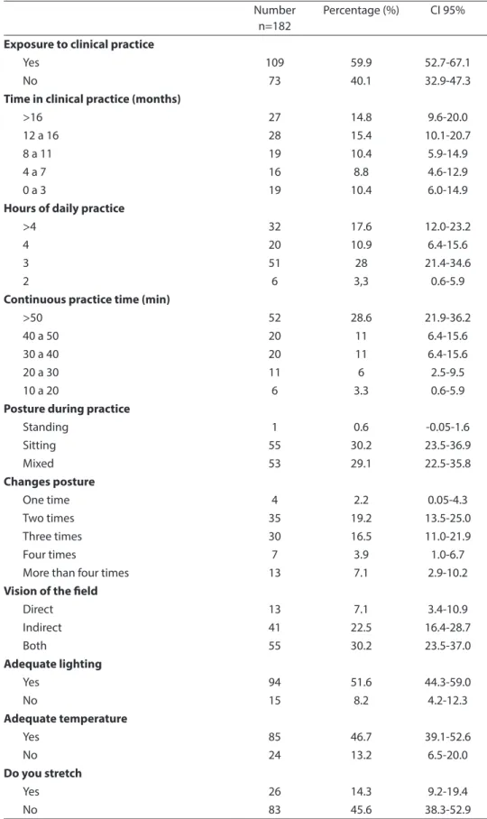
Table 1 -Postural and environmental exposures during clinical dental practice.
Tabla 1 - Exposiciones posturales y ambientales durante la práctica clínica odontológica.
Number n=182
Percentage (%) CI 95%
Exposure to clinical practice
Yes 109 59.9 52.7-67.1
No 73 40.1 32.9-47.3
Time in clinical practice (months)
>16 27 14.8 9.6-20.0
12 a 16 28 15.4 10.1-20.7
8 a 11 19 10.4 5.9-14.9
4 a 7 16 8.8 4.6-12.9
0 a 3 19 10.4 6.0-14.9
Hours of daily practice
>4 32 17.6 12.0-23.2
4 20 10.9 6.4-15.6
3 51 28 21.4-34.6
2 6 3,3 0.6-5.9
Continuous practice time (min)
>50 52 28.6 21.9-36.2
40 a 50 20 11 6.4-15.6
30 a 40 20 11 6.4-15.6
20 a 30 11 6 2.5-9.5
10 a 20 6 3.3 0.6-5.9
Posture during practice
Standing 1 0.6 -0.05-1.6
Sitting 55 30.2 23.5-36.9
Mixed 53 29.1 22.5-35.8
Changes posture
One time 4 2.2 0.05-4.3
Two times 35 19.2 13.5-25.0
Three times 30 16.5 11.0-21.9
Four times 7 3.9 1.0-6.7
More than four times 13 7.1 2.9-10.2
Vision of the ield
Direct 13 7.1 3.4-10.9
Indirect 41 22.5 16.4-28.7
Both 55 30.2 23.5-37.0
Adequate lighting
Yes 94 51.6 44.3-59.0
No 15 8.2 4.2-12.3
Adequate temperature
Yes 85 46.7 39.1-52.6
No 24 13.2 6.5-20.0
Do you stretch
Yes 26 14.3 9.2-19.4
postural exposure ranged between 12 to 16 months, with a work period of three hours a day. The most frequently used position by participants during their clinical practice, was sitting down on a stool. Regarding en-vironmental exposure, most of the students used both types of vision of the operative ield (direct and indirect) and a high percen-tage of them reported adequate lighting and temperature in the work environment, but almost half of them reported not stretching after their clinical practice.
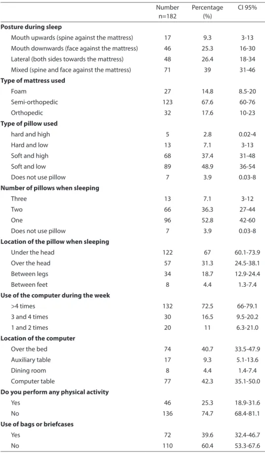
Postural exposures and physical activity unrelated to clinical practice
The most frequently described position used to sleep was mixed (backwards and in front of the mattress). The semi-orthopedic is the most used type of mattress, while the most commonly used pillow is soft and placed under the head. Also, a large number of participants use the computer more than four times a week, mostly in bed. Regarding physical activity, most of the surveyed participants reported no activity at all (table 2).
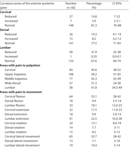
Physical assessment of the musculoskeletal system
Table 3 shows the reduction or increase of the anterior-posterior curvature of the spine, and important occurrences were observed only in the lumbar zone. Also, pain to palpation was found in the upper trapezius with VAS codes between 1 and 4 in about half of the participants, and pain during cervical lateral and lexion move-ments was seen with VAS codes between 1 and 3 in a lower proportion. Regarding the intensity of the pain according to the VAS scale, since most of the participants perceived slight levels of pain during palpation and movement, we decided to dichotomize the values to the following indicators in order to carry out the nomi-nal bivariate anomi-nalysis; no pain perceived (category 1) and pain perceived between 1 and 4 (category 2).
Posture during clinical work with the RULA visual method
When analyzing work posture during the student´s clinical practice, it was observed to be deicient in 43.1 percent (CI; 33-52) (inal value 7), which indicates need for an urgent change in the work area. While 34.8 percent (CI; 25-43) require a rapid change in posture, (inal value 5-6).
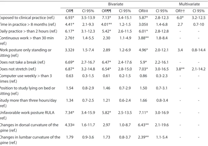
Bivariate and multivariate analysis for pain to palpation
Table 4 shows the bivariate findings obtained when the academic cycle and age estimators were adjusted, resulting in 10 exposures with statistical signiicance. For the multivariate analysis, the best model showed a statistically signiicant relation for four exposures (p=0.000; χ2=30.7); the levels of pain to palpation increased in the partici-pants of the higher semesters and in those with clinical practice for a period of time longer than 8 months. Besides, the students that do not stretch after clinical practice and those who practice in one position during three continuous hours are more likely to develop pain (table 4).
Bivariate and multivariate analysis for pain to movement
Table 5 shows the bivariate results for the adjusted estimators by age and aca-demic cycle, observing 9 exposures with statistical signiicance. For the multivariate analysis, the best model showed a statisti-cally signiicant relation with 3 exposures (p=0.000; 2=35.36), as pain levels increased
in the students with clinical practice. Also, it is 1.2 times more likely for students who practice more than two hours a day in the dental unit and for those that do not take a break between patients, to experience pain.
Discussion
Table 2 -Postural exposures and physical activity than dental practice. Tabla 2 - Exposiciones posturales y actividad física diferente a la práctica.
Number n=182
Percentage (%)
CI 95%
Posture during sleep
Mouth upwards (spine against the mattress) 17 9.3 3-13
Mouth downwards (face against the mattress) 46 25.3 16-30
Lateral (both sides towards the mattress) 48 26.4 18-34
Mixed (spine and face against the mattress) 71 39 31-46
Type of mattress used
Foam 27 14.8 8.5-20
Semi-orthopedic 123 67.6 60-76
Orthopedic 32 17.6 10-23
Type of pillow used
hard and high 5 2.8 0.02-4
Hard and low 13 7.1 3-13
Soft and high 68 37.4 31-48
Soft and low 89 48.9 36-54
Does not use pillow 7 3.9 0.03-8
Number of pillows when sleeping
Three 13 7.1 3-12
Two 66 36.3 27-44
One 96 52.8 42-60
Does not use pillow 7 3.9 0.03-8
Location of the pillow when sleeping
Under the head 122 67 60.1-73.9
Over the head 57 31.3 24.5-38.1
Between legs 34 18.7 12.9-24.4
Between feet 8 4.4 1.3-7.4
Use of the computer during the week
>4 times 132 72.5 66-79.1
3 and 4 times 30 16.5 9.5-20.2
1 and 2 times 20 11 6.3-21.0
Location of the computer
Over the bed 74 40.7 33.5-47.9
Auxiliary table 17 9.3 5.1-13.6
Dining room 8 4.4 1.4-7.4
Computer table 77 42.3 35.1-50.0
Do you perform any physical activity
Yes 46 25.3 18.9-31.6
No 136 74.7 68.4-81.1
Use of bags or briefcases
Yes 72 39.6 32.4-46.7
Table 3 - Physical assessment of musculoskeletal system.
Tabla 3 - Valoración física del sistema osteo-muscular.
Curvature zones of the anterior posterior spine
Number n=182
Percentage (%)
CI 95%
Cervical
Reduced 27 14.8 7-22
Increased 7 3.9 2-5.1
Normal 148 81.3 76-88
Dorsal
Reduced 26 14.3 9.1-19
Increased 15 8.2 4.2-12
Normal 141 77.5 71-83
Lumbar
Reduced 58 31.9 25-38
Increased 1 0.55 0.0-0.1
Normal 123 67.6 60-74
Areas with pain to palpation
Cervical 83 45.6 38-52
Upper trapezius 106 58.2 51-65
Middle trapezius 77 42.3 35-49
Wide dorsal 57 31.3 24-38
Lumbar 58 41.8 34.5-49
Areas with pain to movement
Cervical lexion 64 35.1 28-42
Dorsal lexion 18 9.9 5.5-14
Lumbar lexion 33 18.1 12.0-23
Cervical extension 32 17.5 11.0-23
Dorsal extension 18 9.9 5.0-14
Lumbar extension 41 22.5 16.0-28
Cervical rotation 24 13.1 8.0-18
Dorsal rotation 14 7.7 3-11
Lumbar rotation 15 8.2 4-12
Cervical lateral movement 65 35.7 28-42
Dorsal lateral movement 13 7.1 3-10
Lumbar lateral movement 19 10.4 5-14
musculoskeletal disorders was based on the need to obtain a risk model for occu-pational diseases and test it in future lon-gitudinal studies. These results are useful in the decision making process at the time of presenting preventive strategies to avoid uncomfortable positions during clinical practice, both for the students as well as for the dentist, and at the same time propose modiications to the design of dental chairs and initiate discussions regarding the labor demands for dentists, related with the time spent in clinical practice, continuous work and lack of rest.
The results of this study indicate that a high percentage of students going through their clinical practice presented neck and back pain related to factors of the dental practice and a few to external factors such as daily habits and postures unrelated to clinical practice. Also, studies done by Westgarrd et al.11 and Lehto et al.12 report
that the presence of these disorders may be related to multiple clinical factors such as: static postures, poor lighting, poor work posture, time in practice and not enough resting time.
Table 4 -Bivariate analysis and logistic regression for the presence of muscle palpation pain and factors related to clinical dental practice.
Tabla 4 - Análisis bivariado y regresión logística para la presencia de dolor a la palpación y factores relacionados.
Bivariate Multivariate
OR¶ CI 95% OR¶¶ CI 95% OR‡‡ CI 95% OR†† CI 95%
Exposed to clinical practice (ref.) 6.93* 3.5-13.9 7.13* 3.4-15.1 5.87* 2.8-12.3 6.0* 3.2-12.5
Time in practice > 8 months (ref.) 4.41* 2.1-9.3 4.01** 1.2-1.5 3.05‡ 1.4-6.8 2.7 0.7-10
Daily practice > than 2 hours (ref.) 6.17* 3.1-12.3 5.42* 2.6-11.5 6.01* 2.8-12.8 - -Continuous work > than 30 min
(ref.)
2.76† 1.4-5.5 2.30 1.1-4.9 3.88** 1.8-8.4 -
-Work posture only standing or sitting (ref.)
3.32‡ 1.5-7.4 2.89 1.2-6.9 4.96* 2.0-12.1 3.4 0.8-14.4
Does not take a break (ref.) 6.69* 2.7-16.7 6.47* 2.4-17.6 5.9* 2.2-16.1 -
-Does not stretch (ref.) 6.87* 3.2-14.8 6.54* 2.8-15.0 7.03* 3.0-16.5 3.8** 2.1-14.2
Computer use weekly > than 3 times (ref.)
0.63 0.3-1.5 0.61 0.2-1.5 0.86 0.3-2.3 -
-Position to study lying on bed or sitting (ref.)
1.54 0.8-2.9 1.46 0.7-2.9 1.50 0.7-3.1 -
-Study more than three hours/day (ref.)
1.34 0.7-2.5 1.21 0.6-2.4 1.66 0.8-3.4 -
-Unfavorable work posture RULA (ref.)
7.34* 3.4-15.9 5.82* 2.5-13.5 7.11* 3.0-16.9 -
-Changes in dorsal curvature of the spine (ref.)
4.33¤ 1.6-11.7 2.97 1.0-8.7 6.43** 2.1-19.6 -
-Changes in lumbar curvature of the spine (ref.)
1.79 0.9-3.6 1.73 0.8-3.7 2.39** 1.1-5.4 -
-(ref.) = categoría tomada como referencia en el análisis; valores de probabilidad con significancia estadística; *p = 0.000; †p = 0.007; ‡p = 0.003; ¤p = 0.004 **p = 0.001; ¶estimador sin ajustar; ¶¶estimador ajustado por edad; ‡‡estimador ajustado por semestre; ††estimador ajustado por regresión (chi: 30,7; p = 0000).
(ref.)=category taken as reference in the analysis; probability values with statistical significance; *p=0.000; †p=0.007; ‡p=0.003; ¤p=0.004 **p=0.001; ¶ estimator not adjusted; ¶¶estimator adjusted by age; ‡‡ estimator adjusted by semester; †† estimator adjusted by regression (χ2: 30.7; p=0000).
study analyzed the presence or absence of pain to palpation, being the most affected zones the areas of the upper trapezius and lower trapezius, while pain to movement was most frequent during cervical lateral movements and cervical flexion. Similar findings were reported by Hayes et al.13
in a systematic review, evidencing a high prevalence of muscular pain which varied between 64 and 93 percent, with the spine and the neck as the most affected regions. On the other hand, Dajpratham et al.14
found that this frequency is higher in neck and shoulders in postgraduate students; this could be related with the older age of the individuals and longer time of exposure. The evaluation of factors related with posture during clinical practice, using the
RULA visual method, revealed that deicient posture was the most frequent, which indi-cates need for an urgent change in the site of clinical practice by participants. A study carried out by Malker et al.15, 2007, indicated that there are serious consequences for the musculoskeletal system if the same posture is maintained for prolonged periods of time. This was observed in a large number of dentists, who had bad postures during work, and who need to change in order to avoid future musculoskeletal lesions.
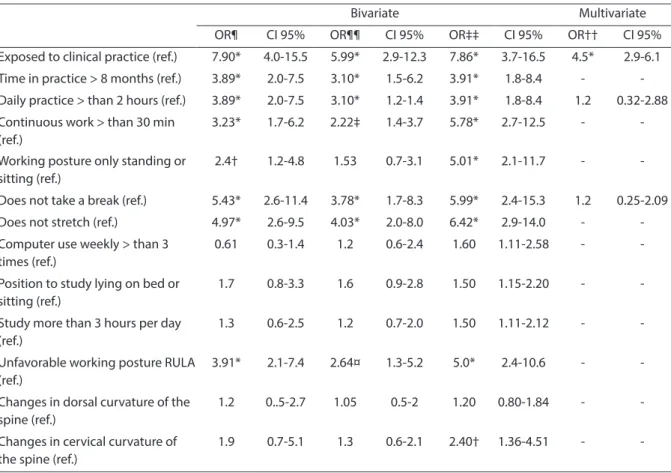
Table 5 - Bivariate analysis and logistic regression for the presence of muscle movement pain and factors related to clinical dental practice.
Tabla 5 - Análisis bivariado y regresión logística para la presencia de dolor al movimiento y factores relacionados.
Bivariate Multivariate
OR¶ CI 95% OR¶¶ CI 95% OR‡‡ CI 95% OR†† CI 95%
Exposed to clinical practice (ref.) 7.90* 4.0-15.5 5.99* 2.9-12.3 7.86* 3.7-16.5 4.5* 2.9-6.1
Time in practice > 8 months (ref.) 3.89* 2.0-7.5 3.10* 1.5-6.2 3.91* 1.8-8.4 - -Daily practice > than 2 hours (ref.) 3.89* 2.0-7.5 3.10* 1.2-1.4 3.91* 1.8-8.4 1.2 0.32-2.88
Continuous work > than 30 min (ref.)
3.23* 1.7-6.2 2.22‡ 1.4-3.7 5.78* 2.7-12.5 -
-Working posture only standing or sitting (ref.)
2.4† 1.2-4.8 1.53 0.7-3.1 5.01* 2.1-11.7 -
-Does not take a break (ref.) 5.43* 2.6-11.4 3.78* 1.7-8.3 5.99* 2.4-15.3 1.2 0.25-2.09
Does not stretch (ref.) 4.97* 2.6-9.5 4.03* 2.0-8.0 6.42* 2.9-14.0 -
-Computer use weekly > than 3 times (ref.)
0.61 0.3-1.4 1.2 0.6-2.4 1.60 1.11-2.58 -
-Position to study lying on bed or sitting (ref.)
1.7 0.8-3.3 1.6 0.9-2.8 1.50 1.15-2.20 -
-Study more than 3 hours per day (ref.)
1.3 0.6-2.5 1.2 0.7-2.0 1.50 1.11-2.12 -
-Unfavorable working posture RULA (ref.)
3.91* 2.1-7.4 2.64¤ 1.3-5.2 5.0* 2.4-10.6 -
-Changes in dorsal curvature of the spine (ref.)
1.2 0..5-2.7 1.05 0.5-2 1.20 0.80-1.84 -
-Changes in cervical curvature of the spine (ref.)
1.9 0.7-5.1 1.3 0.6-2.1 2.40† 1.36-4.51 -
-(ref.)=categoría tomada como referencia en el análisis; valores de probabilidad con significancia estadística; *p = 0.000; †p = 0.01; ‡p = 0.002; ¤p = 0.005; ¶es-timador sin ajustar; ¶¶es¶es-timador ajustado por edad; ‡‡es¶es-timador ajustado por semestre; ††es¶es-timador ajustado por regresión (chi: 35,36); p = 0000).
(ref.)=category taken as reference in the analysis; probability values with statistical significance; *p=0.000; †p=0.01; ‡p=0.002; ¤p=0.005; ¶ estimator not adjusted; ¶¶estimator adjusted by age; ‡‡ estimator adjusted by semester; †† estimator adjusted by regression (χ2: 35,36); p=0000).
also, in this group of students, there was more exposure unrelated to clinical dental practice in comparison to the total sample (p=0.000). Similar results were obtained by Harutunian et al.16 in 2010, who reported
the presence of musculoskeletal symptoms in third year students (p <0.001), associated more with environmental factors than with clinical factors. On the other hand, in this study, the students that did not stretch after clinical practice were 3.8 times more likely to present pain to palpation (p=0.000) and to movement in the spine and neck (p=0.000). Hayes et al.13 demonstrated that dentists
who do not stretch after clinical practice are 4.8 times more likely to present pain in the lumbar area. Besides, in relation to hours of study, those who study more than 16
continuous hours are more prone to develop neck pain, although, this variable was not signiicant in the present study.
Regarding the multivariate analysis for pain to muscular palpation and to move-ment, there was an increase in the asso-ciation and statistical signiicance with the variables of clinical dental practice. These results are similar to the ones reported by Hayes et al.17, 2009 who evaluated a group of
not take a break between patients. Similar results were obtained by Alexopoulos et al.18 who found a direct relationship for the
presence of pain in the neck and lower spine in students that do not take a break after providing dental service.
In accordance with the results of this stu-dy, it can be concluded that some practices and inadequate postures related with dental practice are directly related with the muscu-loskeletal disorders of the participants. This allows to identify the possible disorders that can be developed by dental students and dentists in general, and contribute to reduce the negative impact that can be caused by dental practice in two manners: in the irst place, epidemiological surveillance progra-ms could be developed by the personnel in charge of occupational health to promote the use of preventive protocols to acquire comfortable postures. On the other hand,
the problem is even more complex when seen from the occupational perspective, by involving the demands led by the in-ternational labor organization which has manifested the importance of discussing labor legislation in those countries where workers do not have beneits regarding the time of daily practice, and are subject to continuous working hours and no breaks. In this sense, it would be important for dental personnel to generate discussions to permit modiications in the stools used in their clinical practice, making them even more comfortable, and also, to reduce the conti-nuous time of clinical practice, the hours of practice during the day and the increase of time for breaks in order to permit stretching and a new musculoskeletal and mental organization. This would be reflected in better quality of life for dentists and less musculoskeletal symptoms in the future.
References
1. Giglioli S. Visión Educativa del Lenguaje Ergonómico.
Odous Científica 2008; 9: 19.
2. Evangelos C. Prevalence of musculoskeletal disorders in dentist. BMC Musculoskelet Disord 2004; 5: 1-8.
3. Mandel ID. Occupational risks in dentistry: comforts and concerns. J Am Dent Assoc 1993; 124: 40-9.
4. Caruso C, Waters R. A review of work schedule issues and musculoskeletal disorders with an emphasis on the healthcare sector. Ind health 2008; 4: 523-534.
5. Finkbeiner Bl. Selecting equipment for the ergonomic four-handed dental practice. J Contemp Dent Pract 2001; 4: 44-52.
6. Chaikumarn M. Differences in Dentists’ Working Postures When Adopting Proprioceptive Derivation vs. Conventional Concept. Int J Occup Saf Ergon 2005; 11: 441-9.
7. Valachi B, Valachi K. Mechanisms leading to
musculoskeletal disorders in dentistry. J Am Dent Assoc
2003; 134: 1344-50.
8. Leggat PA, Smith DR. Musculoskeletal disorders self-reported by dentists in Queensland, Australia. Aust Dent J 2006; 51: 324-7.
9. Díaz AJ, Gómez IP, Díaz S. Ergonomic factors that cause the presence of pain muscle in students of dentistry.
Med Oral Patol Oral Cir Bucal 2010; 15: 906-11.
10. Mcatamney L, Corlett E. RULA: a survey method for the. investigation of world-related upper limb disorders.
Applied Ergonomics 1993; 24: 91-9.
11. Westgaard RH. Effects of physical and mental stressors on muscle pain. Scand J Work Environ Health 1999; 25: 19-24.
12. Lehto TU, Helenius HY, Alaranta HT. Musculoskeletal symptoms of dentists assessed by a multidisciplinary approach. Community Dent Oral Epidemiol 1991; 19: 38-44.
13. Hayes M, Cockrell D, Smith DR. A systematic review of musculoskeletal disorders among dental professionals.
Int J Dent Hyg 2009; 7: 159-65.
14. Dajpratham, P, Ploypetch, T, Kiattavorncharoen S, Boonsiriseth, K. Prevalence and associated factors of musculoskeletal pain among the dental personnel in a dental school. J Med Assoc Thai 2010; 93: 714-21.
15. Malker, H, Musculoskeletal disorders (MSDS)
Consequences of prolonged Static postures. J Exper Med
Surg Res 2007; 167-172.
17. Hayes MJ, Smith D. Prevalence and correlates of musculoskeletal disorders among Australian dental hygiene students. Int J Dent Hyg 2009; 7: 176-81.
18. Alexopoulos EC, Stathi IC, Charizini F. Prevalence of musculoskeletal disorders in dentists. BMC
Musculoskelet Disord 2004; 9: 1-8.