ISSN 0102-695X http://dx.doi.org/10.1590/S0102-695X2012005000044 Received 7 Dec 2011 Accepted 20 Feb 2012 Available online 12 Apr 2012
olivaceum
and
A. polyneuron
, Apocynaceae
Fernanda Lisot Krentkowski,
1Márcia do Rocio Duarte
*,21Programa de Pós-graduação em Ciências Farmacêuticas, Universidade Federal do Paraná , Brazil,
2Laboratório de Farmacognosia, Departamento de Farmácia, Universidade Federal do Paraná, Brazil.
Abstract: The family Apocynaceae includes many medicinal species, such as Aspidosperma olivaceum Müll. Arg. and A. polyneuron Müll. Arg. The first, popularly known as “peroba-branca”, is a source of indole alkaloids that are recognized for their antitumor activity. The second, known as “peroba-rosa”, is used by some people for treating malaria. To contribute to the knowledge about Brazilian medicinal plants and potential vegetal drugs, a morpho-anatomical analysis of the leaves and stems of these tree species was carried out. The botanical material was fixed, sectioned and stained using standard techniques. Histochemical tests were performed and the material was also analyzed using a scanning electron microscope. Aspidosperma olivaceum and A. polyneuron are morphologically similar, both presenting alternate, simple, elliptic-lanceolate leaves. The leaves are hypostomatic with anomocytic stomata and have non-glandular trichomes. In the stem, there are peripheral phellogen, lignified fibers and some stone cells in the cortex and pith, as well as numerous gelatinous fibers near the phloem. Prismatic calcium oxalate crystals and laticifers are present in the leaves and stems. Unlike A. olivaceum, A. polyneuron also has cyclocytic stomata, predominantly unicellular non-glandular trichomes and a biconvex midrib with a sharper curve on the adaxial side. In addition, A. olivaceum differs because it has some idioblasts with phenolic content.
Keywords: Aspidosperma Brazilian species laticifers medicinal plants morpho-anatomical diagnosis
Introduction
Apocynaceae are among the ten largest families of Angiosperms, with approximately 400 genera in two subfamilies, Apocynoideae and Plumerioideae, and 3700 species. They are predominantly distributed in the tropics and subtropics, but also occur in temperate regions (Barroso, 1991; Joly, 1998; Souza & Lorenzi, 2005).
The genus Aspidosperma Mart. & Zucc., which is native to the Americas, is found in Brazil, Mexico and Argentina (Corrêa, 1984; Souza & Lorenzi, 2005), includes trees with latex (Markgraf, 1968) and is widely known for its high quality wood and use in traditional medicine (Botsaris, 2007; Oliveira et al., 2009). In Brazil, Aspidosperma olivaceum Müll. Arg., popularly known as “guatambu”, “peroba” or “peroba-branca”, is found in the Atlantic Forest from Minas Gerais to Santa Catarina (Flora Brasiliensis, 1906). This tree is a source of the indole alkaloids olivacine and ellipticine that are known for their antitumor activity (Ohashi & Oki, 1996; Oliveira, 1999; Barbosa et al., 2003; Costa, 2005; Pereira et al., 2007). Aspidosperma polyneuron Müll. Arg., a related species that is commonly known as “peroba-rosa”,
is a tree that is locally used as astringent, antipyretic, anti-diarrheal and especially as anti-malarial agent (Botsaris, 2007), and studies have proven the antimicrobial effect of its leaves, stems and roots (Ferreira et al., 2003; Granato et al., 2005).
Based on the promising phytochemical and pharmacological results of studies about A. olivaceum and A. polyneuron, the objective of the present work was to elucidate the morpho-anatomy of the leaves and stems of these medicinal plants to contribute to what is known
about the Brazilian l ora and to characterize them macro-
and microscopically because of their potential use as vegetal drugs.
Material and Methods
Plant material
Samples of leaves and stems of Aspidosperma olivaceum Müll. Arg. and A. polyneuron Müll. Arg., Apocynaceae, were collected from cultivated plants growing in the sun at the Horto Municipal da Barreirinha, in Curitiba-PR (25º36’S, 49º26’W; 930 m elevation), in
June 2009. These species were identiied by taxonomists
at the Museu Botânico Municipal de Curitiba, in the Jardim Botânico Francisca Maria Garfunkel Richbieter in Curitiba-PR (voucher numbers MBM 352341 and MBM 352342, respectively).
Methodology
Morphological descriptions of the leaves were
based on the classiication proposed by Hickey (1974)
and measurements were made from at least ten leaves, to calculate the average dimensions.
Fragments of mature leaves and young stems, obtained between 5 and 25 cm from the caulinar apex,
were ixed in FAA 70 (Johansen, 1940) and stored in 70%
(v/v) ethanol (Berlyn & Miksche, 1976). Freehand sections (transverse and longitudinal, including paradermal) of the
ixed material were made into semipermanent slides. The
dyes used were astra blue and basic fuchsine (Roeser,
1972). Permanent slides of the ixed material were also
made by dehydrating the material in an ethanol series and embedding it in glycol methacrylate (Feder & O’Brien, 1968). The material was sectioned using a rotary microtome and the sections were stained with toluidine blue (O’Brien et al., 1964). Histochemical tests were carried out with Wagner's reagent for alkaloids (Furr & Mahlberg, 1981), ferric chloride for phenolic compounds (Johansen, 1940), Sudan III for lipophilic substances (Sass, 1951), hydrochloric phloroglucin for lignin (Foster, 1949), lugol solution for starch (Berlyn & Miksche, 1976) and diluted sulfuric acid to reveal the chemical nature of the calcium crystals (Oliveira et al., 1991).
The ultrastructural analysis of the leaf surface (Souza, 1998) was performed using a scanning electron
microscope. Samples were ixed, dehydrated in an
ascending ethanol series, critical point dried with CO2, attached to stubs and sputter coated with gold.
Results
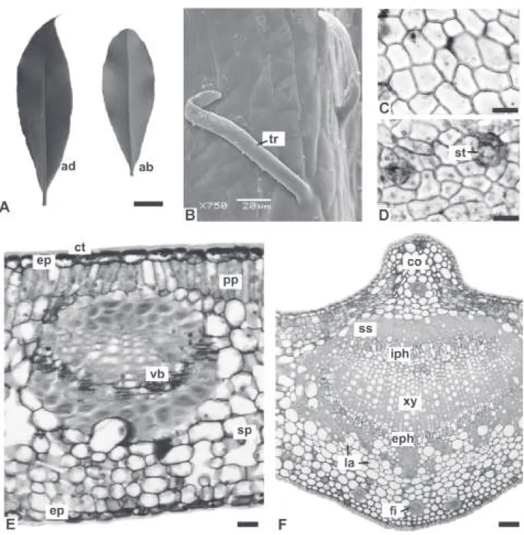
Both species have similar morpho-anatomical characters that consist of alternate, simple leaves, which are symmetrical, coriaceous, elliptic-lanceolate, with acute apex, acute and decurrent base, and entire margin (Figures 1A, 3A). The adaxial surface is bright dark green in comparison with the clearer, opaque abaxial surface. The average size of the leaves of A. olivaceum is 7.5 (4.5-9.5) × 2.3 (1.6-2.8) cm and the average size of the leaves of A. polyneuron is 5 (4.5-5.5) × 1.3 (1-1.5) cm. The average petiole lengths are 0.9 cm and 0.5 cm, respectively.
The venation is pinnate, with a midrib that
gives rise to the secondary veins, and is subclassiied as
camptodromous-brochidodromous, which means that the secondary veins form a series of prominent arcs instead of terminating at the margin.
In face view, the epidermis has cells with practically straight anticlinal walls on both surfaces and is coated with a smooth cuticle (Figures 1B-D, 3B-D). The leaves are hypostomatic, with anomocytic stomata (Figures 1C, 3D). In A. polyneuron, cyclocytic stomata are also found. On the leaves, there are simple non-glandular trichomes that are unicellular or multicellular and have a slightly granular cuticle, thick cell walls and acute apex (Figures 1D, 3B). The distribution of the trichomes is denser on the petiole than on the blade. In A. olivaceum, some multicellular trichomes have a wider apical cell in relation to the basal one. The trichomes of A. polyneuron are predominantly unicellular.
Transverse sections show that the epidermis is uniseriate and coated with a thick cuticle that forms
cuticular langes. The cells of the adaxial surface are
comparatively larger than the cells of the abaxial side and the stomata occur on the same level as the other epidermal cells (Figures 1E, 3E).
The mesophyll is dorsiventral and comprised of one to three strata of palisade cells and approximately
ten layers of spongy parenchyma, making it about 70%
of the height of the chlorenchyma (Figures 1E, 3E). Minor collateral vascular bundles with sclerenchymatic caps adjoining the xylem and phloem are distributed in the mesophyll (Figures 1E, 3E). Laticifers containing alkaloids, lipophilic and phenolic substances are present, and in A. olivaceum it is found few parenchymatic cells with phenolic content.
The midrib is biconvex, and the curvature on the adaxial surface is sharper in A. polyneuron (Figures 1F, 3F). The epidermis of this region consists of one stratum of cells with convex outer periclinal walls. The cuticle is thick and inserted among the cells forming the same
cuticular langes (Figure 1G). The palisade parenchyma is
gradually interrupted and then there is annular collenchyma (Figure 1G) composed of about six layers near the adaxial surface and four strata on the abaxial side (Figures 1F, 3F). In the collenchyma and ground parenchyma, it is
encountered laticifers, ibers with thick cell walls and
reduced lumen, some stone cells and prismatic calcium oxalate crystals (Figures 1G, H, 3F). There is only one bicollateral vascular bundle, which forms an open arc and is surrounded by a practically continuous sclerenchymatic sheath (Figures 1F, 3F). The midrib of A. olivaceum also has some idioblasts with phenolic compounds.
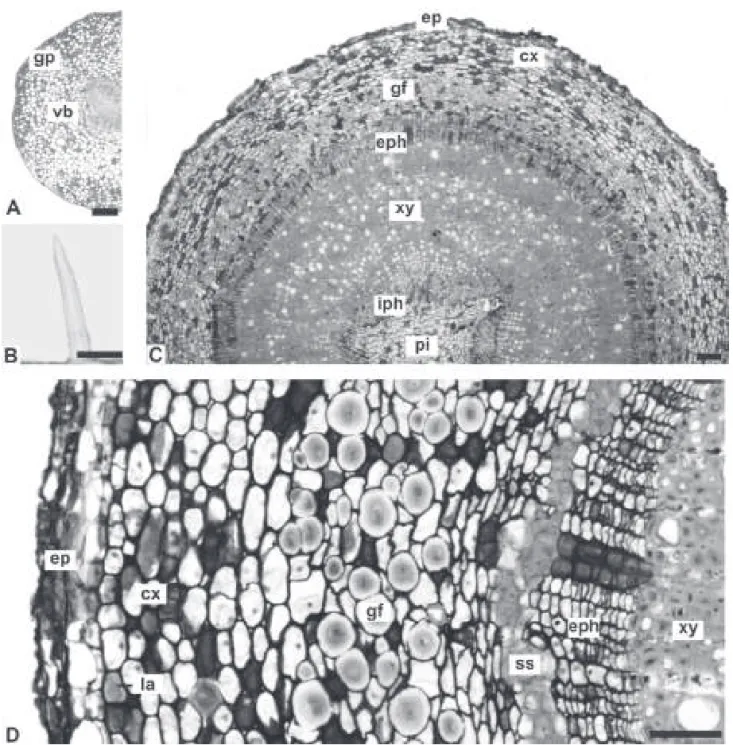
The petiole, in transverse section, is circular to biconvex with a uniseriate epidermis and non-glandular trichomes similar to those previously described (Figures 2A, C, 4A, B). The vascular bundle is bicollateral and forms an open arc (Figures 2A, 4A). In A. olivaceum,
there are ibers in different stages of ligniication (Figure
2B) and a starch sheath encircling the vascular system.
Laticifers, ibers, stone cells and prismatic calcium
In A. olivaceum, few idioblasts with phenolic compounds are also noted.
The stem is circular in transverse section (Figure 4C). At the level analyzed, it has a persistent epidermis and peripheral phellogen (Figures 2F, 4D). Some non-glandular trichomes that are similar to those on the
leaves are also present. In the cortex, it is found annular collenchyma, cortical parenchyma and stone cells. Fibers
with thick ligniied cell walls and reduced lumen occur in
the outer cortex and pith (Figure 2F). Numerous gelatinous
ibers (Figures 2D, E, 4C, D) are encountered in the
vicinity of the vascular system, which consists of groups
Figure 1. Aspidosperma olivaceum Müll. Arg., Apocynaceae - leaf: A. General aspect; B, C. Face view of the adaxial and abaxial sides of the epidermis, respectively; D. Abaxial side of the epidermis in SEM, revealing smooth cuticle, stomata and non-glandular trichomes; E. Transverse section of the intervein region of the blade, showing dorsiventral mesophyll; F. Transverse section of the midrib; G. Detail
of the midrib, indicating collenchyma and laticifers; H. Pormenor of the previous igure. Abbreviations: ab: abaxial side; ad: adaxial side; co: collenchyma; ct: cuticle; ep: epidermis; i: iber; la: laticifer; pp: palisade parenchyma; SEM: scanning electron microscopy; sp:
Figure 2. Aspidosperma olivaceum Müll. Arg., Apocynaceae - petiole and stem: A. Transverse section of the petiole; B, C. Details
of the previous igure, showing iber, prismatic calcium oxalate crystals and multicellular non-glandular trichome; D. Overall organization of the stem in transverse section; E, F. Pormenor of the previous igure; Abbreviations: cx: cortex; ep: epidermis; eph: external phloem; i: iber; gf: gelatinous iber; gp: ground parenchyma; iph: internal phloem; la: laticifer; pi: pith; pr: prismatic crystal;
Figure 3. Aspidosperma polyneuron Müll. Arg., Apocynaceae - leaf: A. General aspect; B. Non-glandular trichomes and smooth cuticle on the adaxial surface of the epidermis, in SEM; C, D. Face view of the adaxial and abaxial sides of the epidermis, respectively; E. Transverse section of the blade showing dorsiventral mesophyll and a minor collateral vascular bundle; F. Transverse section of the midrib revealing the sharp curvature of the adaxial side. Abbreviations: ab: abaxial side; ad: adaxial side; co: collenchyma; ct: cuticle;
ep: epidermis; eph: external phloem; i: iber; iph: internal phloem; la: laticifer; pp: palisade parenchyma; SEM: scanning electron
Figure 4. Aspidosperma polyneuron Müll. Arg., Apocynaceae – petiole and stem: A. Transverse section of the petiole; B. Unicellular,
non-glandular trichome of the petiole; C. Overall aspect of the stem in transverse section; D. Detail of the previous igure. Abbreviations: cx: cortex; ep: epidermis; eph: external phloem; gf: gelatinous iber; gp: ground parenchyma; iph: internal phloem; la: laticifer; pi:
pith; ss: sclerenchymatic sheath; vb: vascular bundle; xy: xylem. Bar: 20 µm (D), 50 µm (B), 100 µm (A, C).
of ligniied ibers, external phloem, xylem and internal
phloem (Figures 2D, E, 4C, D). Laticifers (Figure 2F) and prismatic calcium oxalate crystals are also present. In A. olivaceum there are some phenolic idioblasts as well.
Discussion
Considering the external morphological appearance of leaves of Apocynaceae, Aspidosperma olivaceum and A. polyneuron have aspects of those
found within the family and genus (Markgraf, 1968), such as leaves that are simple and alternate, and blades that are symmetrical, elliptic-lanceolate, and coriaceous with entire margin. In comparison, similar characters were found by Albuquerque (1971) for A. carapanauba Pichon, A. marcgravianum Woodson and A. oblongum A. DC.
found in the family is normally pinnate and subclassiied
as camptodromous-brochidodromous (Mohan & Inamdar,
1982). This type was veriied in the two species studied.
In another study, Quinet & Andreata (2005) noted that A. parvifolium A. DC. and A. pyricollum Müll. Arg have the camptodromous pattern in contrast to craspedodromous venation found in A. compactinervium Kuhlm.
As for the remaining morpho-anatomical aspects investigated, A. olivaceum and A. polyneuron have common characters found in other representatives of the genus, such as leaf epidermal cells with practically straight anticlinal walls, thick and smooth cuticle that forms
cuticular langes, anomocytic stomata (or predominantly
this type) restricted to the abaxial surface, non-glandular trichomes, dorsiventral mesophyll, bicollateral vascular bundle, sclereids, prismatic calcium oxalate crystals and laticifers. In general, these characters are present in A. carapanauba, A. marcgravianum, A. oblongum, A. excelsum Benth. (Albuquerque, 1971), A. parvifolium, A. pyricollum (Quinet & Andreata, 2005), A. spruceanum Benth. ex Müll. Arg., A. desmanthum Benth. ex Müll. Arg. (Reis, 2008) and A. nitidum Benth. (Añez, 2009).
Some differences occur among Aspidosperma species, for example, A. carapanauba and A. excelsum have striated cuticle, A. desmanthum has calcium oxalate druses (Reis, 2008), A. nitidum possesses paracytic stomata (Añez, 2009) and A. quebracho Griseb. has isobilateral mesophyll (Metcalfe & Chalk, 1950). However, structural variations should be evaluated carefully because morphology can be environmentally
inluenced, such as the shape of the epidermal cells,
cuticular ornamentation and the organization of the mesophyll (Cutter, 1986).
In species of Apocynaceae, trichomes can be glandular or non-glandular, simple or branched, and uniseriate or shortly unicellular (Metcalfe & Chalk, 1988; Werker, 2000). Reis (2008) found multicellular,
non-glandular, iliform trichomes which were ornamented with
spines in A. carapanauba, A. desmanthum, A. excelsum and A. spruceanum, whereas Añez (2009) described the presence of short, non-glandular trichomes in A. nitidum. This data is similar to the results of the present study where unicellular and multicellular trichomes were found.
According to Metcalfe & Chalk (1950), non-articulated laticifers, with diversely colored exudates and associated with the vascular system, are typical for
the family. Albuquerque (1971) conirmed this by noting
that the leaves of A. marcgravianum and A. oblongum had non-articulated laticifers. However, subsequent studies included data that contradict this pattern, for example, Demarco et al. (2006) found the presence of articulated laticifers, with thick cell walls, in A. australe Müll. Arg. In addition, Reis (2008) described articulated laticifers, which had thin walls and varied in diameter,
along the vascular bundles in the leaves of A. excelsum, A. desmanthum and A. spruceanum. In relation to A. carapanauba, contradictory results were published because Albuquerque (1971) found that this species had articulated laticifers and Reis (2008) found that the internal secretory structures of this species were not articulated. Although the present work did not determine the type of laticifers, the location of these structures near the vascular system in the two species studied is in accordance with the family. Based on various researches on latex, its composition is varied, consisting of alkaloids, proteins, phytosterols and fatty acids, and may be associated with pharmacological activities (Yoder & Mahlberg, 1976; Furr & Mahlberg, 1981; Endress & Bruyns, 2000).
The young stems of A. olivaceum and A. polyneuron showed incipient secondary growth, and it was possible to distinguish the persistent epidermis
and the supericial periderm that was being formed. In
Apocynaceae, phellogen is generally peripheral (Metcalfe & Chalk, 1950; Esau, 1974). The presence of prismatic
calcium oxalate crystals, ibers and stone cells was also
found in other species of the genus, such as A. quebracho (Metcalfe & Chalk, 1950).
Although there are many similarities between A. olivaceum and A. polyneuron, the results of this study show that they can be distinguished based on the contour of the midrib and the type of predominant trichome. Aspidosperma polyneuron has a midrib in transverse section that is more acute on the adaxial surface, and also has a higher frequency of unicellular trichomes when compared with A. olivaceum. In addition, A. olivaceum has idioblasts with phenolic content. According to Scalbert (1991) and Beckman (2000), these cells can be part of the defense mechanisms of plants because they
are toxic and astringent, which makes it dificult for
herbivores to feed on the plants and makes the plants resistant to microorganisms. These metabolites also interfere on the taste and color of several vegetal species,
being used in food industry for lavoring and dyeing.
Besides, different therapeutic effects are attributed to them, such as expectorant, hepatoprotective, choleretic, anticholesteremic and analgesics, proving their pharmacological importance (Carvalho et al. 2007).
Aknowledgements
The authors express their gratitude to the Centro de Microscopia Eletrônica (UFPR) for the micrographs, the taxonomists of the Museu Botânico Municipal de
Curitiba for the species identiication and to the
REUNI-UFPR for the scholarship to FLK.
References
Aspidosperma da Amazônia Brasileira (Apocynaceae): Aspidosperma carapanauba Pichon, A. marcgravianun Woodson e A. oblongum A. DC. Acta Amaz 1: 9-20. Añez RBS 2009. Análise morfoanatômica das folhas e casca
de Aspidosperma nitidum Benth. e Aspidosperma marcgravianum Woodson (Apocynaceae) com abordagem farmacognóstica e etnofarmacológica. Manaus, 55p. Tese de Doutorado em Ciências Biológicas, Instituto Nacional de Pesquisas da Amazônia, Universidade Federal do Amazonas. Barbosa WLR, Tavares ICC, Soares DC 2003. Alcalóides de
Aspidosperma auriculatum Standl. Rev Bras Farmacogn 13: 6-8.
Barroso GM 1991. Sistemática de Angiospermas no Brasil. Viçosa: UFV Imprensa Universitária.
Beckman CH 2000. Phenolic-storing cells: keys to programmed cell death and periderm formation in wilt disease resistance and in general defence responses in plants. Physiol Mol Plant Pathol 7: 101-110.
Berlyn GP, Miksche JP 1976. Botanical microtechnique and cytochemistry. Ames: Iowa State University.
Botsaris AS 2007. Plants used traditionally to treat malaria
in Brazil: the archives of lora medicinal. J Ethnobiol Ethnomed 3: 13-18.
Carvalho JCT, Gosmann G, Schenkel EP. Compostos fenólicos simples e heterosídicos. In Simões CMO, Schenkel EP, Gosmann G, Mello JCP, Mentz LA, Petrovick PR (Org.) Farmacognosia: da planta ao medicamento 6. ed. Porto Alegre/Florianópolis: UFRGS/UFSC, p. 519-535. Corrêa MP 1984. Dicionário das plantas úteis do Brasil. Rio
de Janeiro: Instituto Brasileiro de Desenvolvimento Florestal.
Costa FJC 2005. Estudo itoquímico e busca de substâncias bioativas em Aspidosperma olivaceum (Müll.) Arg. e Malouetia arborea (Vell.) Miers. (Apocynaceae). Araraquara, 231p. Tese de Doutorado em Química, Instituto de Química, Universidade Estadual Paulista. Cutter EG 1986. Anatomia vegetal: células e tecidos. 2 ed. São
Paulo: Roca.
Demarco D, Kinoshita LS, Castro MM 2006. Laticíferos articulados anastomosados - novos registros para Apocynaceae. Rev Bras Bot 29: 133-144.
Endress ME, Bruyns PV 2000. A revised classiication of
Apocynaceae. Bot Rev 66: 1-56.
Esau K 1974. Anatomia das plantas com sementes. São Paulo: E. Blücher.
Feder N, O' Brien TP 1968. Plant microtechnique: some principles and new methods. Am J Bot 55: 123-142. Ferreira DT, Silva-Junior JV, Soeira LS, Zanolli-Filho LA,
Ishikawa NK, Barbosa AM, Yassumoto L 2003. Avaliação da atividade antifúngica dos extratos etanólicos de raiz, caule e folhas de Aspidosperma
polyneuron. XI Encontro de Química da Região Sul.
Pelotas, Brasil.
Flora Brasiliensis 1906. http://lorabrasiliensis.cria.org.br,
accessed Jul 2010.
Foster AS 1949. Practical plant anatomy. Princeton: D. Van Nostrand.
Furr M, Mahlberg PG 1981. Histochemical analyses of laticifers and glandular trichomes in Cannabis sativa. J Nat Prod 44: 153-159.
Granato D, Nunes DS, Mattos PP, Rios EM, Glinski A, Rodrigues LC, Zanusso-Junior G 2005. Chemical and biological evaluation of rejects from the wood industry. Braz Arch Biol Techn 48: 237-241.
Hickey LJ 1974. Clasiicación de la arquitectura de las hojas de
dicotiledóneas. Bol Soc Argent Bot 16: 1-26.
Johansen DA 1940. Plant microtechnique. New York: McGraw-Hill Book.
Joly AB 1998. Botânica: introdução à taxonomia vegetal. 12 ed. São Paulo: Imprensa Nacional.
Markgraf F 1968. Apocináceas. In Reitz PR Flora Ilustrada Catarinense Itajaí: Herbário Barbosa Rodrigues, p. 1-112.
Mauseth JD 1988. Plant anatomy. Menlo Park: Benjamin/ Cummings.
Metcalfe CR, Chalk L 1950. Anatomy of the dicotyledons: leaves, stem, and wood in relation to taxonomy with notes on economic uses. Oxford: Clarendon.
Metcalfe CR, Chalk L 1988. Anatomy of dicotyledons. Oxford: Clarendon Press.
Mohan JSS, Inamdar JA 1982. Leaf architecture of Apocynaceae. Proc Indian Acad Sci 91: 189-200.
O'Brien TP, Feder N, McCully ME 1964. Polychromatic staining of plants cell walls by toluidine blue O. Protoplasma 56: 368-373.
Ohashi M, Oki T 1996. Ellipticine and related anticancer agents. Expert Opin Ther Pat 6: 1285-1294.
Oliveira AJB 1999. Estudo de seis espécies do gênero Aspidosperma utilizando GC, GC/MS e HPLC: análise qualitativa e quantitativa. Teste autobiográico; cultura de tecidos e células vegetais e rota de preparação de compostos diméricos ramilorina A e ramilorina B. Campinas, 371p. Tese de Doutorado em Química Orgânica, Instituto de Química, Universidade Estadual de Campinas.
Oliveira F, Akisue G, Akisue MK 1991. Farmacognosia. São Paulo: Atheneu.
Oliveira VB, Freitas MSM, Mathias L, Braz-Filho R, Vieira IJC 2009. Atividade biológica e alcaloides indólicos do gênero Aspidosperma (Apocynaceae): uma revisão. Rev Bras Pl Med 11: 92-99.
Pereira MM, Jácome RLRP, Alcântara AFC, Alves RB, Raslan DS 2007. Alcalóides indólicos isolados de espécies do gênero Aspidosperma (Apocynaceae). Quím Nova 30: 970-983.
Reis ARS 2008. Anatomia foliar e o xilema secundário de espécies de Aspidosperma Mart. & Zucc. (Apocynaceae). Belém, 93p. Dissertação de Mestrado em Botânica Tropical, Universidade Federal Rural da Amazônia. Roeser KR 1972. Die Nadel der Schwarzkiefer-Massenprodukt
und Kunstwerk der Natur. Mikrokosmos 61: 33-36. Sass JE 1951. Botanical microtechnique. Ames: Iowa State
College.
Scalbert A 1991. Antimicrobial properties of tannins.
Phytochemistry 30: 3875-3883.
Souza VC, Lorenzi H 2005. Botânica sistemática. São Paulo: Nova Odessa.
Souza W 1998. Técnicas básicas de microscopia eletrônica
aplicadas às Ciências Biológicas. Rio de Janeiro:
Sociedade Brasileira de Microscopia Eletrônica. Werker E 2000. Trichome diversity and development. Adv Bot
Res 31: 1-35.
Yoder LR, Mahlberg PG 1976. Reactions of alkaloid and histochemical indicators in laticifers and specialized parenchyma cells of Catharanthus roseus (Apocynaceae). Am J Bot 63: 1167-1173.
*Correspondence
Márcia do Rocio Duarte
Programa de Pós-graduação em Ciências Farmacêuticas, Universidade Federal do Paraná, Setor de Ciências da Saúde, Sede Botânico
Av. Pref. Lothário Meissner, 632, Jardim Botânico, 80210-170 Curitiba-PR, Brazil