Radiol Bras vol.50 número6
Texto
Imagem


Documentos relacionados
The authors presented their pioneering work emphasizing that after appropriate validation this new magnetic resonance imaging (MRI) and MRI / magnetic resonance spectroscopic
Objectives: To evaluate the use of magnetic resonance imaging in patients with β -thalassemia and to compare T2* magnetic resonance imaging results with
Inclusion criteria for selecting articles were: » Studies based on magnetic resonance imag- ing (MRI), computed tomography (CT) and/ or volumetric cone-beam tomography, which assessed
Proton magnetic resonance spectroscopic imaging and magnetic resonance imaging volumetry in the lateralization of temporal lobe epilepsy: a series of 100 patients. Connelly A,
Published in this issue of Radiologia Brasileira , the article “Predictive performance of BI-RADS magnetic resonance imaging descriptors in the context of suspicious (category
Objective: To assess the role of diffusion-weighted imaging (DWI) in the evaluation of breast lesions classiied as suspicious on magnetic resonance imaging (MRI), correlating
Role of magnetic resonance imag- ing in the planning of breast cancer treatment strategies: comparison with con- ventional imaging techniques. Turnbull L, Brown S, Harvey I,
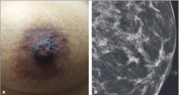
B: Axial MRI sequence of the breasts, with digital subtraction, showing an area of heterogeneous non-nodular enhance- ment with segmental distribution and minimal
