ORIGINAL ARTICLE
Oxidative Stress Tolerance by Calcium and Histidine in Two
Tomato Cultivars Under Nickel Stress
Mozafari H.*
1, Asrar Z.
1, Rezanejad F.
1, Pourseyedi S.
2and
Yaghoobi M.M.
31
Biology Department, Shahid Bahonar University of Kerman, Iran
2
Agronomy Department, Shahid Bahonar University of Kerman, Iran
3
Environmental Sciences Institute, International Center for Sciences, High Technology and Environmental Sciences Mahan, Iran
*Tel: +983413222032, +989131971873
Fax: +983413222032
*E-Mail:
Mozafari.Hossein@gmail.com
Received December 31, 2013
We investigated calcium (Ca) and L-histidine (His) interaction on nickel (Ni)-induced oxidative stress tolerance in two tomato (Solanum lycopersicum Mill.) cultivars including Cal-J N3 and PetoearlyCH. CaCl2 (0 and 300 µM) and L-histidine (0 and 300 µM) effects on the oxidative
responses in these cultivars cultured were compared in the hydroponic media under Ni stress (NiSO4; 0,150 and 300 µM). The activities of antioxidative enzymes including catalase (CAT),
guaiacol peroxidase (GPX), ascorbate peroxidase (APX), superoxide dismutase (SOD) and total content of proteins, malondialdehyde (MDA), other aldehydes, H2O2, Ca2+, Ni2+, ascorbate
(ASC), dehydroascorbate (DHA) and electrolytes leakage (EL) were determined. The obtained results indicated that the application of Ca and His generally reduced oxidative markers such as the contents of EL, H2O2, MDA and activity of CAT as well as the Ni2+content of root and shoot
organs under nickel toxicity, while application of Ni treatment without Ca+His increased these oxidative parameters and accumulation of Ni2+,compared to the control. Applying Ni without
Ca and His has resulted in reduction of GPX, APX and SOD activities as well as concentrations of root and shoot Ca2+and ASC in the two mentioned cultivars. Application of Ca and His lead to
the elevated contents of Ca2+ and ASC, increased activities of GPX, APX and SOD as well as
inhibition of Ni2+ accumulation differently in both cultivars. Ca and His also alleviated the
adverse effects of Ni stress on the selected investigated parameters especially in Petoearly CH cultivar. Thus, interaction of Ca and His appeared to improve adaptive responses to Ni stress leading to decreasing Ni-induced oxidative stress in the tomato plants. Therefore, our results suggest that Ca+His alleviated nickel-induced oxidative stress by uptake and inhibition of translocation of Ni2+ plus Ni chelating mechanism improvement in the tomato cultivars.
ORIGINAL ARTICLE
Oxidative Stress Tolerance by Calcium and Histidine in Two
Tomato Cultivars Under Nickel Stress
Mozafari H.*
1, Asrar Z.
1, Rezanejad F.
1, Pourseyedi S.
2and
Yaghoobi M.M.
31 Biology Department, Shahid Bahonar University of Kerman, Iran 2
Agronomy Department, Shahid Bahonar University of Kerman, Iran
3
Environmental Sciences Institute, International Center for Sciences, High Technology and Environmental Sciences Mahan, Iran
*Tel: +983413222032, +989131971873
Fax: +983413222032
*E-Mail:
Mozafari.Hossein@gmail.com
Received December 31, 2013
We investigated calcium (Ca) and L-histidine (His) interaction on nickel (Ni)-induced oxidative stress tolerance in two tomato (Solanum lycopersicum Mill.) cultivars including Cal-J N3 and PetoearlyCH. CaCl2 (0 and 300 µM) and L-histidine (0 and 300 µM) effects on the oxidative
responses in these cultivars cultured were compared in the hydroponic media under Ni stress (NiSO4; 0,150 and 300 µM). The activities of antioxidative enzymes including catalase (CAT),
guaiacol peroxidase (GPX), ascorbate peroxidase (APX), superoxide dismutase (SOD) and total content of proteins, malondialdehyde (MDA), other aldehydes, H2O2, Ca2+, Ni2+, ascorbate
(ASC), dehydroascorbate (DHA) and electrolytes leakage (EL) were determined. The obtained results indicated that the application of Ca and His generally reduced oxidative markers such as the contents of EL, H2O2, MDA and activity of CAT as well as the Ni2+content of root and shoot
organs under nickel toxicity, while application of Ni treatment without Ca+His increased these oxidative parameters and accumulation of Ni2+,compared to the control. Applying Ni without
Ca and His has resulted in reduction of GPX, APX and SOD activities as well as concentrations of root and shoot Ca2+and ASC in the two mentioned cultivars. Application of Ca and His lead to
the elevated contents of Ca2+ and ASC, increased activities of GPX, APX and SOD as well as
inhibition of Ni2+ accumulation differently in both cultivars. Ca and His also alleviated the
adverse effects of Ni stress on the selected investigated parameters especially in Petoearly CH cultivar. Thus, interaction of Ca and His appeared to improve adaptive responses to Ni stress leading to decreasing Ni-induced oxidative stress in the tomato plants. Therefore, our results suggest that Ca+His alleviated nickel-induced oxidative stress by uptake and inhibition of translocation of Ni2+ plus Ni chelating mechanism improvement in the tomato cultivars.
Key words: Cal-J N3, Petoearly CH, antioxidative enzymes, malondialdehyde, electrolytes leakage
Soil pollution by heavy metals (HMs) including
Cd, Cu, Ni, Pb, and Zn occur by industrial wastes
containing toxic heavy metals in environment
contain heavy metals in an amount enough to cause
toxicity to the crop plants (Whitby and Huchinson,
1974). Therefore, abiotic stresses like heavy metal
stress, air pollutants stress and others negatively
affect processes associated with biomass
production and seed yield in almost all major field
grown crops. Every metal and plant interacts in a
specific way, which depends on several factors such
as type of soil, growth conditions, and the presence
of other ions (Felix-Henningsen, 2010; Ghani, 2010;
Dalvi and Bhalerao, 2013).
Heavy metal toxicity as one of the major abiotic
stresses induces hazardous effects in crop plants. A
common consequence of HM toxicity is the
excessive accumulation of reactive oxygen species
(ROS) and methylglyoxal (MG), both of which can
cause peroxidation of lipids, oxidation of protein,
inactivation of enzymes, DNA damage and/or
interact with other vital constituents of plant cells.
Higher plants have evolved antioxidant defense
systems to scavenge ROS and MG. In addition, HMs
may be sequestered by organic acids, glutathione
(GSH), or by specific metal-binding ligands in cells.
GSH as a central molecule of antioxidant defense
systems is involved in either direct or indirect
control of ROS and MG and their reaction products
in plant cells, thus protecting the plant from HM
induced oxidative damage (Hossain et al., 2012).
Nickel is naturally present in soil and water,
usually in low concentrations. Most agricultural
soils contain an average of 0.005 μg of Ni g-1 dry
weight (DW), and symptoms of phytotoxicity often
become apparent at Ni concentrations as low as
25-30 μg g-l soil (Khalid and Tinsley, 1980). Growth
inhibition, chlorosis and reduced water content of
tissues are commonly observed in plants exposed
to phytotoxics amounts of Ni (Pandolfini et al.,
1992). Despite his harmful effect, several plants
nutritionally require Ni for various metabolic
activities (Welch, 1981) in very low concentrations.
Physiological role of Ni and its toxic effects on
higher plants have been observed (Temple and
Bisessar, 1981).
At cellular and molecular levels, oxidative stress
is widely studied as a key marker of plant stress
especially metal toxicity. Studies have reported that
toxic levels of Cu, Ni and Zn result in a stimulation
of lipid peroxidation leading to a loss of membrane
function. It is documented that excessive heavy
metal such as Ni exposure may increase reactive
oxygen species (ROS) accumulation in plants, and
oxidative stress would arise if the balance between
ROS generation and destruction were broken
(Gajewska and Sklodowska, 2007). Membrane
peroxidation has been observed in plants that
treated with excess Ni amounts (Gajewska and
Sklodowska, 2007). Cells have evolved an elaborate
system of enzymatic and non enzymatic
antioxidants which help to scavenge these
indigenously generated ROS. Various enzymes
involved in ROS-scavenging have been manipulated,
over expressed or down regulated to add to the
present knowledge and understanding the role of
the antioxidant systems. (Ahmad et al., 2010). At
the time the first mechanisms of chloroplast redox
regulation were being discovered, ROS were
regarded as by-products of potentially beneficial
reactions. Indeed, it remains the case that
reduction of O2 to superoxide by the thylakoid
electron transport chain can prevent over reduction
(redox poising) and contribute to chloroplast ATP
pools via pseudo cyclic phosphorylation.
Nevertheless, as they became implicated in diverse
stress responses, ROS subsequently gained the
reputation as damaging molecules (Foyer and
Superoxide Dismutase (SOD) and catalase (CAT)
are enzymes that represent the first line of
antioxidant defense in plant cells. Whereas SOD is
responsible for the removal of superoxide radical
(O2•−), CAT metabolizes H2O2 into H2O and O2. Both
enzymes not only prevent formation of more toxic
ROS, but play an essential role in cellular H2O2
signaling. Since there are a number of ROS
producing sites, both enzymes were found to be
present in different isoforms localized in ROS
generating organelles. Whereas CAT has been
identified in three different isoforms that could be
localized in both cytosol and peroxisomes based on
their amino-acid targeting sequence, SOD is present
in three metallic forms each occupying a specified
organelle. Fe2+ SODs are located in the chloroplasts,
the Mn2+ isoform in the mitochondria and Cu2+/Zn2+
isoforms are present in the chloroplast, cytosol and
possibly in extracellular spaces as well (Skyba et al.,
2012).
Plants possess an enzymatic protective
mechanism including peroxidases such as GPX and
APX, besides CAT and SOD. These anti oxidative
enzymes have an important role in radicals and
peroxides control that are produced under metals
stress such as Ni (Tanyolac et al., 2007). Kumar et
al. (2007) showed that nickel treatment, 100 μM
NiSO4, caused increase in the H2O2 concentration
concomitantly with antioxidant enzyme activities in
maize. Therefore, SOD catalyzes disproportion of
superoxide radicals (O2•-) into H2O2 for decreasing
the hydroxyl radical (OH•) content (Foyer, 1994).
CAT, GPX and APX are contributed in decreasing the
content of plant H2O2. In leaf tissues, H2O2 is
transformed in chloroplasts through the ascorbate
dependent H2O2 scavenging pathway which involves
oxidations and re-reductions of ascorbate,
glutathione and NADPH (Baccouch et al., 1998).
The prevention of toxic metal accumulation in
the cytoplasm and organelles is one of the
important mechanisms for plant tolerance to heavy
metals. In tolerant plants, heavy metals as Ni2+, Cd2+
and Cu2+ are very often chelated or precipitated
inside the cell vacuoles that this indicates a
transport of heavy metals through the cytoplasm
(Howden et al., 1995). In plants with ability for Ni
hyper accumulation, the low molecular chelators
including histidine (Kramer et al., 1996), malonate
or malate (Brooks et al., 1979) and citrate (Lee et
al., 1977) have been contributed for Ni
detoxification. It is approved that yeast tolerance
(Saccharomyces cerevisiae) to Ni2+ toxicity was
depending on high cellular His concentrations (Joho
et al., 1992). Increasing of free histidine and the
chelating of the heavy metals ions were found for
the Ni transport by the imidazole nitrogen of the
amino acid (Kramer et al., 1996). In addition, metal
chelators such as phytosiderophores (Von Wiren et
al., 1996) or organic acids (Ma et al., 2001) regulate
the transportable nutrients availability for metal
uptake by plant cells. Phytosiderophores have been
identified in the barley (Hordeum vulgare) xylem
sap by Shah et al. (2001).
Calcium is an essential macronutrient with
diverse functions in plants (Wu and Hendershot,
2010). Calcium plays an important regulatory role in
cell division, cell extension, cell wall and membrane
synthesis and function (Hanson, 1984; McLaughlin
and Wimmer, 1999; Girija et al., 2002). Calcium also
functions as a second messenger at low
concentrations in the signal transduction between
environmental factors and plant responses
(Marschner, 1995). The concentration of Ca in the
cytosol is very low as 1µM approximately, since Ca
is cytotoxic in higher concentrations at milimolar
plant systems depend on its unique chemical
properties that allowed it to exist in a wide variety
of binding states (Hepler and Wayne, 1985). In the
cell wall, Ca is mainly bound to exchangeable sites
in the middle lamella. By binding to carboxylic
groups of the poly galacturonic acids (pectins) and
cross linking the pectic chains of the middle lamella,
Ca strengthens the cell wall and controls its rigidity
(Demarty et al., 1984). The cell wall selectivity
property correlates with optimum calcium content
in plant tissues and rhizosphere (Girija et al., 2002).
The cell wall is important chelator of toxic metals
such as nickel, as far as the metal is transported
apoplastically especially in root tissue. Pectic sites,
histidyl groups and extracellular carbohydrates
present in plants cell wall causes immobilization of
heavy metals and prevent their uptake in to cytosol.
The heavy metals are bound to carboxylic groups of
poly galacturonic acids restricting the plant uptake.
Recent studies indicated that the chemical
properties of the cell wall might modulate metal
uptake and consequently metal tolerance (Dalvi
and Bhalerao, 2013). The cell walls inevitably come
into direct contact with the heavy-metal-containing
medium in soil and act as a cation exchanger in
plant roots (Küpper et al., 2001). It has been
described that calcium may decrease the uptake,
translocation and accumulation of heavy metals in
plants (Osteras and Greger, 2006).
There is little information about the sensitivity
and physiological responses of different tomato
cultivars to Ni toxicity, although tomato rank is the
second important vegetables in terms of planting
areas and production. Moreover, results from effect
of Ni toxicity on the antioxidant enzymes are
contradictory and both increase and decrease in
their activities have been observed under different
treatments. This warrants the further study of
Ni-induced antioxidant systems in other plants such as
tomato cultivars. The objectives of the present
study are to evaluate the effects of Ca, His and Ni
treatments on the activities of four anti oxidative
enzymes (CAT, GPX, APX, SOD), and accumulation
of protein, MDA, other aldehydes, H2O2, Ca2+, Ni2+,
ASA, DHA and membrane ion leakage in the two
probable Ni sensitive tomato cultivars in
hydroponic culture. Thus, the aim of this research is
to determine the Ca and His role in the alleviation
of oxidative stress and improvements of the
antioxidant enzyme activities in the two mentioned
tomato cultivars from Iran. Moreover, we evaluated
and compared Ni stress sensitivity and tomato
cultivars response to Ca, His and Ni treatments
between these two important tomato cultivars.
MATERIALS AND METHODS
Plant Growth: Two tomato (Solanum
lycopersicum Mill.) cultivars including Cal-J N3and
Petoearly CH, being widely planted in southeastern
of Iran, were used in this study. Seeds of recent
tomato cultivars were placed on two sheets of filter
paper in petri dishes (90 mm) containing Hoagland
solution for optimum seedling growth (Hoagland
and Arnon, 1950). After 7 d of culture, uniform
seedlings of tomato were selected and transferred
into dark polyethylene vessels (two plants per
vessel), each supplied with 50 mL of a modified
one-tenth-strength Hoagland solution containing
0.5 mM KNO3, 0.4 mM Ca(NO3)2, 10 µM Fe-EDTA,
0.2 mM MgSO4, 0.1 mM KH2PO4, 10 µM H3BO3, 2
µM MnCl2, 2 µM ZnSO4, 0.1 µM Na2MoO4 and 0.2
µM CuSO4 buffered pH 5.8±0.1. The growth
medium was continuously aerated and nutrient
solutions were exchanged once a day. Seedlings
and plants were grown in a greenhouse with
supplementary light provided by sodium vapor
photoperiod of 16/8 h and temperature of 25°C and
22°C (day/night), respectively, and 60% constant
relative humidity.
Experimental design: We evaluated several
treatments (containing excess Ni, Ca and His)
effects on seed germination and plantlet growth
parameters at independent experiment in tomato
cultivars for 20 d. Thus, we observed primitive
response of tomato plants to the Ni, Ca and His
concentrations and the final Ni, Ca and His levels
were selected for main project (data not shown in
results and discussion parts). After this stage, the
optimum time (3 weeks) and concentrations of Ca,
His and Ni resulted for treatments application on
the tomato plants.
Therefore, tomato plants were grown in a
modified one-tenth-strength Hoagland solution for
3 weeks after transplanting. After this period, the
effect of Ni exposure and other compound
(L-histidine and CaCl2) on growth parameters,
membrane properties and oxidative parameters
were investigated by replacing the nutrient with a
fresh solution containing the respective added
compounds for 10 d. The indicated total treatments
concentrations were supplied as NiSO4 (0, 150 or
300 µM) and Ca as CaCl2 (0 or 300 µM) and
L-histidine (0 or 300 µM) (extra pure, Merck Co.,
Germany) that adjusted to pH 5.8±0.1 with 0.1 M
KOH or 0.1 M HCl as required. The tomato plants
under Hoagland media without adding NiSO4, CaCl2
and L-histidinewere presented as “Control” in our
results description. At the end of the treatment
period, 3rd leaf, whole root and shoot organs were
fresh weighted (FW) and length per plant in both
shoot and root organs were determined. After that,
these organs were washed in deionized water,
blotted dry with tissue paper, weighted, frozen in
liquid nitrogen and stored at -70°C until analysis.
Electrolyte leakage: The electrolyte leakage was
determined as described by Ben Hamed et al.
(2007) in leaves tissue of the tomato plants. The
leave samples (0.2 g) were placed in test tubes
containing 10 ml of double distilled water. The
leaves were cut into discs of uniform size (5 mm
length). The tubes were incubated in a water bath
at 32°C for 2 h and the initial electrical conductivity
of the medium (EC1) measured. The samples were
autoclaved at 121°C for 20 min to release all the
electrolytes, cooled to 25°C and the final electrical
conductivity (EC2) measured. The electrolyte
leakage (EL) was calculated by using the formula: EL
= 100 (EC1/ EC2).
H2O2 content: For determination of H2O2
concentration, leaf fresh tissue (0.1 g) was
extracted with 3 ml TCA (0.1 %, w/v) in an ice bath
and centrifuged at 12,000 x g for 15 min (Velikova
et al., 2000). The content of H2O2 was expressed as
μM g-1 FW using the extinction coefficient (ε=0.028
mM-1 mm-1) at 390 nm.
Analysis of ASC and DHA: ASC and DHA of
leaves were measured as described by de Pinto
(1999). Briefly, total ascorbate was determined
after reduction of DHA to ASC with DTT, and the
concentration of DHA was estimated from the
difference between total ascorbate pool (ASC plus
DHA) and ASC. A standard curve was developed
based on ASC in the range of 0-20 mg L-1.
MDA and other aldehydes assay: The level of
lipid peroxidation in tomato leaves was determined
as the amount of 2-thiobarbituric acid-reactive
substances (TBARS) by means of malondialdehyde
(MDA) and other aldehydes content formed as
described by Heath and Packer (1969) and Meirs et
al. (1992) respectively. The concentration of MDA
and other aldehydes was calculated using an
0.00457 mM-1 mm-1 expressed as
Μmol g-1 FW.
Enzyme activity determinations: The total
protein content in leaves and roots was assayed
according to the method of Bradford (1976) using
bovine serum albumin as a standard. All
spectrophotometric analyses of enzyme were
conducted in a total volume of 3 mL in a Carry
UV/visible light spectrophotometer at 25°C. CAT
(E.C 1 .11.1.6) activity of leaves was measured
according to the modified method of Dhindsa et al.
(1981). The activity was calculated using an
extinction coefficient of 4 mM-1mm-1. The unit (U) of
CAT activity was defined as the amount of enzyme
that decomposed 1 mM H2O2 per minute per mg
protein in 100 µL enzyme extract are given in U mg-1
protein. The activities of peroxidases were assayed
using two different electron donors guaiacol and
ascorbate. The GPX (EC 1.11.1.7) activity of leaves
was determined by using method derived from
Plewa et al. (1991). The increase of absorbance, due
to tetra guaiacol formation, was recorded at 470
nm and enzyme activity was determined using an
extinction coefficient of 2.55 mM-1mm-1. The unit
(U) of GPX activity was defined as the amount of
enzyme that produced 1 mM tetra guaiacol per
minute per mg protein in 20 µL enzyme extract that
given in U mg-1 protein. The APX (EC 1.11.1.11)
activity of leaves was assayed by monitoring the
decrease in absorbance at 290 nm due to ascorbate
oxidation (Nakano and Asada, 1987). The activity of
APX was calculated using the extinction coefficient
of 0.28 mM-1mm-1. The unit (U) of APX activity was
defined as the amount of enzyme that decomposed
1 mM ascorbate per minute per mg protein in 150
µL enzyme extract that given in U mg-1 protein.
Total SOD (EC 1.15.1.1) activity of leaves was
determined by the ability of SOD to inhibit
reduction of Nitro Blue Tetra zolium (NBT) to
furmazone at pH=7.0 (Giannopolitis and Ries,
1977). The unit (U) of SOD activity was defined as
the amount of enzyme that inhibits the NBT photo
reduction by 50 %. SOD activity values that given in
U mg-1 protein.
Ca and Ni accumulation Analysis: Samples of
whole root and shoot were oven dried at 70°C for
72 h and after determination of dry biomass. Dry
root and shoot biomass (0.5 g was digested in an
acid mixture of nitric acid:sulphuric acid:perchloric
acid (2:1:1, v/v). Acid mixture was allowed to
evaporate and the metal residues were dissolved in
10 ml of 0.1 N HCl. Ni and Ca concentrations in the
plant digest was measured using coupled plasma
atomic emission spectroscopy (ICP, OES, Varian CO)
on dry weight basis (Ganje and Page, 1974).
Experimental Design and statistical data
analysis: The experimental design was a completely
randomized design with 12 treatments, 2 cultivars
and 4 replications per treatments. Samples were
collected from two plants per culture vessel. Data
were analyzed by using one-way analysis of
variance (ANOVA). Differences between means
were considers significant at confidence level of
P≤0.05. All statistical analyses were done using the
software SPSS package, Version 18.0. The Duncan
test analysis was done to determine the significant
difference between treatments.
RESULTS
In general, the interactive effects between Ca
and His were significant to alleviation of most
growth and oxidative stress markers under Ni 300
µM especially in Petoearly CH cultivar compared to
Cal-J N3. Although, application of excess Ca or His in
hydroponic media also improved some growth
parameters and oxidative markers especially in Cal-J
N3 under Ni stress. Whenever external histidine
from 0 to 150 and 300 µM nickel concentrations
significantly decreases length and FW of shoot and
root in the two studied cultivars compared to the
control plants. A further increase in Ca2+ and His
concentration from 0 to 300 µM, separately or
together, had positive effects on shoot and root
length especially in Petoearly CH cultivar. However,
histidine, root FW was more in the treatments
containing only than calcium treatments in this
Petoearly CH cultivar (Table 1).
Effect of exogenous Ca, His and Ni on total
protein, ASC and DHA content
As it is shown in Table 2, nickel treatments
without application of histidine and calcium,
resulted in significant increase and decrease of total
leaf protein in Cal-J N3 and Petoearly CH cultivars
respectively, compared to the control, but the
application of histidine adversely declined leaf
protein content. Furthermore, addition of Ca plus
Ni 150 µM decreased root total protein significantly
and comparable to stress conditions.
Moreover, the interaction of Ca and His had
significant effect on decreasing the total protein
content markedly under high Ni 300 µM conditions
at 0.05 levels in Cal-J N3 cultivar compared to
Petoearly CH. Exogenous His without addition of
Ca2+ was effective in tomato cultivar Petoearly CH
(Table 2). Generally, exogenous application of Ca
and His increased leave ASC content under non-Ni
conditions compared to the control samples
especially in Petoearly CH cultivar (Table 2).
Utilization of Ca and His increased levels of ASC
significantly under 300 µM Ni stress in Petoearly CH
cultivar and we determined highest concentration
of ASC in this condition. The interaction of Ca and
His had no significant effect on ASC content in the
absent of Ni. Under high Ni stress conditions (300
µM), histidine elevated ASC concentration in Cal-J
N3 cultivar.
Ni treatment (150 µM) led to asignificant
decrease in DHA content by separate applications
of Ca and His compared to this stress condition
without Ca and His. 300 µM of nickel treatment
along with 300 µM of Ca and His, DHA content was
substantially reduced more than stress condition
while this interactive effect of Ca and His was not
observed in Petoearly CH cultivar under the
mentioned toxic nickel level (Table 2).
H2O2, EL and TBA reactive substances
The applied toxic nickel levels increased EL
percentage compared to the control in both
cultivars of tomato. Exogenous application of His
alone in Cal-J N3 cultivar and also Ca with or
without His in Petoearly CH cultivar under 150 µM
of Ni improved the membrane integrity. In 300 µM
of nickel treatment containing both components, EL
was significantly decreased compared to stress
conditions in Petoearly CH cultivar (Table 3).
It is generally regarded that H2O2 and other
aldehydes content in both cultivars decreased at
the concentrations of 150 and 300 μM of Ni
containing Ca and His compared to the control. The
Ni levels of 150 and 300 μM containing separate
treatments of Ca and His decreased the MDA
content only in Petoearly CH cultivar. The
interactive effects between Ca and His was
significant in decreasing of H2O2 and other
aldehydes contents in the Petoearly CH cultivar
under 300 μM of Ni at the 95% confidence level (P ≤
0.05) (Table 3).
Response of antioxidant enzymes
The results illustrate that both Ca and His could
up-regulate the antioxidant mechanisms in the
tomato cultivars under unstressed and Ni toxicity
and/or His increased CAT activity in both cultivars
of tomato (Table 4). CAT activity decreased under
300 µM of Ni treatment with applications of both
Ca and His but APX activity increased under similar
conditions compared to the toxic level (300 µM) of
Ni in both cultivars of tomato. Under high Ni stress
condition (300 µM), SOD activity decreased
comparing to the control in Cal-J N3 cultivar (Table
4). Excessive histidine increased the activity of GPX
and SOD under Ni treatments (150 and 300 µM)
compared to the control in Cal-J N3 cultivar. In this
cultivar, the interaction effects of Ca and His
stimulated APX activity in Ni applications that was
significant statistically. In Petoearly CH cultivar, Ca
increased GPX and SOD activities under Ni 150 µM
(Table 4).
Ca and Ni accumulation in root and shoot of
the cultivars
The results from ICP, OES showed that high
concentrations of Ca in shoots and roots of the
tomato cultivars under CaCl2 treatments and non
stress conditions (Figures 1A-D). The treatments
containing NiSO4 withoutCa and His, increased Ni
accumulation in root and shoot of the two cultivars
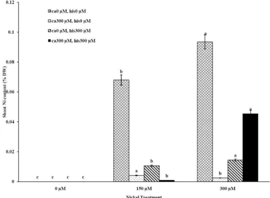
similarly (Figures 2A-D). Figures 2A and 2B show
that after treatment with Ni 150 μM, Ni content in
the shoots was about 1.28-fold higher in cv. Cal-J
N3 compared to cv. Petoearly CH (cv. Cal-J N3:
approximately 0.068 %DW, cv. Petoearly CH:
approximately 0.053 %DW). Furthermore, there
were considerable differences between the
cultivars in terms of Ni accumulation of shoots after
treatment with 300 μM of nickel. Accumulation of
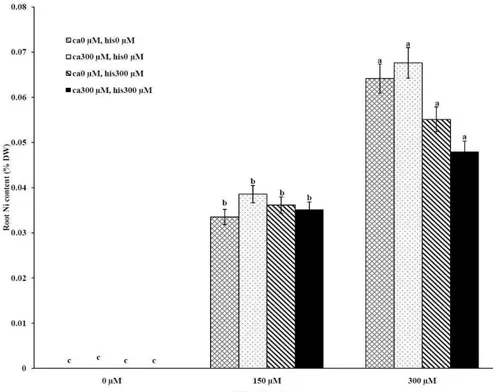
Ni in the roots also differed between the cultivars.
Assimilation of Ni induces a greater decrease in the
Ca uptake of root and shoot in both cultivars of
tomato (Figures 1A-D). The application of Ca and
His in hydroponic media increased calcium content
of shoot and root in Petoearly CH and Cal-J N3
cultivars compared with the control plants (Figures
1A-D). In root tissues, Ca uptake declined slightly
with the increase in Ca and His concentration and
Ca concentration increased in shoot under similar
conditions. The relationship between Ca content of
root and shoot was inversed with respect to Ca
accumulation in the tomato plants especially in
Petoearly CH cultivar under treatments with 300
μM of Ni (Figures 1A-D). However, Ca content of
roots improved under both nickel toxic levels 150
µM and 300 µM containing CaCl2 without His in
Cal-J N3 and Petoearly CH, respectively compared to
same stress conditions (Figures 1C and D).
Our data showed that Ni accumulation
decreased significantly in shoot of the tomato
cultivars under 150 and 300 μM of nickel
treatments containing Ca+His (Figures 2 A and B).
Furthermore, the interactive effect of Ca and His on
Ni content was observed in roots of the cultivars
treated by 300 μM of Ni. We also observed this
interaction effect only about decrease of Ni content
of root only in Petoearly CH cultivar under 150 μM
of Ni treatment (Figure 2D). The application of Ca
with His led to high decreasing in shoot nickel
accumulation in Cal-J N3 cultivar treated by150 μM
toxic level. On other hand, our data from root and
shoot Ni analysis showed that nickel translocation
from roots to shoots was declined. This was
observed specially under Ni stress (150 μM) in both
cultivars and also in Cal-J N3 cultivar treated by Ni
300 μM (Figures 2A-D). According to Table 1, it
seems that Petoearly CH cultivar was more
Ni-sensitive compared to other tomato cultivar. Thus,
we observed significant effects for interaction of Ca
and His in Petoearly CH cultivar compared to Cal-J
N3 under Ni stress at 0.05 level. In other hand,the
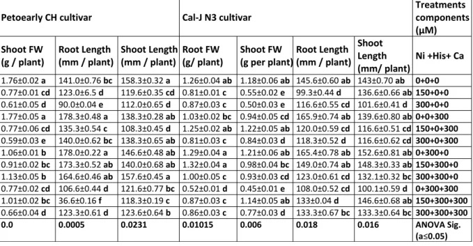
Tab. 1 clearly show that Ni-induced growth
inhibition especially in the case of roots was more
pronounced in cv. Petoearly CH. In the latter cultivar
treatment with Ni 300 μM, root length and fresh
weight were reduced by 94% and 66%, respectively,
comparing to the control (length control: 141 mm,
Ni 300: 9 mm; FW control: 1.42 g, Ni 300: 0.49 g),
while the same Ni dose decreased root length and
weight by 20% and 31%, respectively (length
control: 145.6 mm, Ni 300: 116.6 mm; FW control:
1.26 g, Ni 300: 0.87 g) in cv. Cal-J N3.
Figure 1A. The Mean of shoot calcium accumulation determined and three-way ANOVA with multiple but
equal number of observations per test tube for the effects of individual treatments and their interactive effects on Ca content changes in Cal-J N3 cultivar of tomato treated with a nutrient solution containing different concentrations of nickel, calcium and histidine. Vertical bars indicate the mean of four replications ± SD (n=4). Different letters indicate significantly different values at a particular treatment component containing calcium and histidinde (P ≤ 0.05).
Figure 1B. The Mean of shoot calcium accumulation determined and three-way ANOVA with multiple but
Figure 1C. The Mean of root calcium content determined and three-way ANOVA with multiple but equal number of observations per test tube for the effects of individual treatments and their interactive effects on Root Ca content changes in Cal-J N3 cultivar of tomato treated with a nutrient solution containing different concentrations of nickel, calcium and histidine. Vertical bars indicate the mean of four replications ± SD (n=4). Different letters indicate significantly different values at a particular treatment component containing calcium and histidinde (P ≤ 0.05).
Figure 1D. The Mean of root calcium content determined and three-way ANOVA with multiple but equal
Figure 2A. The Mean of shoot nickel accumulation determined and three-way ANOVA with multiple but equal number of observations per test tube for the effects of individual treatments and their interactive effects on Ni content changes in Cal-J N3cultivar of tomato treated with a nutrient solution containing different concentrations of nickel, calcium and histidine. Vertical bars indicate the mean of four replications ± SD (n=4). Different letters indicate significantly different values at a particular treatment component containing calcium and histidinde (P ≤ 0.05).
Figure 2B. The Mean of shoot nickel accumulation determined and three-way ANOVA with multiple but equal
Figure 2C. The Mean of root nickel content determined and three-way ANOVA with multiple but equal number of observations per test tube for the effects of individual treatments and their interactive effects on
Root Ni content changes in Cal-J N3 cultivar of tomato treated with a nutrient solution containing
different concentrations of nickel, calcium and histidine. Vertical bars indicate the mean of four replications ± SD (n=4). Different letters indicate significantly different values at a particular treatment component containing calcium and histidinde (P ≤ 0.05).
Figure 2D. The Mean of root nickel content determined and three-way ANOVA with multiple but equal number
of observations per test tube for the effects of individual treatments and their interactive effects on
Root Ni content changes in Petoearly CH cultivar of tomato treated with a nutrient solution
Table 1. Mean±SE of the Length and FW from shoot and root organs determined in our research and three-way ANOVA with multiple but equal number of observations per test tube for the effects of individual treatments and their interactive effects on parameters changes in the two cultivars of tomato including Cal-J N3 and Petoearly CH. Means followed by different letter within columns are significantly different at P ≤ 0.05 according to Duncan’s Multiple Range Test.
Treatments components (µM) Cal-J N3 cultivar
Petoearly CH cultivar
Ni +His+ Ca Shoot
Length (mm/ plant) Root Length
(mm / plant) Shoot FW
(g per plant) Root FW
(g/ plant) Shoot Length
(mm / plant) Root Length
(mm / plant) Shoot FW
(g / plant)
0+0+0
143±0.70 ab
145.6±0.60 ab
1.18±0.06 ab
1.26±0.04 ab
158.3±0.32 a
141.0±0.76 bc
1.76±0.02 a
150+0+0
136.6±0.66 ab
99.3±0.44 d
0.55±0.02 e
0.81±0.01 c
119.6±0.35 cd
123.0±6.5 d
0.77±0.01 cd
300+0+0
101.6±0.41 d
116.6±0.55 cd
0.50±0.03 e
0.87±0.03 c
112.0±0.65 d
90.0±0.04 e
0.61±0.05 d
0+0+300
139.6±0.80 ab
165.9±0.74 ab
0.94±0.05 cd
1.03±0.02 bc
138.3±0.28 ab
178.3±0.48 a
1.77±0.05 a
150+0+300
116.6±0.51 cd
120.0±0.59 cd
1.22±0.05 ab
1.25±0.02 ab
108.3±0.45 d
135.3±0.54 c
0.77±0.06 cd
300+0+300
116.6±0.62 cd
118.3±0.52 d
0.84±0.03 d
0.81±0.03 c
138.3±0.65 ab
140.0±0.62 bc
0.59±0.03 e
0+300+0
152.6±0.81 ab
165.4±0.78 ab
1.21±0.06 ab
1.29±0.04 a
146.6±0.48 ab
178.0±0.22 a
1.06±0.01 b
150+300+0
148.3±0.33 ab
149.0±0.74 ab
0.98±0.04 bc
1.32±0.04 a
140.0±0.68 ab
173.3±0.52 ab
0.91±0.02 bc
300+300+0
132.1±0.32 bc
123.0±0.61 cd
0.93±0.03 cd
1.00±0.05 c
157.6±0.45 a
164.6±0.46 ab
1.13±0.05 b
0+300+300
100.1±0.59 d
108.0±0.52 cd
0.45±0.01 e
0.52±0.01 d
121.6±0.77 bc
106.6±0.44 d
0.77±0.02 cd
150+300+300
146.6±0.68 ab
133±0.04 d
1.14±0.05 ab
0.87±0.03 c
118.3±0.19 c
36.6±0.16 f
1.01±0.02 bc
300+300+300
133.3±0.64 bc
133.3±0.67 bc
0.77±0.03 d
0.86±0.03 c
123.6±0.64 b
123.3±0.61 d
0.66±0.04 d
ANOVA Sig. (a≤0.05) 0.016 0.018 0.006 0.01015 0.0231 0.0005 0.0
Table 2. Mean±SE of the total protein, ASC and DHA parameters determined in our research and
three-way ANOVA with multiple but equal number of observations per test tube for the effects of individual treatments and their interactive effects on parameters changes in the two cultivars of tomato including Cal-J N3 and PetoearlyCH. Means followed by different letter within columns are significantly different at P ≤ 0.05 according to Duncan’s Multiple Range Test.
Treatments components (µM) Cal-J N3 cultivar
Petoearly CH cultivar
Ni +His+ Ca Leaf protein
(mg/ g FW) Root protein
(mg/ g FW) Leaf ASC
(mg/ g FW) Leaf DHA
(mg/ g FW) Leaf protein
(mg/ g FW) Root protein
(mg/ g FW) Leaf ASC
(mg/ g FW)
0+0+0
5.61±0.24 d
2.51±0.05 cd
3.93±0.19 d
4.57±0.25 b
7.51±0.31 a
1.73±0.04 de
2.08±0.12 f
150+0+0
5.16±0.23 d
4.04±0.16 b
3.55±0.16 d
1.70±0.04 c
7.14±0.32 a
3.64±0.13 bc
2.25±0.11 f
300+0+0
8.62±0.74 ab
7.16±0.34 a
5.04±0.20 cd
4.63±0.21 b
4.51±0.22 c
5.09±0.24 a
8.15±0.41 bc
0+0+300
5.57±0.15 d
1.22±0.04 d
8.76±0.26 a
4.18±0.26 b
4.86±0.23 c
0.55±0.01 f
2.71±0.11 f
150+0+300
9.01±0.44 ab
5.47±0.27 b
2.89±0.10 d
0.93±0.01 de
6.13±0.28 ab
0.52±0.02 f
2.48±0.15 f
300+0+300
6.39±0.31 cd
7.64±0.31 a
4.18±0.18 d
1.48±0.02 d
7.70±0.29 a
3.11±0.12 c
2.33±0.14 f
0+300+0
1.70±0.26 f
0.75±0.01 e
7.05±0.28 b
2.83±0.13 c
5.50±0.11 bc
2.37±0.13 cd
9.27±0.34 bc
150+300+0
5.09±0.11 d
3.19±0.12 bc
3.51±0.21 d
0.56±0.01 e
7.26±0.23 a
5.80±0.24 a
2.62±0.16 f
300+300+0
2.58±0.10 f
2.01±0.13 cd
6.36±0.29 c
1.00±0.04 c
4.97±0.24 c
1.19±0.03 ef
5.76±0.24 d
0+300+300
4.73±0.18 de
4.08±0.24 b
4.26±0.17 d
4.40±0.17 b
5.36±0.26 bc
0.76±0.01 f
1.61±0.01 g
150+300+300
6.16±0.27 c
7.70±0.25 a
2.09±0.11 de
7.66±0.28 a
6.48±0.31 ab
2.89±0.12 cd
16.20±0.74 a
300+300+300
7.43±0.32 c
2.80±0.15 cd
1.63±0.04 e
0.69±0.01 e
3.65±0.11 d
4.83±0.21 ab
4.76±0.26 de
Table 3. Mean±SE of the some of leafs biochemical parameters determined in our research and three-way ANOVA with multiple but equal number of observations per test tube for the effects of individual treatments and their interactive effects on parameters changes in the two cultivars of tomato including Cal-J N3 and Petoearly CH. Means followed by different letter within columns are significantly different at P ≤ 0.05 according to Duncan’s Multiple Range Test.
Treatments components (µM) Cal-J N3 cultivar
Petoearly CH cultivar
Ni +His+ Ca Leaf H2O2
content (µM/ g FW) Leaf EL (%)
Leaf MDA (µM/ g FW) Leaf
aldehydes (µM/ g FW) Leaf H2O2
content (µM/ gFW) Leaf EL (%)
Leaf MDA (µM/ g FW)
0+0+0
61.10±2.88 e
8.11±0.42 d
0.07±0.01 f
2.22±0.12 c
13.92±0.64 g
6.86±0.24 e
0.16±0.02 cd
150+0+0
127.0±5.54 a
27.69±1.28 ab
0.19±0.01 e
2.80±0.11 a
191.36±5.14 a
27.59±1.24 bc
0.43±0.02 b
300+0+0
101.1±4.11 b
27.14±1.33 ab
0.28±0.01 c
3.08±0.13 b
152.13±6.24 b
41.38±2.36 a
0.49±0.03 a
0+0+300
87.26±3.21 c
10.97±0.54 c
0.27±0.01 c
1.69±0.02 d
128.54±5.88 d
31.19±1.08 b
0.13±0.01 f
150+0+300
78.03±2.19 d
33.69±1.66 ab
0.22±0.01 d
1.60±0.01 d
165.72±8.47 b
23.04±1.14 c
0.27±0.01 c
300+0+300
86.74±2.45 c
25.41±0.97 ab
0.40±0.02 b
4.31±0.18 a
140.08±7.24 c
40.15±2.99 a
0.20±0.01 d
0+300+0
70.85±3.02 d
18.95±0.95 ab
0.28±0.01 c
2.18±0.19 bc
40.85±2.08 f
14.51±0.48 d
0.16±0.01 e
150+300+0
82.38±1.02 c
19.52±1.05 ab
0.28±0.01 c
2.19±0.21 bc
42.38±2.56 f
45.43±2.54 a
0.16±0.01 e
300+300+0
53.92±2.05 f
37.74±1.98 a
0.38±0.02 b
2.62±0.10 a
43.67±2.33 f
42.20±1.26 a
0.21±0.02 d
0+300+300
79.05±3.54 d
15.31±0.74 bc
0.27±0.01 c
2.39±0.12 a
48.54±2.47 f
41.55±2.17 a
0.19±0.01 d
150+300+300
124.1±4.56 a
33.74±1.44 ab
0.26±0.01 c
3.34±0.14 ab
193.67±6.75 a
21.85±1.45 c
0.28±0.03 c
300+300+300
61.10±3.21 e
26.41±1.11 ab
0.54±0.03 a
2.81±0.16 a
82.90±4.12 e
32.08±1.67 b
0.30±0.02 c
ANOVA Sig. (a≤0.05) 0.004. 0.028 0.026 0.005 0.001 0.0 0.004
Table 4. Mean±SE of the antioxidative enzymes activity of leaf determined in our research and three-way
ANOVA with multiple but equal number of observations per test tube for the effects of individual treatments and their interactive effects on parameters changes in the two cultivars of tomato including Cal-J N3 and Petoearly CH. Means followed by different letter within columns are significantly different at P ≤ 0.05 according to Duncan’s Multiple Range Test.
Treatments components (µM) Cal-J N3cultivar Leaf
Petoearly CH cultivar Leaf
Ni +His+ Ca GPX (U/mg protein) CAT (U/mg protein) APX (U/ mg protein) SOD (U/ mg protein) GPX (U/mg protein) CAT (U/mg protein) APX (U/mg protein) 0+0+0
6.55±0.28 g
2.46±0.13 d
1.30±0.03 de
8.67±0.34 c
18.39±0.87 d
10.97±0.42 e
3.50±0.14 de
150+0+0
9.76±0.51 f
7.44±0.28 c
0.98±0.02 e
8.46±0.41 c
17.85±0.56 e
7.19±0.34 f
2.11±0.16 e
300+0+0
17.72±0.32 d
13.05±0.1 b
1.93±0.06 d
5.81±0.31 e
68.95±2.77 a
16.26±0.75 c
9.05±0.33 c
0+0+300
14.70±0.65 de
2.92±0.17 d
4.42±0.26 a
8.52±0.34 c
21.26±0.99 d
26.21±1.04 b
0.97±0.02 f
150+0+300
9.84±0.39 f
9.17±0.37 bc
1.76±0.03 d
5.37±0.24 e
70.40±2.11 a
1.51±0.03 h
12.91±0.24 b
300+0+300
8.38±0.33 f
9.85±0.15 bc
3.84±0.25 a
7.77±0.34 d
17.55±0.64 e
5.50±0.18 f
1.76±0.04 f
0+300+0
52.36±1.13 b
2.03±0.13 d
2.27±0.06 c
28.08±1.5 a
34.61±1.33 b
5.53±0.24 f
4.06±0.15 de
150+300+0
26.35±1.02 c
24.34±1.3 a
2.19±0.05 c
13.35±0.54 b
27.20±1.24 c
14.47±0.41 c
1.49±0.03 f
300+300+0
58.50±2.34 a
6.51±0.22 c
4.03±0.14 a
26.70±1.24 a
19.44±0.57 d
10.13±0.62 e
1.66±0.03 f
0+300+300
7.79±0.28 g
2.97±0.17 d
1.98±0.05 d
11.61±0.64 bc
18.11±0.84 d
41.83±1.24 a
0.41±0.01 g
150+300+300
24.86±1.18 c
9.09±0.45 bc
2.85±0.05 ab
8.70±0.4 6c
26.09±1.29 c
4.85±0.19 g
6.44±0.28 cd
300+300+300
19.42±0.88 d
6.88±0.52 c
2.81±0.06 ab
6.60±0.34 d
27.85±1.18 c
12.14±0.55 d
16.88±0.71 a
DISCUSSION
Although the Ca and Ni accumulation in roots
are usually inversely correlated, root Ca
accumulation is more often accounted for a higher
share of variability in root elongation compare with
Ni accumulation in root tissue at high concentration
of Ca compared with the control conditions (Wu
and Hendershot, 2010). Therefore, the evaluation
of root elongation requires including Ni and Ca as
predictor whenever environmental conditions (low
Ca concentrations) significantly affect the amount
of Ca accumulated. Root elongation is highly
positively correlated with total Ca content in the
roots (Wu and Hendershot, 2010). In this study our
data showed that Ca plus Ni increased length and
FW of root in the tomato cultivars significantly in
comparison to Ni treatments without excess CaCl2.
Thus, the treatments containing excess Ca (300 µM)
has positive effects on shoot and root length
elongation (Table 1, Figures 1A-D). However, Ca and
His interaction effect on alleviation of growth and
Ca accumulation of tomato plants were significant
especially in Petoearly CH cultivar under Ni stress.
Additionally, Ca plus His alleviated calcium content
of shoots and roots, more than control plants in
both cultivars (Figures 1A-D). In root tissue of the
cultivars, Ca uptake declined slightly with the
increase in Ca and His concentration and Ca
concentration increased in shoot under similar
conditions. Therefore, the relationship between
root and shoot Ca was inversed because of higher
Ca accumulation in shoot especially in Petoearly CH
cultivar under Ni 300 μM treatments (Figures 1A-D).
However, Ca content of roots increased under
nickel levels 150 µM and 300 µM containing CaCl2
without His in Cal-J N3 and Petoearly CH,
respectively which is compared to same stress
conditions (Figures 1C and D).
In this investigation, the MDA and other
aldehydes as lipid peroxidation markers were higher
than the control under Ni toxic levels (Table 3).
Membrane degradation was reported as a
consequence of Ni stress in roots of Triticum
aestivum (Pandolfini et al., 1992). Highly reactive
oxygen species could be responsible for Ni-induced
membrane damage that increasing ROS content has
been reported for roots and leaves of wheat, hairy
roots of Alyssum bertolonii and Nicotiana tabacum
(Gajewska and Sklodowska, 2007). Thompson et al.
(1987) documented that lipoxygenase activation
precedes lipid peroxidation. This mechanism might
underlie Ni induced peroxidation as seen in primary
leaves of Phaseolus vulgaris under Cu and Zn stress
(Weckx et al., 1997). In addition, Pandolfini et al.
(1992) showed that Ni causes membrane
degradation leading to a marked K+ and other cation
leakage probably. In our research, electrolytes
leakage percentage (as an important membrane
damage index) increased in both cultivars under
nickel stress compared to the control plants (Table
3). It has been suggested that the extent of lipid
peroxidation and membrane permeability were
closely related to higher levels of ROS during either
senescence or under stress conditions (Weckx and
Clijsters, 1997).
Our findings shown that Ca and His interaction
inhibited MDA and other aldihydes generation
compared to stress condition in the tomato
cultivars especially in Petoearly CH compare with
other cultivar (Table 3).
Application of His alone in Cal-J N3 cultivar and
also Ca and/or His in Petoearly CH cultivar during
150 µM Ni, declined EL percentage. EL was
significantly decreased, compared to stress
300 µM of nickel treatment containing both Ca and
His (Table 3).
We evaluated the activities of SOD, CAT, GPX
and APX that involved in free radicals and H2O2
detoxification under the experiment treatments.
Higher activity of the SOD led to production of H2O2.
Subsequently, increasing in activity of CAT, GPX and
APX can remove high H2O2 significantly in the
tomato plants (Table 4).
In the present study, a significant increase in leaf
SOD activity was observed in both tomato cultivars
treated with Ca and His treatments during different
levels of nickel (Table 4), suggesting that SOD may
function as a ROS scavenger (Alscher et al., 2002).
Recent studies have demonstrated that over
expression of chloroplastic Cu/Zn-SOD in transgenic
Nicotiana tabacum (Badawi et al., 2004) can
provide enhanced ion toxicity tolerance. Even
though a high SOD activity (compared in equal total
protein content of tissue) protects plants against
the superoxide radical, it cannot be considered
solely responsible for membrane protection against
peroxidation. This ROS should be then scavenged by
other enzymes such as catalase and peroxidases.
The fact that leave CAT activity of the Cal-J N3
cultivar in the control and Ni stress treatments was
lower than Petoearly CH cultivar could be the result
of genetic differences and higher Ni sensitivity of
Cal-J N3 cultivar to Ni toxicity. However, when the
tomato plants were subjected to NiSO4, the
increases in CAT activity were higher under Ni stress
300 μM than the control but Ca and His stimulated
its activity during Ni stress (Table 4).
It seems that up regulation of CAT is a
requirement for the removal of H2O2 in roots and
leaves of stressed plants or decreasing in CAT
activity may result from enzyme deactivation (Dat
et al., 2000). The contradiction between CAT
activation and suppression in the presence of
metals may depend upon the element, its
concentration and the plant species and cultivars
(Gratao et al., 2005).
Higher activity of APX was recorded after
application of Ni, Ca and His treatments in the
tomato cultivars. The higher level in APX activity
was observed in the tomato cultivar Petoearly CH
than other cultivar (Table 4). Further more GPX
activity stimulation was considerably higher than
APX in our research (Table 4). Consequently, APX
may remain in a saturated state for its substrate. In
addition, the increase in activities of APX and GPX
has been found by Jocsak et al., (2008) in barley
seedlings. However, the activities of APX and GPX in
both tomato cultivars were also affected by Ca and
His treatments compared to Ni 150 µM lacking of
Ca and His. The similar results were found for other
plant species by Mittova et al. 2002 and also
elevated APX activity in plants exposed to toxic
levels of Ni has been reported earlier in wheat and
rice (Gajewska and Skłodowska, 2008). It has been
speculated that under stress conditions, H2O2 acts
as a systemic intracellular signal for the induction of
APX (Hernandez et al., 2004). Therefore, H2O2 is
considered to playing a central role in physiological
functions such as plant signaling, regulating plant
development and adaptation to biotic and abiotic
stresses. Increased availability of H2O2 is a
commonly observed feature of plant stress
response signature. Stresses such as high light, heat,
salinity, cold and pathogen attack, besides others,
are all considered to trigger H2O2 accumulation that
contributes to the regulation the expression of
many genes. H2O2-mediated regulation of cell
division, differentiation and growth must be viewed
chemistry of the plant cell that is governed by
photosynthesis and respiration but also within the
bigger picture of the physiological environment. The
physiological context involves a continuous supply
of environmental stimuli that can trigger
intracellular H2O2 accumulation or modulate the
response to such accumulation (Foyer and Noctor,
2012; Chakraborty and Pradhan, 2012).
These antioxidative changes could be related to
complexity of the ascorbate-glutathione cycle that
have enzymes coded by multigenic families whose
products are localized in different cell
compartments and are regulated differently by
stress conditions. For instance, in the cytosolic
fraction of sorghum leaves subjected to water
stress there was a decrease in monodehydro
ascorbate reductase (MDHAR) activity, and
increased dehydro ascorbate reductase (DHAR)
activity (Mittova et al., 2002). Our data also showed
that under high Ni stress conditions (300 µM),
application of histidine resulted in more increased
concentration of ASC than the control in Cal-J N3
cultivar while Ca and His interaction on increasing
ASC content was significant at 0.05 Level in
Petoearly CH cultivar plants (Table 2).
Generally, the obtained data (Figures 2A-D)
indicated that Ca and His inhibited Ni accumulation
and translocation in root and shoot in the tomato
cultivars under Ni stress via ion chelating
mechanisms. However, heavy metal detoxification
in plants is linked to the synthesis of cysteine-rich
poly- peptides, so called phytochelatins (PCs) (Grill
et al., 1985). Similar to these compounds,
L-histidine are able to chelate Ni2+ indifferent parts of
plants (Howden et al., 1995). The function of
calcium in preventing toxic effects of HMs during
the transport across the cell wall is controversially
discussed, very often the optimum Ca content in
plant tissue correlate with HM tolerance (de Knecht
et al., 1994).
The tomato plants accumulated appreciable
amounts of Ni in both roots and shoots under Ni
stress without Ca and His. Our results shown that
inhibition of shoot and root Ca uptake was detected
after lipid peroxidation began (Figures 1A-D and
Table 3). This latter phenomenon appears to be a
primary effect of Ni stress that may result in
inhibition of tomato plants growth and nutrition
especially in Cal-J N3 cultivar that this cultivar was
less Ni sensitive compared to the other cultivar in
our work (Table 1). Similar to the results obtained
by the earlier studies (Madhava and Sresty, 2000),
we found that the Ni uptake in the two cultivars
(especially Petoearly CH cultivar) is dependent upon
nickel concentration in treatments (Figures 2A-D).
Accumulated calcium in root tissue increased
tolerance of plants to Ni toxicity and also decrease
Ni uptake and its accumulation especially in tomato
cultivar Petoearly CH (Figures 1A-D and 2A-D).
The Ni and Ca internalization process within the
root is physiologically controlled and root growth is
a complicated process (Table 1). Our study showed
that the measured accumulations of Ni and Ca
correlate best with the treatments whenever data
collected under varied concentration Ca and His
treatments. This suggests that uptake of Ni and Ca
is affected by influence of Ca and His on Ni uptake
and translocation under the stress conditions (Wu
and Hendershot, 2010).
Generally, based on the obtained results it can
be suggested that Ni toxicity induced oxidative
stress in the Ni sensitive cultivars of tomato
resulting in increased generation of H2O2 and
activation of antioxidant enzymes. The different
increases in CAT, APX, SOD and GPX activities under
ROS in tomato plants especially in Petoearly CH
cultivar. The data shown that Ca and His elevated
antioxidative responses in studies on tomato
cultivars under Ni toxicity especially Petoearly CH
through inhibition the Ni toxic effects by decreasing
nickel uptake and its translocation. For the first time
our results showed that the interaction effects
between Ca and His could alleviate nickel stress and
growth promoting in sensitive tomato cultivar
plants such as Petoearly CH. Finally, exogenous
Ca+His improved tolerance of this Ni-sensitive
tomato cultivar under oxidative stress and higher
accumulated Ni of shoots compared to Cal-J N3
cultivar. Furthermore, new researches to
understand the basic mechanism under Ca
interaction effects are essential in cell wall with
heavy metal ligands such as His for alleviation of
oxidative stress. Also, determination of the
differences in activity among the isoforms of SOD,
CAT, GPX and other antioxiative enzymes is our
next aim in future researches through the PAGE
under Ca, His and Ni-induced oxidative stress in the
tomato plants. Thus, field experiments are
necessary at contaminated soils with nickel
pollution for oxidative stress decreasing and plants
tolerance in these conditions. In conclusion, the
application of Ca+His as chelators was beneficial to
higher oxidative stress tolerance in the tomato
cultivars (especially Petoearly CH comparing to Cal-J
N3) and increasing growth of the tomato plants
under Ni stress conditions.
ACKNOWLEDGMENTS
This Originalresearch Paper is a part of research
project in Ph.D thesis (for Ph.D candidate) that
funded by Biology Department, Shahid Bahonar
University of Kerman, Iran. We thank MSc Elnaz
Ghotbi for her coordination in text English editing of
this MS.
REFERENCES
Ahmad, P., Abdul Jaleel, C., Salem, M.A., Nabi, G.,
Sharma, S. (2010) Roles of enzymatic and
nonenzymatic antioxidants in plants during
abiotic stress.Crit Rev Biotech., 30(3), 161–175.
Alscher, R.G., Erturk, N., Heath, L.S. (2002) Role of
superoxide dismutases (SODs) in controlling
oxidative stress in plants. J Exp Bot., 53,
1331-1341.
Baccouch, S., Chaoui, A., Ferjani, E. (1998)
Nickel-induced oxidative damage and antioxidant
responses in Zea mays shoots. Plant Physiol.
Biochem., 36, 689-694.
Badawi, G.H., Yamauchi, Y., Shimada, E. (2004)
Enhanced tolerance to salt stress and water
deficit by over expressing superoxide dismutase
in tobacco (Nicotiana tabacum) chloroplasts.
Plant Sci., 166, 919-928.
Ben Hamed, K., Castagna, A., Salem, E. (2007) Sea
fennel (Crithmum maritimum L.) under salinity
conditions: a comparison of leaf and root
antioxidant responses. Plant Growth Regul., 53,
185-194.
Bradford, M.M. (1976) A rapid and sensitive method
for the quantification of microgram quantities
of protein utilizing the principle of protein-dye
binding. Analyt Bioch., 72, 248-254.
Brooks, R.R., Morrison, R.S., Reeves, R.D. (1979)
Hyperaccumulation of nickel by Alyssum
Linnaeus (Cruciferae). Proceedings of the Royal
Society. London Ser. B, Biol Sci., 203, 387-403.
Dalvi, A.A., Bhalerao, S.A. (2013) Response of Plants
towards Heavy Metal Toxicity: An overview of
Avoidance, Tolerance and Uptake Mechanism.
Annal. Plant Sci., 2(9), 362-368.
Dat, J., Vandenabeele, S., Vranova, E. (2000) Dual