The Bactofilin Cytoskeleton Protein BacM
of
Myxococcus xanthus
Forms an Extended
β-Sheet Structure Likely Mediated by
Hydrophobic Interactions
David M. Zuckerman1☯, Lauren E. Boucher2☯, Kefang Xie1, Harald Engelhardt3,
Jürgen Bosch2, Egbert Hoiczyk1*
1W. Harry Feinstone Department of Molecular Microbiology and Immunology, Johns Hopkins Bloomberg School of Public Health, Baltimore, Maryland, United States of America,2Department of Biochemistry and Molecular Biology, Johns Hopkins Bloomberg School of Public Health and Johns Hopkins Malaria Research Institute, Baltimore, Maryland, United States of America,3Department of Structural Biology, Max Planck Institute of Biochemistry, Am Klopferspitz 18, Martinsried, Germany
☯These authors contributed equally to this work.
Abstract
Bactofilins are novel cytoskeleton proteins that are widespread in Gram-negative bacteria.
Myxococcus xanthus, an important predatory soil bacterium, possesses four bactofilins of which one, BacM (Mxan_7475) plays an important role in cell shape maintenance. Electron and fluorescence light microscopy, as well as studies using over-expressed, purified BacM, indicate that this protein polymerizesin vivoandin vitrointo ~3 nm wide filaments that fur-ther associate into higher ordered fibers of about 10 nm. Here we use a multipronged ap-proach combining secondary structure determination, molecular modeling, biochemistry, and genetics to identify and characterize critical molecular elements that enable BacM to polymerize. Our results indicate that the bactofilin-determining domain DUF583 folds into an extendedβ-sheet structure, and we hypothesize a left-handedβ-helix with polymeriza-tion into 3 nm filaments primarilyviapatches of hydrophobic amino acid residues. These patches form the interface allowing head-to-tail polymerization during filament formation. Biochemical analyses of these processes show that folding and polymerization occur across a wide variety of conditions and even in the presence of chaotropic agents such as one molar urea. Together, these data suggest that bactofilins are comprised of a structure unique to cytoskeleton proteins, which enables robust polymerization.
Introduction
All living cells organize their cytoplasm to ensure efficient growth and cell division. This orga-nization relies in part on cytoskeleton proteins, versatile scaffolds that are crucial for essential processes ranging from cell shape maintenance to morphogenesis, polarity determination, cell OPEN ACCESS
Citation:Zuckerman DM, Boucher LE, Xie K, Engelhardt H, Bosch J, Hoiczyk E (2015) The Bactofilin Cytoskeleton Protein BacM ofMyxococcus xanthusForms an Extendedβ-Sheet Structure Likely Mediated by Hydrophobic Interactions. PLoS ONE 10 (3): e0121074. doi:10.1371/journal.pone.0121074
Academic Editor:John R. Kirby, University of Iowa, UNITED STATES
Received:October 8, 2014
Accepted:January 29, 2015
Published:March 24, 2015
Copyright:© 2015 Zuckerman et al. This is an open access article distributed under the terms of the
Creative Commons Attribution License, which permits unrestricted use, distribution, and reproduction in any medium, provided the original author and source are credited.
Data Availability Statement:All relevant data are within the paper and its Supporting Information files.
growth and division, chromosomal segregation, and transport of cellular cargoes (for reviews see [1–8]). While research has focused for decades on the eukaryotic cytoskeleton proteins actin, tubulin, and intermediate filaments [9–11], it has only recently been realized that bacte-ria possess structural homologues of these proteins, as well as a number of uniquely bactebacte-rial cytoskeletal proteins thus far not described for eukaryotes [12,13].
While sequence homology searches initially failed to identify prokaryotic homologues to eu-karyotic cytoskeletal proteins, determination of the structure of the cell-division protein FtsZ revealed that it is a close structural homologue to eukaryotic tubulin, showing particularly high similarity in the GTP-binding domain [5,14]. In contrast, the bacterial actin protein MreB was initially identified based on sequence homology mapped to the eukaryotic actin ATPase do-main [15] and later confirmed as an actin-like cytoskeletal protein through sequence homology in regions responsible for actin’s overall structure [16]. Outside of these regions, MreB lacks se-quence homology to actin, but was found to possess a strikingly similar tertiary structure [17]. Like its many homologues and paralogs, MreB controls the cellular morphology of non-spheri-cal bacteria. Finally, bacterial intermediate filament-like proteins were identified in bacteria with the discovery of crescentin inCaulobacter crescentus[18]. Like their eukaryotic counter-parts, bacterial intermediate filament (IF) assembly is non-polarvialong coiled-coil domains, structural elements that are found in many bacterial proteins, indicating that IF-like cytoskele-tons are potentially widespread in bacteria [19–23]. Together, these discoveries underscore the necessity of determining the structure of cytoskeletal proteins, as this helps not only to under-stand the assembly and function of these structures, but also to identify evolutionarily distant homologues and relatives. This is particularly true for cytoskeleton protein families that are found uniquely in bacteria and lack obvious eukaryotic counterparts. One such uniquely bacte-rial cytoskeletal system is the Walker A cytoskeletal ATPases (WACAs), a family of proteins defined by their ATP-binding domain [5]. The best-studied example of a WACA is the ParA protein, which forms a rudimentary mitotic apparatus that partitions chromosomes or plas-mids into daughter cells during cell division [24].
Another recently discovered family of uniquely bacterial cytoskeletal proteins is the bactofi-lins (reviewed in [13]). Bactofilins are small proteins, nearly ubiquitous in Gram-negative bac-teria, which are defined by the presence of the highly conserved domain DUF583 [25]. The first member of this family to be identified, the protein CcmA ofProteus mirabilis, was discov-ered in a transposon-based genetic screen [26]. Insertion of the transposon at the C-terminal region of the ORF led to expression of a truncated CcmA, and resulted in mechanically fragile cells with severe morphological deformations. Later, homologues of CcmA were discovered in
C.crescentus,Helicobacter pylori,and M.xanthusand, in all cases, found to be important for cell shape maintenance [27–29]. Deletion of the bactofilin genebacAinC.crescentusled to a reduction in stalk length, while overexpression of eitherbacAorbacBfused to a large fluores-cent protein caused an increase in the curvature of the cell [27]. Similarly, deletion of the bacto-filin geneccmAinH.pyloriresulted in the loss of the characteristic helical cell shape,
dramatically straightening the cells [28]. Finally, lack of BacM in the normally rod-shapedM.
xanthusproduced aberrant morphologies ranging from mildly bent to severely crooked cells [29]. The cytoskeletal nature of bactofilins was further confirmed by immunofluorescence mi-croscopy, which revealed that BacM has a filament-like staining pattern inM.xanthus, and, when biochemically isolated, is recovered as bundle-forming fiber [29]. Similarly, whenbacA
fromC.crescentusor theM.xanthusbactofilins were exogenously over-expressed inE.coli, fil-amentous forms of the proteins were recovered, indicating that these proteins have a high pro-pensity to polymerize [27,29]. Based on bioinformatics [28], protein-protein interaction studies [27], and increased sensitivity of bactofilin mutants to cell-wall targeting enzymes [29], it appears that bactofilins may exert their influence over the cell morphology by contributing to
Hopkins Malaria Research Institute. The funders had no role in study design, data collection and analysis, decision to publish, or preparation of the manuscript.
proper peptidoglycan (PG) maintenance. In addition to morphology, bactofilins may affect other cellular functions. In a recent report, a BacM paralog inM.xanthus, BacP, was found to recruit and localize SofG, a small GTPase important to cell motility, to the cell pole [30]. Addi-tionally, an mCherry fusion of SO1662, the only bactofilin identified inShewanella oneidensis, assembled as a fluorescent band at midcell suggesting a possible involvement in cell division [27]. Thus, bactofilins may be versatile scaffolds that recruit and localize enzymes (i.e. PG syn-thesis and remodeling enzymes) and structural proteins (i.e. components of the type IV pilus and cell division machineries) to specific cellular locations.
Other than electron microscopic analyses of negatively stained, purified filaments, no other structural information has been reported for any of the bactofilins [27,29]. Since these proteins do not share sequence homology to other structurally analyzed cytoskeletal or filament-form-ing proteins and have not yet been subjected to further ultrastructural analysis, no data exist about the molecular basis for their ability to polymerize. Detailed analyses of their amino acid sequences suggest a three-domain structure in which N- and C-terminal domains with broad sequence variability flank the highly conserved bactofilin domain DUF583. The N-terminus of bactofilins from several species appears to be used to anchor the protein to membranes [26,29], and appears to play no role in polymerization [29]. No information has yet been reported for the function of the C-terminus, although one plausible idea is that this part of the protein is in-volved in the recruitment of interaction partners. Moreover, given the extreme variability of the amino acid sequences of the N- and C-termini it can currently not be ruled out that for some bactofilins, the roles of the two termini are reversed.
In this study, we express the bactofilin domain and C-terminus of BacM and find that poly-mers form spontaneously in the absence of nucleotides under a wide variety of conditions. To determine the basis of this polymerization, we sought to analyze the structural features of BacM. Circular dichroism and infrared spectroscopic analysis of the protein indicate that the bactofilin domain contains noα-helices, but an extendedβ-sheet. Bioinformatic analysis of bactofilin homologs reveals a repeat region containing highly conserved glycines and hydro-phobic residues. We use these data to evaluatein silico3D models of the bactofilin domain of BacM and predict a left-handedβ-helix-like fold, a structural motif not previously
demonstrat-ed for any bacterial cytoskeleton proteins. Using thisin silicomodel, we examine potential pro-tein-protein interactions that would result in the extendedβ-sheet structure we measured, and
could result in spontaneous filament formation. We hypothesize that homo-dimer interactions occurviahead-to-tail stacking of individual subunits. Systematic mutations of predicted im-portant amino acids abrogate the function of the proteinin vivoand disrupt filament formation
in vitro, in support of the model. In summary, these results enhance our understanding of this important family of cytoskeletal proteins and pave the way towards assigning individual do-mains and amino acids to specific functional and structural aspects of the protein.
Materials and Methods
Bacterial Strains and Culture Conditions
AllM.xanthusstrains used in this study are listed inTable 1and are derived from the wild type strain DK1622 [31]. Cells were grown either on CTT medium (1% casitone, 10 mM Tris-HCl pH 8.0, 8 mM MgSO4, 1 mM KH2PO4) or CTT agar (CTT medium solidified with 1.5%
Plasmid and Strain Construction
All strain constructions were performed as previously described [29]. To generateM.xanthus
strains possessing mutant versions of BacM, plasmidpMKK224containing thebacMgene under the control of theOarpromoter [29] was used as a template for site-directed mutagene-sis. Pfu turbo (Stratagene, LaJolla, CA) together with appropriate primers (Table 2) introduced the desired mutations, which were confirmed by sequencing of the recovered plasmids. The plasmids were then introduced into anM.xanthusΔbacMstrain (EH301)viaelectroporation, and integration into the chromosome was selected by resistance to oxytetracycline. Expression of the mutated versions of BacM was confirmed by immunoblotting with an BacM anti-body as described [29]. For expression inE.coli, plasmidpTET151-7475 TR[29] was used as a
template for all mutagenesis reactions.
Protein Expression in
E
.
coli
Plasmids containing either N-terminally truncated wild typebacMorbacMmutant sequences generated through site-directed mutagenesis were transformed intoE.colistrain BL21 (DE3). Transformants were selected on LB agar containing ampicillin. To induce protein expression, overnight cultures were inoculated into fresh LB media and grown at 37°C to an OD600nm~0.6.
1 mM isopropylβ–D-1-thiogalactopyranoside was added to the cultures and the cells were
cul-tivated at 16°C overnight. Cells were harvested and stored at -80°C. For protein purification, cells were thawed, re-suspended in lysis buffer (100 mM NaH2PO4, 10 mM Tris-HCl pH 8.0,
8 M urea) and incubated at RT for 1 h. Cell debris was removed by centrifugation (10 min 10,000 x g) and the BacM-containing supernatant was added to equilibrated Ni-NTA agarose beads (Invitrogen, Carlsbad, CA) and incubated for 1 h at RT. Beads were washed with wash buffer (100 mM NaH2PO4, 10 mM Tris-HCl pH 6.4, 8 M urea) and eluted with elution buffer
(100 mM NaH2PO4, 10 mM Tris-HCl pH 5.8, 8 M urea). Purified protein aliquots were stored
at 4°C.
Table 1. Bacterial strains used in this study.
Strain Relevant description Source or
reference
M.xanthus
DK1622 Wild type [31]
EH301 ΔbacM [29]
EH344 [EH301] Poar:bacM. Complementation at the chromosomalattBsite
under control of theoarpromoter. TetR. [29]
EH106 [EH301] Poar:bacMΔC-term. Complementation at the chromosomal
attBsite under the control of theoarpromoter. TetR. This study
EH171 [EH301] Poar:bacMI124D/F125R. Complementation at the
chromosomalattBsite under control of theoarpromoter. TetR. This study
EH175 [EH301] Poar:bacML35E. Complementation at the chromosomalattB
site under control of theoarpromoter. TetR. This study
E.coli
BL 21 Star (DE3)
E.colihost for protein expression. Invitrogen
TOP10 E.colihost for plasmid maintenance. Invitrogen
BacM Polymerization Assay
Electron microscopic visualization was used to assess the polymerization of wild type BacM and various mutants. Purified BacM in 8 M urea was diluted to ~10μM (unless otherwise indicated) and dialyzed overnight against various buffers and solutions. For wild type BacM the following conditions were used: 20 mM Tris-HCl, pH 7.5 (control); 20 mM glycine-HCl, pH 3.5 and 4.5; 20 mM glycine-NaOH, pH 8.5, 9.5 and 10.5; 20 mM Na2HPO4-NaOH, pH
11.5; 20 mM NaH2PO4, pH 6.0 (pH adjusted with citric acid); 20 mM Tris-HCl, pH 7.5
con-taining 0.25, 0.5, 1 or 2 M NaCl; or 1, 2, 3 or 4 M urea. To test the specific effects of glycine, BacM was dialyzed against 20 mM Tris-HCl, pH 7.5, CHES, pH 9.5 or CAPS, pH 10.5 with or without 20 mM glycine (pH adjusted after the addition of glycine to final pH). For the re-con-stitution of fibers from various mutant BacM, protein solutions were diluted to 10μM with 20 mM Tris-HCl, pH 7.5 and dialyzed against this buffer overnight.
Table 2. Plasmids and primers used in this study.
Plasmids Relevant description Source or
reference
pMKK224 pSWU30 with Poar:bacM. Template for site-directed mutagenesis
[29]
pDMZ106 pSWU30 with Poar:bacMwith a stop codon at aa 131. For construction of EH106
This study
pDMZ171 pSWU30 with Poar:bacMI124D/F125R. For construction of EH171
This study
pDMZ175 pSWU30 with Poar:bacML35E. For construction of EH175
This study
pTET151–7475 TR Expression plasmid for His-tagged BacM protein lacking the N-terminus. Template for site-directed mutagenesis.
[29]
pTET151–7475 N-Hydro Expression plasmid for His-tagged BacM protein lacking the N-terminus with an L35E mutation.
This study
pTET151–7475 C-Hydro Expression plasmid for His-tagged BacM protein lacking the N-terminus with I124D/F125R mutations.
This study
pTET151–7475Δ-Cterm Expression plasmid for His-tagged BacM protein lacking the N-terminus with a stop codon at aa 131.
This study
Primers Sequencea
BacM L35E CCACACGCTCGAGGGCAAGGGGAG
GC—BacM L35E CTCCCCTTGCCCTCGAGCGTGTGG
BacM I124D/F125R GACCGCGGTGTCGACCGCGAGGG
CTCGCTG
GC—BacM I124D/F125R CAGCGAGCCCTCGCGGTCGACACC
GCGGTC
BacM 131 stop GGCTCGCTGAAGTAGGAGAACCTG
GGC
GC—BacM 131 stop GCCCAGGTTCTCCTACTTCAGCGAG
CC
a.Boldedresidues are mismatches for site-directed mutagenesis.
Circular Dichroism (CD) Spectroscopy
Wild type or BacM mutant proteins (as indicated in the text) were purified fromE.coliand dia-lyzed against 20 mM Tris-HCl, pH 7.5 to a final concentration of 0.5–1 mg/ml. Under these conditions, wildtype BacM, but not mutant BacM, formed polymers. CD spectra were recorded from 320–180 nm in continuous mode at a scanning speed of 50 nm/min using a Jasco J-810 Spectropolarimeter and the Spectra Manager Software (Jasco Inc., Easton, MD). For compari-son, theα-helix-only myosin A-tail interacting protein, MTIP was used [32,33]. Data points
were subtracted from a background reading of the buffer, and the molar ellipticity was calculat-ed. Data are presented as the molar ellipticity (deg cm2/dmol) at each wavelength (nm).
Fourier Transform Infrared (FTIR) Spectroscopy
250μg purified BacM fibers were centrifuged at 6°C in a tabletop centrifuge (10,000 x g for 10 min). After washing the pellet with water, the fibers were re-suspended in 100μl de-ionized water and 80μl of the sample was evaporated to dryness on the surface of a germanium crystal under a stream of dry nitrogen gas. A Bruker Vertex 70 FTIR spectrometer (Bruker, Billerica, MA) equipped with a TGS detector was used to collect in the mid-infrared region (4000 to 800 cm-1) 1024 scans in ATR mode at a resolution of 2 cm-1. To record the spectrum of the deuteriated BacM fibers, the sample was re-measured after a 30 min continuous exposure to a D2O-saturated nitrogen gas stream in a custom-made chamber containing the crystal. 16 scans
each were used to record the HD exchange kinetics in 2 to 5 min intervals. To enhance absorp-tion bands for analysis, Fourier self-deconvoluabsorp-tion (FSD) of the OPUS software (version 6) was applied to spectra using a noise reduction parameter of 0.25 and a bandwidth of 18.7 cm-1. For
comparison the antiparallelβ-strand-containing porin Omp32 was used [34].
In silico
Modeling of BacM
Homology models of BacM were generated using the online servers QUARK [35], I-TASSER [36,37], Robetta [38], MUFOLD [39], MULTICOM [40,41], and Phyre2 [42]. All six servers were run using the default server settings. I-TASSER, MUFOLD, and MULTICOM each select-ed ten templates for threading of the target sequence, which was then dividselect-ed into fragments. The fragments of the threaded sequence were then combined to build initial models, energy minimized, and clustered to generate five top models for each I-TASSER and MUFOLD pre-diction, and one top model for MULTICOM. The Robetta and Phyre2 servers generated top models based on traditional homology modeling, using a single template for each generated model. SeeS1 Fig.for the templates used by the five template-based servers.
The top I-TASSER model was selected for docking studies, based on its agreement with the repeat sequence prediction (seeResultsfor rationale). The full-length model and a model trun-cated after Lys130, were docked using the ClusPro 2.0 server [43] and default settings with no
restraints. The ClusPro server generated models were grouped into four categories based on the weighting of the interactions calculated: balanced, electrostatic-favored, hydrophobic-fa-vored, and Van der Waals combined with electrostatics. Models were selected for their ability to form head-to-tail interactions that matched within vitromicroscopic observations. The changed solvent accessible surface areas for the various models were calculated using the Pisa server [44].
Light and Fluorescence Microscopy
spotted on a glass slide.Samples were imaged using a Nikon Eclipse 90i microscope with a 100x/NA 1.4 phase-contrast oil immersion objective (Nikon, Melville, NY). Individual images were recorded using an ORCA ER CCD camera (Hamamatsu, Bridgewater, NJ) and processed using the Volocity software package (PerkinElmer, Waltham, MA). For immunofluorescence microscopy, cells were grown in liquid culture and then placed in submerged culture and al-lowed to adhere overnight to autoclaved glass cover slips in 35 mm plastic dishes at 32°C. Cells were rinsed with phosphate magnesium (PM) buffer (20 mM Na-phosphate, 1 mM MgSO4,
pH 7.4) and fixed for 40 min at room temperature with 4% paraformaldehyde diluted in PM buffer. Finally, cells were permeabilized with 0.2% triton X-100, lysozyme-treated (1 mg/ml), washed, blocked with 2% BSA, and probed with an affinity-purified anti-BacM antibody as de-scribed [29]. Samples were imaged as above, using a DAPI or TRITC filter cube.
Electron Microscopy
Polymerized BacM was applied to glow-discharged carbon-coated 400 mesh copper grids (Electron Microscopy Sciences, Hatfield, PA) and negatively stained with un-buffered 2% ura-nyl acetate. The grids were examined in a Philips BioTwin CM120 microscope (FEI, Hillsboro, OR) or a Hitachi 7600 microscope (Hitachi High Technologies America Inc., Schaumburg, IL). Digital images were captured on 4k x 4k CCD cameras (Gatan, Warrendale, PA).
Results
Circular Dichroism and Infrared Spectroscopy of Purified BacM Fibers
indicate a Primarily Parallel
β
-sheet Structure
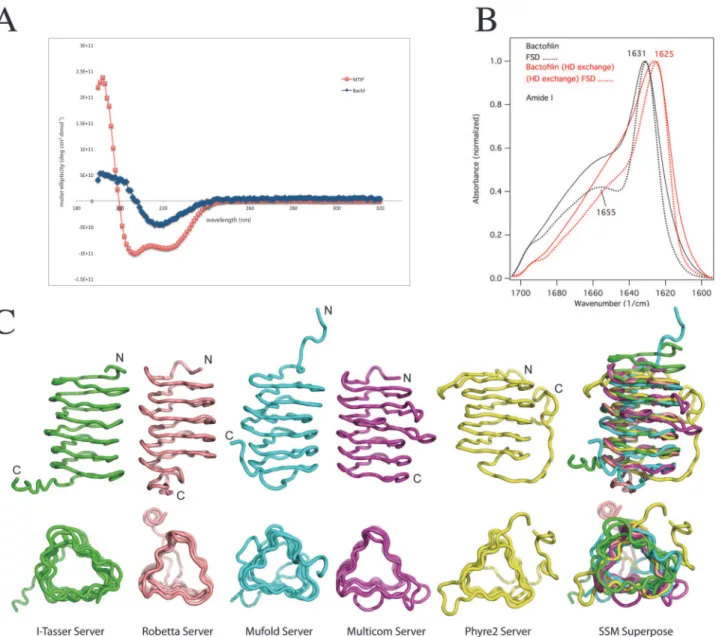
To begin characterization of the structure of the newly discovered bactofilin family of proteins, we purified fibers of exogenously expressed BacM fromE.coliand analyzed them by circular dichroism (CD). The recorded spectrum indicated a minimum at 218 nm that is characteristic for aβ-sheet-only structure [45], showing no resemblance to the CD spectrum of the control α-helix-only protein MTIP ofP.falciparum(Fig. 1A). To confirm this assessment, we
exam-ined the fibers using Fourier transform infrared spectroscopy (FTIR) (Fig. 1B). The amide I re-gion of the recorded spectrum revealed a prominent peak at 1631 cm-1, which is characteristic ofβ-sheets (for assignments of peaks to secondary structure elements see [46,47]). In
agree-ment with the CD results, no indication ofα-helices was found as the peak between 1650 and
1655 cm-1in the spectrum (characteristic ofα-helices or unstructured loops; [46,47])
disap-peared when the protein was deuterated, which is typical for unstructured regions, here pre-sumably coinciding with the proline-containing C-terminus. Moreover, the
FTIR-measurements were consistent withβ-strands forming parallelβ-sheets, since the peak at
1690–1695 cm-1that is characteristic of anti-parallelβ-sheets such as the core structure of
porin Omp32 [34] was very weak. Finally, the significant shift of the majorβ-structure peak
from 1631.9 to1625 cm-1upon deuteration is characteristic of a so-called "extendedβ-sheet".
This structure is the result of intermolecularβ-structure-like interactions of aggregating or sys-tematically polymerizing protein molecules (Fig. 1B, [46–48]). In summary, the results of the CD- and IR-spectroscopy indicated that BacM: (i) lacksα-helices, (ii) contains a small amount
of unstructured secondary structure, and (iii) mainly consists of parallelβ-sheets that through
the formation of fibers (iv) form extendedβ-sheet structures.
Generation of a 3-dimensional
in silico
Molecular Model of Mature BacM
servers were chosen from the top 10 competitors in the latest CASP competition for template-based modeling [49]. We used six servers, which operate by three different modeling ap-proaches: single-template-based modeling; multi-template, threading-based modeling; andab initiomodeling. Reference structures were chosen automatically by the modeling programs, primarily based on percent sequence identity with the target and other scoring criteria unique to each server. For single-template-based modeling the Phyre2 server [42] used the trimeric LpxA-like protein YdcK fromSalmonella choleraas template [DOI:10.2210/pdb2f9c/pdf], while the Robetta server [38] used the human dynactin p27 subunit [PDB 3TV0] [50]. For Fig 1. Generation of anin silicoBacM model.(A) The CD spectrum of isolated purified BacM fibers indicates aβ-structure-only protein devoid ofα-helices. (B) The observed peak at 1631 cm-1of the amide I region of the FTIR spectrum of purified BacM fibers is likewise characteristic ofβ-sheets. No indications of
α-helices are found as the peak between 1650 and 1655 cm-1in the Fourier self-deconvoluted (FSD) spectrum disappeared upon deuteration. This behavior
is indicative for unstructured regions. The shift of the majorβ-structure peak from 1631.9 cm-1to 1625 cm-1upon deuteration is characteristic of an
“extended
β-sheet”which is the result of intermolecularβ-structure-like interactions of the polymerized BacM. (C) Ribbon diagrams of various BacM models generated by five different molecular modeling servers. All models predict that nearly the entire protein is formed by a left-handedβ-helix with the non-structured flexible C-terminus variously oriented with respect to the barrel-shaped molecule. Noα-helices are present in the models of the protein.
multi-template, threading-based modeling, we used 3 servers: I-TASSER [36,37], MUFOLD [39], and MULTICOM [40,41]. These servers selected ten unique templates, threaded the query sequence into the structures, generated fragments, and reassembled the fragments to build models. A sixth server, QUARK, was used to generate anab initiomodel of the entire BacM protein [35]. A comparison of the top models from the template-based modeling servers are presented inFig. 1Cand the sequences of the templates used by the various servers can be found inS1 Fig.
All template-based modeling servers produced top models with a type-T, left-handedβ -helix (LBH) fold, consistent with the CD- and IR-spectroscopy data (Fig. 1A, B). In contrast, the QUARK-generatedab initiomodel (not shown) predicted aβ-sandwich orβ-roll structure containing unstructured regions. As this model disagreed with the spectroscopy data, it was not considered further. A superposition of the top models generated by each of the five, tem-plate-based servers is shown inFig. 1C. All models were essentially in agreement regarding the overall structure of BacM, converging on related structures despite the variety of the templates and modeling methods used. Consistent with our experimental observations, all template-based servers predicted that the majority of the protein is made ofβ-sheets and lacksα-helices. This model of aβ-stranded structure would thereby represent a structural motif that has not
been previously demonstrated in any bacterial cytoskeleton protein. As expected from the se-quence alignments, the highest degree of confidence for this structure prediction lies in the highly conserved bactofilin domain. No good template structure was identified for the short C-terminus due to the highly flexible nature predicted for this region, and the highest variability between the models from the different prediction programs was in this region (Fig. 1C). In summary, the template-based models indicated that BacM (i) lacksα-helices, (ii) contains a small amount of unstructured secondary structure, and (iii) mainly consists of parallelβ-sheets that form a characteristic LBH fold structure.
Sequence Analysis reveals a Repeated Motif Common to Bactofilins,
which predicts the Stacking of Hydrophobic Residues
While all template-based models for BacM are generally similar in that they predict aβ
-sole-noid structure of six stacked repeat-elements, there are differences in the specific details regard-ing alignment and lengths of theβ-strands. In order to determine if any of these models appear
more likely to represent the structure of BacM than the others, we undertook a bioinformatics approach. To identify potential repeating units, we analyzed the sequences of the four bactofi-lin paralogs ofM.xanthususing the HHrepID server [51] to identify conserved repeats. The server used BLAST to search for similar sequences to aid in identification of repeat sequences, and six repeats were identified for each bactofilin. The six repeats of each of the four bactofilins are aligned inFig. 2Aand A sequence logo, graphically representing sequence conservation at different positions in the repeat, was generated using the WebLogo server [52,53]. For clarity, we slightly adjusted the output so that the beginning of each repeat started with aβ-strand and
residue (Val or Phe), followed by a shorter turn with a conserved Gly/Ala, and ending with a longerβ-strand with two conserved hydrophobic residues (varying) and short turn. This
se-quence pattern is clearest and most highly conserved for repeat number 4 (Fig. 2B) and also conserved among bactofilins of multiple species, suggesting it is important to the function or structure of bactofilins (S2 Fig.). The repeat pattern identified by sequence alignment, con-served hydrophobic positions, and repeat length match well with the predictedβ-solenoid
structure output of the modeling servers (Fig. 1C).
Using our bioinformatics analysis, we next evaluated the models we had generated (Fig. 1C); we first evaluated the models for incorporation of 6 repeating units. All single-tem-plate and multi-temsingle-tem-plate-based models contained this feature, supporting the strength of this prediction. We next determined whether theβ-strands and conserved anchoring residues were
Fig 2. Bactofilins contain repeat regions with conserved hydrophobic residues withinβ-strands.(A) Alignment of the repeat regions in the 4 bactofilin paralogs ofM.xanthusidentified by HHrepID reveal that each paralog contains 6 repeats (top); Sequence logo showing conservation of residues at each position in the repeat, generated using the server at weblogo.berkeley.edu (bottom). (B) Repeat regions of BacM are aligned, revealing the highly conserved hydrophobic residues. S:β-strand; Orange box: highly conserved Gly residue; Red box: long second-strand anchor. (C) Conserved hydrophobic residues align along theβ-sheet sides of the solenoid structure. Side and top views of the ribbon diagram of the I-TASSER BacM model with the first two hydrophobic residues of each repeat highlighted in purple. (D) Table of parameters used to judge the quality of the predicted models by the five template-based servers. Models were evaluated based on the number of repeats and their alignment as well as ordered N- and C-termini. Checks and minuses denote that a given model possesses or lacks a given quality, respectively.
aligned, as expected in the core of the model. We found that I-TASSER, Robetta, and Phyre aligned theβ-strands consistent with the prediction from the sequence alignment, while the MULTICOM and MUFOLD models had frame-shifts of the repeats. In the MULTICOM model, the last two repeats were shifted by one turn, with the strand of a preceding repeat shifted into a loop. For the MUFOLD model, the shift occurred due to a loop insertion in a strand of repeat 4, shifting the alignment of subsequent repeats. Our final criteria were to evalu-ate the N- and C-terminal repeats and determine if they were aligned and if they stacked well, or were disordered. The quality of the N- and C-terminal repeats in terms of their order and alignment of the repeats is important because we expected that the monomers dimerize in a head-to-tail manner to form filaments and then laterally associate to form fibers (see below). If the N- and C-terminal repeats are not well ordered or properly aligned, then subsequent dock-ing attempts would likely produce results inconsistent with our electron microscopic observa-tions of BacM forming straight filaments. The N-termini of the I-TASSER, MULTICOM, and Robetta structures were correctly aligned and stacked. Additionally, the Phyre2 model had a well-ordered N-terminus, however, the second strand of the first repeat was shifted into the turns, and therefore was not correctly aligned. The first strand of the MUFOLD N-terminal re-peat did not lay down on top of the second rere-peat and would interfere in docking. Looking at the C-terminus, many models were disordered, likely due to the C-terminal domain with no known homology to any structure. To determine which models to use in subsequent protein docking analysis, we evaluated how well the last repeat of the bactofilin core was ordered and aligned. In the Robetta model, the C-terminal repeat did not lie flat against the other repeats and the rest of the C-terminus was in a position that would block dimer formation. Both the Phyre2 and MUFOLD models had a poorly ordered C-terminal repeat. The MULTICOM and I-TASSER models both had ordered C-termini. In addition to a completely ordered sixth re-peat, the I-TASSER model generated the first strand of what would be an unidentified seventh repeat. It is possible that this partial“pseudo repeat”exists, as there is a Leu and Thr that could fit into the sequence conservation and repeat alignment. However, our analysis using HHrepID did not identify these residues as part of a seventh repeat, and it is not currently possible to tell whether this additional strand is present or if it only is an artifact of the model building process. Despite this uncertainty, however, we chose to proceed with our experiments using the I-TAS-SER model, as this model was superior for accommodating stacking of the hydrophobic resi-dues in the conserved repeat regions, and for having well-ordered N- and C-termini (Fig. 2C). A table comparing the various models in terms of the qualities discussed above is provided in
Fig. 2D.
BacM assembles
in vivo
and
in vitro
into Filaments, Fibers, and Ribbons
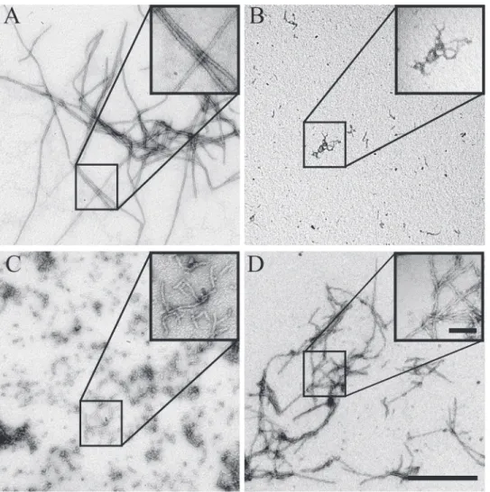
concentrations (>100μM) were used (Fig. 3A). Both fibers and ribbons appear to comprise multiple filaments, and we observed individual filaments of about 3 nm following dialysis against a strongly alkali glycine buffer (S3 Fig., pH 10.5). Under these buffer conditions, BacM assembles exclusively into the 3 nm filaments. When dialyzed against a less alkali buffer (pH 9.5), a mixture of 3 nm filaments and 10 nm fibers was observed, and 10 nm fibers were exclusively observed following dialysis against a pH 8.5 glycine buffer (S3 Fig.). While glycine is an appropriate buffer in this pH range [54], it is also a charged molecule. Thus, glycine may contribute to BacM filament formation by acting as a counterion, leading to a reduction in lon-gitudinal binding. Such an effect was recently reported for the disassembly of microtubules in spermine-containing buffers [55]. To separate out the possible contributions of glycine as a buffer, and glycine as a counterion to BacM, we dialyzed BacM against Tris, pH 7.5, CHES, pH 9.5, and CAPS, pH 10.5 with or without glycine present. At all pH tested, 10 nm fibers were recovered in buffers lacking glycine (Fig. 3). The addition of glycine to each buffer favored re-covery of 3 nm filaments, with exclusively filaments in the pH 10.5 buffer, a mixture in pH 9.5 buffer, and partial“unraveling”of fibers at pH 7.5, similar to the results of glycine alone, indi-cating that glycine is most likely exerting its effect as a counterion. To test if the 3 nm filaments constitute BacM polymers that form due to the presence of glycine, or comprise the elementary polymeric structure of BacM, we generated 10 nm fibers in a Tris, pH 7.5 buffer and subse-quently dialyzed these fibers against an alkali, glycine containing buffer. These 10 nm fibers separated into 3 nm filaments (S4 Fig.). Together, these data suggest that BacM polymerizes into 3 nm filaments and that these filaments are either unstable or possess a high propensity to Fig 3. Structure of BacM fibers, ribbons, and filaments after dialysis against various buffers.Exogenously expressed and purified BacM polymerizes into different structures upon dialysis against different buffers.(A)In a phosphate-citric acid buffer (pH 6.0, top), or at high concentrations (100μM, bottom), large ribbon-like structures were observed. (B)In Tris, CHES or CAPS buffers (pH 7.5, 9.5 or 10.5, respectively), 10 nm fibers resembling those isolated from
M.xanthuscells predominate (top row). The addition of 20 mM glycine to the buffers (bottom row) favors the formation of 3 nm filaments at increasing abundance with increasing pH. Scale bar = 50 nm.
bundle into fibers, and that, under high protein concentrations, this interaction can lead to higher-ordered ribbon-like structures.
The BacM Monomer Model predicts Hydrophobic Patches Important for
Filament Formation and a Charged Surface
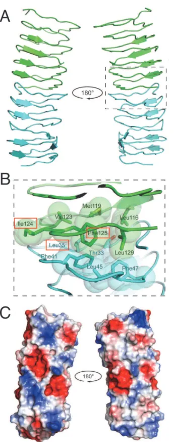
Since BacM forms homo-monomeric filaments and fibersin vivo[29] and, like all bactofilins studied, similar fibersin vitro(see also [27]), we modeled the protein-protein interface respon-sible for dimerization using the ClusPro 2.0 docking server [43] in conjunction with our elec-tron microscopic data (Fig. 4). The BacM model indicated a monomer size of roughly 3x3x3 nm and the electron microscopic data revealed ~3 nm wide filaments, suggesting an end-to-end stacking of monomers. Additionally, the extendedβ-sheet structure observed by FTIR suggest
the most likely arrangement was a head-to-tail dimer. Using the best-predicted model from I-TASSER, we performed docking calculations to attempt to identify residues important to dimer formation.
We proceeded to dock the full-length I-TASSER model, resulting in a head-to-tail dimer that involved the C-terminal residues Met131to Ala139, which included the“pseudo-seventh re-peat.”However, it is known that this C-terminal region of BacM is not necessary for filament formation. Fibers isolated fromM.xanthuscontain a high proportion of proteolyzed BacM [29], which lacks this“pseudo-repeat”region and the C-terminal residues identified by the ClusPro server to be at the interface; therefore, we reasoned that the bactofilin domain alone is sufficient for polymerization. To experimentally test this prediction, we expressed the bactofilin domain alone inE.coliand purified it under denaturing conditions. Upon dialysis into a poly-merization buffer, we observed robust polypoly-merization of the protein, though the architecture of the fiber was twisted and highly kinked, and filaments tended to be shorter relative to the wild type protein (Fig. 5D). This suggests that the C-terminus is indeed dispensable for poly-merization but contributes to the stability of the fiber, and the lateral packing of filaments, which are responsible for the“smooth”appearance of the wild type fibers.
Based on thein vivoandin vitrodata, which indicated that the C-terminus is not essential for polymerization, we used only the bactofilin domain for docking of the I-TASSER model. Using the ClusPro 2.0 server, we generated a best-balanced model that resulted in head-to-tail dimer formation (Fig. 4A). We found that the interface of the head-to-tail dimer is largely me-diated by hydrophobic patches at the N- and C-termini of each monomer: Thr33, Leu35, Leu45, and Phe47at the N-terminus, and Leu116, Met119, Val123, Phe125, and Leu129at the C-terminus
(Fig. 4B). The changed solvent accessible surface area (SASA) was ~1307 Å. Additionally, the top head-to-tail model was arranged so that the repeats continued to stack in order with strand 1, repeat 1 of monomer 1 found on the same side of the solenoid as strand 1, repeat 1 of mono-mer 2, allowing a continued stacking of repeat units, mediated by the conserved hydrophobic residues, from one monomer to the next, throughout the filament.
We next investigated the surfaces of the BacM dimer to predict how they could facilitate lat-eral interactions. As can be seen from the electrostatic surface maps (Fig. 4C), the exposed sur-face of BacM has strips of positively and negatively charged residues that could interact with other filaments to form higher ordered fibers. These oppositely charged surface areas could in-teract laterally, explaining the formation of the 10 nm wide fibers and why the thinner fila-ments are virtually never found. Additionally, when attempting to model a head-to-tail dimer interface using the ClusPro server, a result of two monomers interacting laterallyviathese elec-trostatic patches was a common result (not shown).
Fig 4. Modeling of the dimerization domain of bactofilin predicts hydrophobic interface and charged surfaces.(A) The bactofilin domain from the I-TASSER model was modeled using ClusPro 2.0 and predicts head-to-tail dimerization. Individual BacM monomers are represented as blue and green ribbon structures, and the dimer is presented as rotated 180o. (B) The interface between two bactofilin-domains is predicted to
patches of hydrophobic residues at the N- and C-termini. These filaments are likely further sta-bilized by lateral electrostatic interactions that result in the formation of fibers, creating highly stable structures.
Wild Type BacM forms Stable Filaments
in vitro
and Filament Formation
depends on the Hydrophobic Patches of the Predicted Interface Domain
Sequences containing hydrophobic residues at specific locations on the solenoid are predicted to mediate interactions between repeat subunits within one BacM monomer. In order to test positions to form a hydrophobic pocket. The residues mutated in this study are highlighted with red boxes. (C) An electrostatic surface map of the dimer model reveals patches of charged residues that are solvent exposed. Negatively charged areas are shown in red, positively charged areas in blue, and areas that are charge neutral are shown in white.
doi:10.1371/journal.pone.0121074.g004
Fig 5. Evaluation of polymerization and fiber formation of recombinant mutant forms of BacM.While the wild type (A) and the C-terminal truncation mutant (D) are able to polymerize, the L35E (B) and the I124D/ F125R (C) mutants are no longer able to form fibers and aggregate instead. (D) Despite their ability to polymerize, the C-terminal truncation forms fibers that show a distinct aberrant“braided”morphology when compared with the smooth wild type fibers. The scale bar in the large field is 0.5μm, while the scale bar in the inset is 100 nm.
the prediction of our dimer model that these hydrophobic repeats also mediate interactions be-tweensubunits, we performed anin vitroassembly assay. We expressed His-tagged constructs encoding the bactofilin domain and C-terminus of BacM inE.coli, and purified the protein under denaturing conditions (8 M urea). We expressed wild type BacM and two BacM mutants with hydrophobic residues (within repeat 1 and repeat 6 (Fig. 2B)) mutated to charged residues (L35E and I124D/F125R). These residues were selected for mutation due to their predicted lo-cation at the dimer interface, and for their high degree of conservation. These proteins were pu-rified under denaturing conditions (8 M urea). To ensure that these mutants were properly folded, urea was removed by dialysis, and the proteins were analyzed by CD spectroscopy. The recorded spectra closely matched that of wild type BacM, indicating aβ-strand-only structure (S5 Fig.). To assay for polymerization, the mutants were diluted to ~10μM, and urea was re-moved by dialysis. Spontaneous fiber formation of the dialysate was monitored by negative staining (Fig. 5). Wild type BacM formed relatively uniform, ~10 nm fibers, while filament for-mation was completely abolished for both mutants. Small aggregates of ~3 nm wide particles in preparations of both mutants were observed. This observation is consistent with BacM sub-units that fail to polymerize into filaments, but are still able to laterally associate, or alternative-ly, the formation of short filaments (Fig. 5B and C). To discriminate between these two hypotheses, these mutants were dialyzed against an alkali glycine-containing buffer to reduce lateral interactions (Fig. 3). In this buffer, these small protein aggregates were completely ab-sent, consistent with an interpretation that the aggregates observed are monomers of BacM that are associatingvialateral electrostatic charges, which can be disrupted by addition of gly-cine in an alkali solution (S6 Fig.).
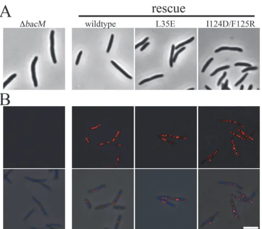
TheΔbacMmutantM.xanthuswas described as having a morphological defect, where the typical rod-shape of the bacterium was crooked or kinked [29]. In order to test if the hydropho-bic interface is required for proper BacM functionin vivo, the L35E and the I124D/F125R mu-tations were introduced separately into aΔbacMmutant ofM.xanthusto test for rescue of the wildtype morhpology (Fig. 6). While introduction of wild typebacMrestored the rod-shaped phenotype, both polymerization-defective mutants failed to rescue, although they were robust-ly expressed, as measured by immunoblot (Figs.6AandS7A). When examined by immunoflu-orescence with an anti-BacM antibody, the overexpressed wild typebacMdisplayed the expected pattern of extended fibers across the length of the cell (Fig. 6Band [29]). BacM con-taining mutations to either interface domain appeared to form fiber-like scon-taining to varying de-grees. Unlike the wild type BacM, however, this staining appeared discontinuous across the cell. This effect was more severe with the I124D/F125R mutant than the L35E mutant, though they both failed to rescue (Fig. 6). BacM lacking the C-terminus was unable to be visualized in
M.xanthus, as steady-state levels of the mutant protein were undetectable by immunoblot, pos-sibly due to instability of the protein (S7B Fig.). Together, these data are consistent with our hy-pothesis that the bactofilin domain alone mediates polymerization of BacM into filaments, and that a hydrophobic interface is required for this interaction.
Wild Type BacM forms Higher-ordered Fibers
in vitro
and Fiber
Formation depends on Charged Surface Areas
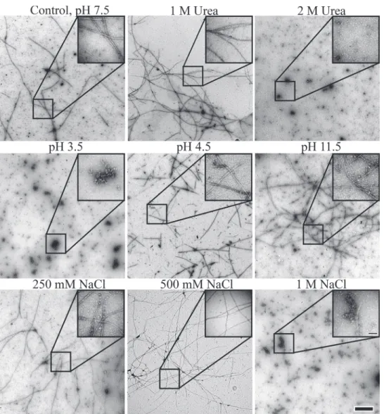
that the isolation of exogenously expressed monomers fromE.coliis only possible in the pres-ence of high concentrations of urea (seeMaterials and Methods). In order to understand the fiber formation process better, we tested various conditions and their influence on fiber forma-tion. For this, recombinant native BacM protein was purified under denaturing conditions (8 M urea) and then dialyzed against buffers with various salt concentrations, chaotropic agents or pH. The ability to form fibers under these conditions was assessed by negative staining and examination under the transmission electron microscope (Fig. 7). Under these conditions, spontaneous formation of native-like BacM fibers was observed when the urea con-centration was reduced to 1 M or lower. In the absence of urea, BacM formed fibers across a wide range of pH in Tris buffer, and was only inhibited at extremes of pH (below pH 4.5 or above pH 11.5). Like urea, salt prevented fiber formation and polymerization at concentrations higher than 0.5 M. Unlike the samples dialyzed against glycine, however, 10 nm fibers were still observed, suggesting that NaCl does not similarly act as a counterion, preventing lateral in-teractions between filaments. Together these data showed that theβ-sheet-based fold of BacM is extraordinarily stable and that lateral association for fiber formation is a robust process that occurs in a wide range of pH and even in the presence of chaotropic agents.
Fig 6. Mutations to putative BacM interface fail to rescue the morphological phenotype of a deletion mutant.(A) Images ofΔbacMcells or the indicated rescue strains were taken by phase contrast light microscopy. (B) The same cell lines were fixed and imaged by immunofluorescence microscopy with an anti-BacM antibody (red) and stained with DAPI (blue). Top panel, anti-anti-BacM. Bottom panel, merge with phase contrast. Scale bar = 5μm.
Discussion
The fact that bacteria contain multiple cytoskeletal systems was realized only recently, and their discovery and rapid characterization owes much to macromolecular structural studies, as there is often low sequence homology to their eukaryotic counterparts. BacM is a newly discov-ered, uniquely bacterial, cytoskeleton protein [27] that forms highly stable, helically arranged polymers inM.xanthus[29]. Unlike bacterial cytoskeleton proteins of the MreB and FtsZ families [17,56], BacM polymerizes independently of nucleotides or cofactors. While this is similar to the bacterial IF homologous CreS, which also assembles in the absence of nucleotides or cofactors, CreS fibers disassemble when the pH is above 8.4, a hallmark of IFs [18]. Addi-tionally, assembly of CreS, as other IF proteins, is mediated by its extensive coiled-coil do-mains. The resilience of BacM fibers at extreme pH, as well as the lack of predicted coiled-coils Fig 7. Evaluation of polymerization and fiber formation of recombinant wild type BacM in the electron microscope after various treatments.Fiber formation is robust and occurs under a wide variety of conditions. In the presence of chaotropic agents, such as urea, the wild type protein polymerizes at concentrations of up to 1 M (upper row), while polymerization is more sensitive to low than to high pH values (middle row) and does not occur at NaCl concentrations of 0.5 M and higher (lower row) indicating the importance of charge for the lateral association of BacM filaments. The scale bar in the large field is 0.5μm, while the scale bar in the inset is 100 nm.
(or experimental evidence ofα-helices at all), strengthens the notion that bactofilins comprise
a novel class of bacterial cytoskeleton.
The propensity of BacM to spontaneously form highly stable fibers presents a great difficul-ty in crystallizing BacM protein for direct structural studies. As an alternative to X-ray crystal-lography, we computed a 3-D model of BacM in an attempt to predict important structural and functional residues and domains within the protein. The proposed model adopts a type-T left-handedβ-helix fold (LBH), a highly regular and symmetrical structure with little variation
in shape and size over the length of the domain [57], and is consistent with our experimental observations. So far all LBH X-ray structures that have been solved belong to bacterial proteins, a vast majority of which have enzymatic, particularly transferase, activities, composing the tri-meric LpxA-like superfamily [58–60]. All but one LBH-containing proteins form homo-tri-mers (trimeric LpxA-like proteins), the only exception being the anti-freeze protein from the spruce budworm, which appears to be monomeric under physiological conditions [61]. Impor-tantly, the LBH domain facilitates the trimerization of these proteins by laterally aligning into a bundle in the center of the structures. Although additional alignments resulting in LBH do-main-mediated fiber formation is thought possible [62], it has so far not been reported for any protein under physiological conditions. The main reason being that the self-association of the LBH domain is likely restricted to trimerization by additional structural elements at the N- or C-termini of the proteins [63]. Consequently, it has been predicted that removal of these struc-tural elements would expose theβ-strands, leading to un-restricted self-assembly [62]. Analysis of the sequence of the N-terminally cleaved mature BacM shows that ~80% of the protein is formed by the LBH fold-containing DUF583 domain. Only 5 and 23 aa at the N- and C-termi-nus, respectively, are not part of this structure, likely limiting their influence to restrict poly-merization. Since BacM forms 3 nm wide filamentsin vivoandin vitro, the individual protein monomers are therefore most likely stacked head-to-tail within these filaments. This interpre-tation is in line with (i) the electron microscopic observations, (ii) ourin silicomodels of the di-merization and filament formation, and (iii) the spectroscopic data that show the presence of extendedβ-sheet structures, which indicate that theβ-sheets within the BacM polymer are highly oriented. Since these predicted 3 nm filaments are rarely observedin vitroand not re-coveredin vivo, it is clear that they either immediately interact with each other by forming the ~10 nm thick fibers or that filament polymerization and fiber bundling are coupled processes. This finding is reminiscent of eukaryotic microtubules, which are assembled by end-to-end in-teractions of dimer subunits to form protofilaments, which form a tubevialongitudinal inter-actions between the protofilaments [64]. Analogous to our finding that lateral interactions of BacM filaments were disrupted by the presence of anionic glycine, it was recently reported that incubation of assembled microtubules with the cation spermine triggered the disassembly of microtubules into protofilaments, presumably by acting as a counterion, weakening the lateral interactions between protofilaments and favoring disassembly [55].
immunofluorescence light microscopy. Light microscopic observation of these fiber-forming mutants showed that they failed to rescue the morphological phenotype of abacMmutant indicating that the altered fiber morphology interferes with the biological function of the cytoskeleton protein.
The spontaneous polymerization of BacM under a wide range of conditions poses a funda-mental problem for the bacterial cell. Namely, how the cells control this process. In earlier work, we had established that BacM is post-translationally cleaved, and proposed that this process is important for the control of polymerization [29]. Here we propose that the concen-tration of the protein is also a factor. During ourin vitropolymerization experiments, we ob-served thick, multi-stranded ribbons of BacM that predominate at high protein concentration (~100μM). These multi-stranded ribbons may explain the rod-like structures observed by immunofluorescence examination of BacM in ~20% of wild type cells [29]. Intriguingly, these thicker BacM rods are observed in virtually all cells upon overexpression ofbacM[29], suggest-ing that at higher concentration, excess BacM filaments continue to bundle with existsuggest-ing fibers eventually forming thicker ribbons.
While the monomer model has high degree of confidence for the structure prediction of the highly conserved bactofilin domain, the structure and arrangement of the C-terminus is cur-rently less clear. This domain most likely also accounts for the small portion of unstructured region measured in the FTIR experiments. This interpretation is in line with the observation that the C-terminus of BacM contains a cluster of five proline residues [29] that are usually in-dicative of unstructured regions of proteins [65,66]. Although these poly-proline-containing regions are“unstructured,”they have been identified as important elements engaging in the re-cruitment, recognition, and binding of other peptides and proteins [67]. Since it has been sug-gested that bactofilins act as scaffolds, exerting their morphogenic effect through the binding of PG-remodeling enzymes [27,28], this region of the protein could be an important binding site for these enzymes. Alternatively, the highly charged surface of BacM that contains negatively and positively charged patches could interact with such enzymes and other BacM-binding pro-teins through electrostatic interactions similar to the lateral BacM-BacM interactions described here. Finally, the C-terminus appears also to play a role in lateral interaction between individu-al fibers, because a truncated version of the protein polymerizes but forms characteristic kinked bundles that look distinctly different from the smoother bundles formed by the wild type protein.
the structural principles guiding polymerization and fiber formation and eventually result in studies that will establish the atomic structure of this important novel class of bacterial cytoskeleton proteins.
Supporting Information
S1 Fig. Sequence-based alignment of templates used for homology modeling.The 30 struc-tures used as templates to generate homology models by five servers were aligned using Clus-talW. The sequences have been aligned with the BacM sequence residues 28–150. ClustalX coloring was applied to sequences in Jalview to denote amino acid conservation [73]. (PDF)
S2 Fig. Bactofilin repeats are conserved between bactofilins of multiple species.4M. xan-thusbactofilin paralogs are aligned with BacA and BacB ofC.crescentus(CC_1873 and CC_3022, respectively), CcmA fromP.mirabilis(PMI1961) and CcmA fromH.pylori
(HPG27_1480). (TIF)
S3 Fig. Structure of BacM fibers, ribbons, and filaments after dialysis in glycine buffers at different pH.Exogenously expressed and purified BacM polymerizes into different structures upon dialysis against 20 mM glycine buffer at various pH. At pH 8.5 10 nm fibers resembling those isolated fromM.xanthuscells predominate (pH 8.5). Upon further increase of the pH, the 10 nm fibers are more and more replaced by 3 nm filaments (pH 9.5), which at pH 10.5 are the only observed form of polymerized BacM (pH 10.5). Scale bar, 50 nm.
(TIF)
S4 Fig. Reconstituted 10 nm BacM fibers can be laterally separated by a pH change into 3 nm filaments.BacM was purified in 8M urea, and subsequently dialyzed against 20 mM gly-cine, pH 8.5. A sample was removed, applied to a copper grid, negative stained, and imaged by transmission electron microscope (left). The remainder of the dialysate was then dialyzed against 20 mM glycine, pH 10.5 and imaged as above (right). Scale bar = 25 nm.
(TIF)
S5 Fig. Non-polymerizing mutants and C-terminal truncation mutant of BacM are proper-ly folded.The indicated BacM mutants were purified as described in Materials and Methods, and dialyzed against 20 mM Tris, pH 7.5 to a final concentration of 0.5–1.0 mg/ml. These dial-ysates were examined by circular dichroism spectroscopy, as described in Materials and Meth-ods. Theα–helix-only protein MTIP was used as a control [32,33].
(TIF)
S6 Fig. Polymerization-defective mutants of BacM form aggregatesvialateral interactions. The indicated mutants of BacM were expressed inE.coliand purified in 8 M urea, and subse-quently dialyzed against 20 mM Tris, pH 7.5 (left) or 20 mM glycine, pH 10.5 (right). Samples were applied to a copper grid, negative stained, and imaged by transmission electron micro-scope. While aggregates were ubiquitously found at pH 7.5, they were absent in samples pre-pared from the glycine buffer. Scale bar = 25 nm.
(TIF)
S7 Fig. BacM L35E and I124D/F125R mutants have normal steady-state levels and theΔ C-terminus mutant fails to express.Lysates from the indicated strains ofM.xanthuswere sepa-rated by SDS-PAGE and immunoblotted with an affinity purified anti-BacM antibody [29].
rescue (EH344); Lane 4:ΔbacMwith I124D/F125RbacMrescue (EH171); Lane 5:ΔbacMwith L35EbacMrescue (EH175).(B)Lane 1: wildtype (DK1622); Lane 2:ΔbacM(EH301); Lane 3 ΔbacMwithbacM-ΔC-termrescue (EH106). Unlabeled lanes are from strains not discussed in this report.
(TIF)
Acknowledgments
We would like to thank the members of the Hoiczyk laboratory for helpful discussions and comments on the manuscript.
Author Contributions
Conceived and designed the experiments: DMZ LEB KX HE JB EH. Performed the experi-ments: DMZ LEB KX HE JB EH. Analyzed the data: DMZ LEB KX HE JB EH. Contributed re-agents/materials/analysis tools: JB EH. Wrote the paper: DMZ LEB KX HE JB EH.
References
1. Shih YL, Rothfield L. The bacterial cytoskeleton. Microbiol Mol Biol Rev. 2006; 70: 729–754. PMID:
16959967
2. Gitai Z. Diversification and specialization of the bacterial cytoskeleton. Curr Opin Cell Biol. 2007; 19: 5– 12. PMID:17178455
3. Thanbichler M, Shapiro L. Getting organized—how bacterial cells move proteins and DNA. Nat Rev Microbiol. 2008; 6: 28–40. PMID:18059290
4. Graumann PL. Dynamics of bacterial cytoskeletal elements. Cell Motil Cytoskeleton. 2009; 66: 909– 914. doi:10.1002/cm.20381PMID:19466751
5. Löwe J, Amos LA. Evolution of cytomotive filaments: the cytoskeleton from prokaryotes to eukaryotes. Int J Biochem Cell Biol. 2009; 41: 323–329. doi:10.1016/j.biocel.2008.08.010PMID:18768164
6. Cabeen MT, Jacobs-Wagner C. The bacterial cytoskeleton. Annu Rev Genet. 2010; 44: 365–392. doi:
10.1146/annurev-genet-102108-134845PMID:21047262
7. Erickson HP, Anderson DE, Osawa M. FtsZ in bacterial cytokinesis: cytoskeleton and force generator all in one. Microbiol Mol Biol Rev. 2010; 74: 504–528. doi:10.1128/MMBR.00021-10PMID:21119015
8. Celler K, Koning RI, Koster AJ, van Wezel GP. Multidimensional view of the bacterial cytoskeleton. J Bacteriol. 2013; 195: 1627–1636. doi:10.1128/JB.02194-12PMID:23417493
9. Popp D, Robinson RC. Many ways to built an actin filament. Mol Microbiol. 2011; 80: 300–308. doi:10. 1111/j.1365-2958.2011.07599.xPMID:21362063
10. Teixidó-Travesa N, Roig J, Lüders J. The where, when and how of microtubule nucleation—one ring to rule them all. J Cell Sci. 2012; 125: 4445–4456. doi:10.1242/jcs.106971PMID:23132930
11. Goldman RD, Cleland MM, Murthy SN, Mahammad S, Kuczmarski ER. Inroads into the structure and function of intermediate filament networks. J Struct Biol. 2012; 177: 14–23. doi:10.1016/j.jsb.2011.11. 017PMID:22120848
12. Ingerson-Mahar M, Gitai Z. A growing family: the expanding universe of the bacterial cytoskeleton. FEMS Microbiol Rev. 2012; 36: 256–266. doi:10.1111/j.1574-6976.2011.00316.xPMID:22092065
13. Lin L, Thanbichler M. Nucleotide-independent cytoskeletal scaffolds in bacteria. Cytoskeleton. 2013; 70: 409–423. doi:10.1002/cm.21126PMID:23852773
14. Nogales E, Wolf SG, Downing KH. Structure of the alpha beta tubulin dimer by electron crystallography. Nature. 1998; 391: 199–203. PMID:9428769
15. Bork P, Sander C, Valencia A. An ATPase domain common to prokaryotic cell cycle proteins, sugar ki-nases, actin, and hsp70 heat shock proteins. Proc Natl Acad Sci USA. 1992; 89: 7290–7294. PMID:
1323828
16. Jones LJ, Carballido-Lopez R, Errington J. Control of cell shape in bacteria: helical, actin-like filaments inBacillus subtilis. Cell. 2001; 104: 913–922. PMID:11290328
18. Ausmees N, Kuhn JR, Jacobs-Wagner C. The bacterial cytoskeleton: an intermediate filament-like function in cell shape. Cell. 2003; 115: 705–713. PMID:14675535
19. Bagchi S, Tomenius H, Belova LM, Ausmees N. Intermediate filament-like proteins in bacteria and a cy-toskeletal function inStreptomyces. Mol Microbiol. 2008; 70: 1037–1050. doi:10.1111/j.1365-2958. 2008.06473.xPMID:18976278
20. Waidner B, Specht M, Dempwolff F, Haeberer K, Schaetzle S, Speth V, et al. A novel system of cyto-skeletal elements in the human pathogenHelicobacter pylori. PLoS Pathog. 2009; 5: e1000669. doi:
10.1371/journal.ppat.1000669PMID:19936218
21. Fiuza M, Letek M, Leiba J, Villadangos AF, Vaquera J, Zanella-Cleon I, et al. Phosphorylation of a novel cytoskeletal protein (RsmP) regulates rod-shaped morphology inCorynebacterium glutamicum. J Biol Chem. 2010; 285: 29387–29397. doi:10.1074/jbc.M110.154427PMID:20622015
22. Fenton AK, Hobley L, Butan C, Subramaniam S, Sockett RE. A coiled-coil repeat protein CcrP in Bdel-lovibrio bacteriovorusprevents cellular indentation, but is non-essential for vibroid cell morphology. FEMS Microbiol Lett. 2010; 313: 89–95. doi:10.1111/j.1574-6968.2010.02125.xPMID:20977494
23. Specht M, Schätzle S, Graumann PL, Waidner B.Helicobacter pyloripossesses four coiled-coil-rich proteins that form extended filamentous structures and control cell shape and motility. J Bacteriol. 2011; 193: 4523–4530. doi:10.1128/JB.00231-11PMID:21642462
24. Gerdes K, Howard M, Szardenings F. Pushing and pulling in prokaryotic DNA segregation. Cell. 2010; 141: 927–942. doi:10.1016/j.cell.2010.05.033PMID:20550930
25. Marchler-Bauer A, Anderson JB, Chitsaz F. Derbyshire MK, DeWeese-Scott C, Fong JH, et al. CDD: specific functional annotation with the Conserved Domain Database. Nucleic Acids Res. 2009; 37: D205–210. doi:10.1093/nar/gkn845PMID:18984618
26. Hay NA, Tipper DJ, Gygi D, Hughes C. A novel membrane protein influencing cell shape and multicellu-lar swarming ofProteus mirabilis. J Bacteriol. 1999; 181: 2008–2016. PMID:10094676
27. Kühn J, Briegel A, Mörschel E, Kahnt J, Leser K, Wick S, et al. Bactofilins, a ubiquitous class of cyto-skeletal proteins mediating polar localization of a cell wall synthase inCaulobacter crescentus. EMBO J. 2010; 29: 327–339. doi:10.1038/emboj.2009.358PMID:19959992
28. Sycuro LK, Pincus Z, Gutierrez KD, Biboy J, Stern CA, Vollmer W, et al. Peptidoglycan crosslinking re-laxation promotesHelicobacter pylori's helical shape and stomach colonization. Cell. 2010; 141: 822– 833. doi:10.1016/j.cell.2010.03.046PMID:20510929
29. Koch MK, McHugh CA, Hoiczyk E. BacM, an N-terminally processed bactofilin ofMyxococcus xanthus, is crucial for proper cell shape. Mol Microbiol. 2011; 80: 1031–1051. doi:10.1111/j.1365-2958.2011. 07629.xPMID:21414039
30. Bulyha I, Lindow S, Lin L, Bolte K, Wuichet K, Kahnt J, et al. Two small GTPases act in concert with the bactofilin cytoskeleton to regulate dynamic bacterial cell polarity. Dev Cell. 2013; 25: 119–131. doi:10. 1016/j.devcel.2013.02.017PMID:23583757
31. Kaiser D. Social gliding is correlated with the presence of pili inMyxococcus xanthus. Proc Natl Acad Sci USA. 1979; 76: 5952–5956. PMID:42906
32. Bosch J, Turley S, Daly TM, Bogh SM, Villasmil ML, Roach CM, et al. Structure of the MTIP-MyoA com-plex, a key component of the malaria parasite invasion motor. Proc Natl Acad Sci USA. 2006; 103: 4852–4857. PMID:16547135
33. Bosch J, Turley S, Roach CM, Daly TM, Bergman LW, Hol WG. The closed MTIP-myosin A-tail com-plex from the malaria parasite invasion machinery. J Mol Biol. 2007; 372: 77–88. PMID:17628590
34. Zeth K, Diederichs K, Welte W, Engelhardt H. Crystal structure of Omp32, the anion-selective porin fromComamonas acidovorans, in complex with aperiplasmic peptide at 2.1Åresolution. Structure. 2000; 8: 981–992. PMID:10986465
35. Xu D, Zhang Y.Ab initioprotein structure assembly using continuous structure fragments and opti-mized knowledge-based force field. Proteins. 2012; 80: 1715–1735. doi:10.1002/prot.24065PMID:
22411565
36. Zhang Y. I-TASSER server for protein 3D structure prediction. BMC Bioinformatics. 2008; 9: 40. doi:
10.1186/1471-2105-9-40PMID:18215316
37. Roy A, Kucukural A, Zhang Y. I-TASSAR: a unified platform for automated protein structure and func-tion predicfunc-tion. Nat Protoc. 2010; 5: 725–738. doi:10.1038/nprot.2010.5PMID:20360767
38. Kim DE, Chivian D, Baker D. Protein structure and analysis using the Robetta Server. Nucleic Acids Res. 2004; 32: W526–W531. PMID:15215442
40. Cheng J. A multi-template combination algorithm for protein comparative modeling. BMC Struct Biol. 2008; 8: 18. doi:10.1186/1472-6807-8-18PMID:18366648
41. Wang Z, Eickholt J, Cheng J. MULTICOM: A multi-level combination approach to protein structure pre-diction and its assessment in CASP8. Bioinformatics. 2010; 26: 882–888. doi:10.1093/bioinformatics/ btq058PMID:20150411
42. Kelley LA, Sternberg MJE. Protein structure prediction on the Web: a case study using the Phyre serv-er. Nat Protoc. 2009; 4: 363–371. doi:10.1038/nprot.2009.2PMID:19247286
43. Kozakov D, Beglov D, Bohnuud T, Mottarella SE, Xia B, Hall DR, et al. How good is automated protein docking? Proteins. 2013; 81: 2159–2166. doi:10.1002/prot.24403PMID:23996272
44. Krissinel E, Henrick K. Inference of macromolecular assemblies from crystalline state. J Mol Biol. 2007; 372: 774–797. PMID:17681537
45. Greenfield NJ. Using circular dichroism to estimate protein secondary structure. Nat Protoc. 2006; 1: 2876–2890. PMID:17406547
46. Arrondo JLR, Muga A, Castesana J, Goñi FM. Quantitative studies of the structure of proteins in solu-tion by Fourier-transform infrared spectroscopy. Prog Biophys Mol Biol. 1993; 59: 23–56. PMID:
8419985
47. Barth A, Zscherp C. What vibrations tell us about proteins. Quarterly Rev Biophys. 2002; 35: 369–430. PMID:12621861
48. Zandomeneghi G, Krebs MRH, McCammon MG, Fändrich M. FTIR reveals structural differences be-tween nativeβ-sheet proteins and amyloid fibrils. Prot Sci. 2004; 13: 3314–3321. PMID:15537750
49. Huang YJ, Mao B, Aramini JM, Montelione GT. Assessment of template-based protein structure predic-tions in CASP10. Proteins. 2014; 82: 43–56. doi:10.1002/prot.24488PMID:24323734
50. Yeh TY, Kowalska AK, Scipioni BR, Cheong FKY, Zheng M, Derewenda U, et al. Dynactin helps target Polo-like kinase 1 to kinetochores via its left-handed beta-helical p27 subunit. EMBO J. 2013; 32: 1023–1035. doi:10.1038/emboj.2013.30PMID:23455152
51. Biegert A, Söding J. HHrepID: de novo protein repeat identification by probabilistic consistency. Bioin-formatics. 2008; 24: 807–814. doi:10.1093/bioinformatics/btn039PMID:18245125
52. Crooks GE, Hon G, Chandonia JM, Brenner SE. WebLogo: a sequence logo generator. Genome Res. 2004; 14: 1188–1190. PMID:15173120
53. Schneider TD, Stephens RM. Sequence logos: a new way to display consensus sequences. Nucleic Acids Res. 1990; 18: 6097–6100. PMID:2172928
54. Ruzin SE. Plant Microtechnique and Microscopy. Oxford University Press; 1999.
55. Ojeda-Lopez MA, Needleman DJ, Song C, Ginsburg A, Kohl PA, Li Y, et al. Transformation of taxol-sta-bilized microtubules into inverted tubulin tubules triggered by a tubulin conformation switch. Nat Mater. 2014; 13: 195–203. doi:10.1038/nmat3858PMID:24441880
56. Löwe J, Amos LA. Crystal structure of the bacterial cell-division protein FtsZ. Nature. 1998; 391: 203– 206. PMID:9428770
57. Zheng J, Zanuy D, Haspel N, Tsai CJ, Alemán C, Nussinov R. Nanostructure design using protein building blocks enhanced by conformationally constrained synthetic residues. Biochem. 2007; 46: 1205–1218.
58. Jenkins J, Pickersgill R. The architecture of parallel beta-helices and related folds. Prog Biophys Mol Biol. 2001; 77: 111–175. PMID:11747907
59. Iengar P, Joshim NV, Balaram P. Conformational and sequence signatures in beta helix proteins. Structure. 2006; 14: 529–542. PMID:16531237
60. Kajava AV, Steven AC. Beta-rolls, beta-helices, and other beta-solenoid proteins. Adv Protein Chem. 2006; 73: 55–96. PMID:17190611
61. Gauthier SY, Kay CM, Sykes BD, Walker VK, Davies PL. Disulfide bond mapping and structural charac-terization of spruce budworm antifreeze protein. Eur J Biochem. 1998; 258: 445–453. PMID:9874210
62. Schuler B, Rachel R, Seckler R. Formation of fibrous aggregates from a non-native intermediate: the isolated P22 tailspike beta-helix domain. J Biol Chem. 1999; 274: 18589–18596. PMID:10373469
63. Bryan AWJr, Starner-Kreinbrink JL, Hosur R, Clark PL, Berger B. Structure-based predictions reveals capping motifs that preventβ-helix aggregation. Proc Natl Acad Sci USA. 2011; 108: 11099–11104. doi:10.1073/pnas.1017504108PMID:21685332
64. Nogales E, Wang H-W, Niederstrasser H. Tubulin rings: which way do they curve? Curr Opin Struct Biol. 2003; 13: 256–261. PMID:12727521
66. Rath A, Davidson AR, Deber CM. The structure of“unstructured”regions in peptides and proteins: role of the polyproline II helix in protein folding and recognition. Biopolymers. 2005; 80: 179–185. PMID:
15700296
67. Freund C, Schmalz HG, Sticht J, Kühne R. Proline-rich sequence recognition domains (PRD): ligands, function and inhibition. Handb Exp Pharmacol. 2008; 186: 407–429. doi:10.1007/978-3-540-72843-6_ 17PMID:18491062
68. Nogales E, Downing KH, Amos A, Löwe J. Tubulin and FtsZ form a distinct family of GTPases. Nat Struct Biol. 1998; 5: 451–458. PMID:9628483
69. Govaerts C, Wille H, Prusiner SB, Cohen FE. Evidence for assembly of prions with left-handed beta-he-lices into trimers. Proc Natl Acad Sci USA. 2004; 101: 8342–8347. PMID:15155909
70. Stork M, Giese A, Kretzschmar HA, Tavan P. Molecular dynamics simulations indicate a possible role of parallel beta-helices in seeded aggregation of poly-Gln. Biophys J. 2005; 88: 2442–2451. PMID:
15665127
71. Langedijk JP, Fuentes G, Boshuizen R, Bonvin AM. Two-rung model of a left-handed beta-helix for pri-ons explains species barriers and strain variation in transmissible spongiform encephalopathies. J Mol Biol. 2006; 360: 907–920. PMID:16782127
72. Merlino A, Esposito L, Vitagliano L. Polyglutamine repeats and beta-helix structure: molecular dynam-ics structure. Proteins. 2006; 63: 918–927. PMID:16514608