REVIEW
The Complexity of a Dengue Vaccine: A
Review of the Human Antibody Response
Jacky Flipse, Jolanda M. Smit*
Department of Medical Microbiology, University Medical Center Groningen, University of Groningen, Groningen, The Netherlands
Abstract
Dengue is the most prevalent mosquito-borne viral disease worldwide. Yet, there are no vaccines or specific antivirals available to prevent or treat the disease. Several dengue vac-cines are currently in clinical or preclinical stages. The most advanced vaccine is the chime-ric tetravalent CYD-TDV vaccine of Sanofi Pasteur. This vaccine has recently cleared Phase III, and efficacy results have been published. Excellent tetravalent seroconversion was seen, yet the protective efficacy against infection was surprisingly low. Here, we will de-scribe the complicating factors involved in the generation of a safe and efficacious dengue vaccine. Furthermore, we will discuss the human antibody responses during infection, in-cluding the epitopes targeted in humans. Also, we will discuss the current understanding of the assays used to evaluate antibody response. We hope this review will aid future dengue vaccine development as well as fundamental research related to the phenomenon of anti-body-dependent enhancement of dengue virus infection.
Introduction
The genusFlavivirusof the familyFlaviviridaecomprises over 50 closely related viruses, in-cluding dengue virus (DENV), Japanese encephalitis virus (JEV), yellow fever virus (YFV), tick-borne encephalitis virus (TBEV), and West Nile virus (WNV) (Fig 1). Flaviviruses are ar-thropod-borne pathogens, and transmission occurs by ticks (TBEV) or mosquitoes (e.g., JEV and DENV). Flaviviruses are present worldwide, ranging from the tropics (JEV and DENV), to moderate climates (DENV and WNV), to near-arctic climate (TBEV) [1].
Infection with a flavivirus can cause a wide range of clinically overt symptoms [1,2], poten-tially resulting in death. For example, JEV is the leading cause of viral encephalitis in Asia, with a 30%–40% case fatality rate [2]. Dengue is the most common arthropod-borne viral infection
occurring worldwide, with an estimated 360 million infections and 96 million symptomatic cases in 2010 [3]. On average, 500,000–1 million individuals develop severe disease, including
hemorrhage and plasma leakage, resulting in 25,000 deaths [4].
Currently, there are vaccines available for YFV, TBEV, and JEV. Yet, there is no vaccine available for the closely related DENV [5]. This is in part due to the existence of four genetically and antigenically distinct DENV serotypes (Fig 1). There is approximately 40% divergence be-tween the amino acid sequences of the serotypes (Fig 1) [6,7] and up to9% mismatch within a serotype (Fig 1) [8]. The diversity of the genotypes of JEV, WNV, and TBEV is much less, OPEN ACCESS
Citation:Flipse J, Smit JM (2015) The Complexity of a Dengue Vaccine: A Review of the Human Antibody Response. PLoS Negl Trop Dis 9(6): e0003749. doi:10.1371/journal.pntd.0003749
Editor:Cameron P. Simmons, Oxford University Clinical Research Unit, VIETNAM
Published:June 11, 2015
Copyright:© 2015 Flipse, Smit. This is an open access article distributed under the terms of the Creative Commons Attribution License, which permits unrestricted use, distribution, and reproduction in any medium, provided the original author and source are credited.
Funding:JF and JMS acknowledge funding of the Dutch Organization for Scientific Research (NWO Vidi grant to JMS). The funders had no role in study design, data collection and analysis, decision to publish, or preparation of the manuscript.
with4.1%,2%, and5.6% difference, respectively [9,10]; therefore, no distinct serotypes exist.
Another factor for the complexity of the DENV vaccine lies in the severity of disease. All four DENV serotypes can cause symptoms ranging from acute febrile illness to severe manifes-tations as hemorrhage or organ impairment. Severe disease is most often seen during second-ary, heterotypic reinfections [11,12]. The incidence of severe disease during secondary, heterologous infection relative to primary infection can be 20-fold to 80-fold higher [12–15].
The observation that disease can be more severe during secondary infections severely ham-pered the development of a vaccine, as it implies the need to simultaneously induce immunity to all four existing DENV serotypes over a prolonged period [16,17].
Multiple vaccine formulations are currently being tested in preclinical and clinical stages, and these have been reviewed before [18]. Here, we will focus on the Sanofi Pasteur live
Fig 1. Close relationship between several flaviviruses (left) and within the species of dengue virus (right).The phylogenetic tree is based on the amino acid sequence of the envelope glycoproteins. The methodology and National Center for Biotechnology Information (NCBI) IDs of all used genotypes for the flaviviruses and dengue viruses are provided inS1 Dataset. The table denominates the percentage of consensus between the serotypes based on the envelope amino acid sequences. Sequence identities were calculated using the Sequence Identity and Similarity (SIAS) calculator (http://imed.med.ucm.es/ Tools/sias.html). Scale bar of 0.1 (flaviviruses) or 10 (dengue virus) denotes 0.1 or 10 (silent) substitutions per amino acid for the flavivirus and dengue sequences, respectively.
attenuated vaccine since this is the most advanced vaccine with known efficacy results. The sults of the trials will be reviewed and discussed within the context of the host immune re-sponse and the assays used to understand and evaluate both the vaccine and the host immune response.
Sanofi Trials
Sanofi Pasteur developed a tetravalent chimeric YFV/DENV vaccine (CYD-TDV). The vaccine was based on the backbone of the attenuated YFV strain 17D in which the structural genes en-coding for the premembrane (prM) and envelope (E) proteins of YFV were replaced with those of DENV [19]. YFV/DENV chimeric viruses were made from all four DENV serotypes. The re-sulting viruses thus have the attenuated replication machinery of YFV and the outer structure of a DENV serotype. Hence, the vaccine induces CD4+T cell and antibody responses against the DENV structural proteins and CD8+T cell responses against the YFV nonstructural (NS) proteins [20–22]. Preclinical in vitro assays showed genomic stability and no toxicity (reviewed
in [19]) and induction of antiviral responses in human dendritic cells [23].
Subsequently, clinical studies were performed using a three-dose regimen containing 105 CCID50of each YFV/DENV chimeric virus. The Phase I and II trials showed that the vaccine is
safe and tolerable in humans [19,24], which was the primary end point. Additionally, the au-thors of the Phase II trials also determined the seroconversion and the efficacy against virologi-cally confirmed DENV. In one study, excellent tetravalent seroconversion against DENV was noted, as 95%–100% of the individuals seroconverted [25]. Yet, in the same study, the efficacy
was surprisingly low, being 30%, whilst another study reported near 64% efficacy (Table 1). These Phase II trials were conducted with relatively low numbers of participants. Next, large Phase III trials were conducted in Asia and Latin America to determine the efficacy of the vac-cine. However, the recent reports of these trials were quite enigmatic. The Phase III studies in Southeast Asia and South America reported an efficacy range of 51.1%–79% and 31.3%–77.5%,
respectively. Overall, the vaccine was shown to be efficacious, as the 95% CI was higher than 25% (primary end point). It should be noted, however, that the reported efficacies varied per
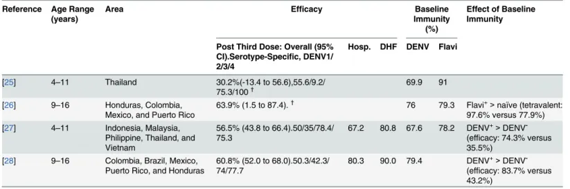
Table 1. An overview of the results from the CYD-TDV vaccine trials.
Reference Age Range (years)
Area Efficacy Baseline
Immunity (%)
Effect of Baseline Immunity
Post Third Dose: Overall (95% CI).Serotype-Specific, DENV1/ 2/3/4
Hosp. DHF DENV Flavi
[25] 4–11 Thailand 30.2%(-13.4 to 56.6),55.6/9.2/
75.3/100† 69.9 91
[26] 9–16 Honduras, Colombia, Mexico, and Puerto Rico
63.9% (1.5 to 87.4).†
76 79.3 Flavi+>naïve (tetravalent: 97.6% versus 77.9%) [27] 4–11 Indonesia, Malaysia,
Philippine, Thailand, and Vietnam
56.5% (43.8 to 66.4).50/35/78.4/ 75.3
67.2 80.8 67.6 78.2 DENV+>DENV -(efficacy: 74.3% versus 35.5%)
[28] 9–16 Colombia, Brazil, Mexico, Puerto Rico, and Honduras
60.8% (52.0 to 68.0).50.3/42.3/ 74/77.7
80.3 90.0 79.4 DENV+>DENV -(efficacy: 83.7% versus 43.2%)
95% CI, 95% confidence interval; Hosp., hospitalization; DHF, dengue hemorrhagic fever.
†
Study was a Phase II clinical trial, with a relatively low number of participants.
country and per study. Additionally, when the serotype specific efficacy was calculated, the lowest efficacy was consistently seen for DENV2 (Table 1).
Strikingly, the vaccine cohort had significantly lower incidence of dengue hemorrhagic fever (80%–90% efficacy) and hospitalization (67%–80% efficacy) [27,28]. Baseline immunity seems
to be beneficial in terms of developing tetravalent seroconversion and overall efficacy against symptomatic DENV (Table 1).
While the protection against hemorrhagic fever is encouraging, these trials also taught us that seroconversion alone does not predict protective efficacy. Clearly, more research is re-quired to identify the correlate of protection [29]. Furthermore, it showed us that we need to have a better understanding of the immune response to DENV infection. Hence, below we will discuss what is known about the function of T and B cells in immunity against DENV. Most at-tention has been directed towards the role of antibodies in immunity against DENV, and there-fore, these will be the primary focus of this review.
Human Immune Response and Disease
After a primary DENV infection, individuals are protected against disease upon reinfection with the homologous serotype. Cross-protection against other serotypes is limited and exists only for 1–2 months post–primary infection, while disease severity was found to be alleviated
for 2–9 months thereafter [30,31]. Recent information suggests that cross-protection against
severe disease lasts up to 2 years [32–35]. Intriguingly, after the cross-protective period,
indi-viduals are at risk of developing more severe dengue upon secondary infection with a hetero-typic serotype. Moreover, the chance to develop severe disease increases with the time between the primary and the secondary infection [33,34].
The increased chance of severe disease can be explained by original antigenic sin, a phenom-enon in which the human immune system preferentially activates memory T and B cells against the original antigen rather than instructing naïve T and B cells against the current anti-gen [36,37]. Indeed, it was found that upon a secondary heterotypic DENV infection, the acute T cell response is mostly directed towards the previous infecting serotype [38,39]. Over time, the T cells against conserved, cross-reactive epitopes are preferentially expanded, resulting in a DENV-broad [20,38,40] and potentially flavivirus-broad response [39,41]. As for B cells, a pre-dominant monotypic response with high avidity against the infecting serotype is observed 6–9
days after disease onset [42,43]. Yet, within 6 months of infection, a broad cross-reactive B cell repertoire is seen [43]. Indeed, cross-reactive B cells are predominantly present at the time of secondary infection [42]. These cells have been speculated to contribute to enhanced severity of dengue disease severity [44] (discussed below). After a secondary heterotypic infection, sta-ble populations of DENV-broad cross-reactive B cells are seen [42,43], and these cells secrete high levels of high-avidity antibodies [42,45,46].
Antibodies are suggested to be more important than T cells in triggering the onset of severe disease. This was suggested because infants born to dengue immune mothers were noted to have a higher risk for severe disease development during primary infection [47]. Halstead and others found that waning antibody titers can enhance DENV infectivity in vitro and in vivo [48–50] and developed the theory of antibody-dependent enhancement (ADE) of disease
[48,51]. During ADE, the pre-existing cross-reactive antibodies bind to the newly infecting DENV serotype and specifically target the immune complexes to Fc-receptor-expressing cells, cells that are highly permissive to DENV. The high viral burden triggers the immune system, which at the end is responsible for the onset of severe signs like plasma leakage [51–53].
upon reinfection with another DENV serotype, these antibodies can contribute to severe dis-ease development. Hence, we wished to gather information on the human antibody epitopes and their relative contributions to the human antibody repertoire after DENV vaccination and infection. Although we primarily focus on antibody epitopes, we also included a brief descrip-tion of the role of T cells in connecdescrip-tion with the CYD vaccine.
Human Antibody Responses
We first reviewed the antibody responses in the sera of primary and secondary DENV cases (S1 Table). The majority of antibodies are raised against the E protein, and a small fraction tar-get the prM and the NS proteins. This is not very surprising as E and prM are exposed on the viral surface and soluble NS1 is secreted by infected cells [54]. The higher fraction of E protein antibodies suggests that the human antibody response predominantly targets DENV particles (structural proteins) rather than NS1-positive cells, i.e., infected cells or cells having bound soluble NS1 [55,56]. Interestingly, we see that during secondary infection the antibody reper-toire broadens as higher responses against the prM and NS1 proteins are seen. This implies that antibodies against E, prM, and NS1 are differentially induced between primary and sec-ondary infection (discussed further below). A detailed insight in the specific antibody reper-toire may therefore help us to better understand the contribution of distinct epitopes to infection neutralization.
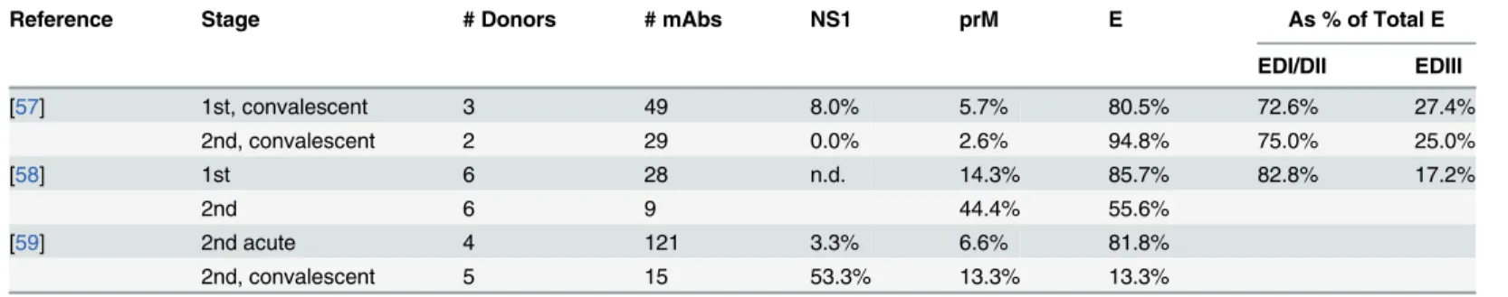
Indeed, several elegant studies have used immortalized B cells from human blood samples to generate monoclonal antibodies of these cultures. Unfortunately, the studies conducted so far show considerable variability in numbers and epitopes of antibodies isolated from individu-al patients (S2 Table). This is likely due to differences in donor backgrounds and immortalizing method used. Therefore, we next focused on those studies in which primary and secondary an-tibody responses or acute and convalescent samples are compared (Table 2). Even then, the re-sults are highly variable: e.g., the prM response strongly expands in two studies but decreased in one study. The latter study also showed a stable E response between primary and secondary responses, while the others reported a reduction thereof. Yet, when we looked at both sera and monoclonals (S1andS2Tables), overall, the E antibodies are dominant during the primary re-sponse. The results for secondary responses are more variable (Table 2), but in sera prM and NS antibodies are particularly detected in secondary cases (S1 Table).
Table 2. Temporal evaluation of human B cell-derived monoclonal antibodies against DENV.
Reference Stage # Donors # mAbs NS1 prM E As % of Total E
EDI/DII EDIII
[57] 1st, convalescent 3 49 8.0% 5.7% 80.5% 72.6% 27.4%
2nd, convalescent 2 29 0.0% 2.6% 94.8% 75.0% 25.0%
[58] 1st 6 28 n.d. 14.3% 85.7% 82.8% 17.2%
2nd 6 9 44.4% 55.6%
[59] 2nd acute 4 121 3.3% 6.6% 81.8%
2nd, convalescent 5 15 53.3% 13.3% 13.3%
To generate the monoclonal antibodies (mAbs) listed in this table, peripheral blood mononuclear cells (PBMCs) had been taken after primary (1st) and secondary (2nd) infection or between the acute and convalescent phases. Note to table: in reports in which multiple donors had been used, all percentages werefirst calculated as % per donor and then averaged over all donors. Hence, some percentages in this table can differ from those in the reports in which the value is reported as the % of experiment rather than per donor. Not all antibodies were characterized; hence, values may be lower than 100%. n.d.: not determined. EDI/DII and DIII refer to the structural domains within the E ectodomain.
Furthermore, since binding of one epitope can enhance or diminish binding of antibodies against other epitopes [60–62], it would be interesting to see whether shifts in these ratios
influ-ence neutralization of DENV particles by antibodies against specific epitopes. Based on the ta-bles, we tried to estimate the balance between the various targeted epitopes. For primary convalescent sera, a ratio of approximately 3 E antibodies to 1 prM antibody was found. In sec-ondary convalescent cases, this was near 1 on 1.
Furthermore, the E protein consists of three ectodomains (D): E DI–DIII. In humans, DI
and DII are immunodominant domains relative to DIII, as 3-fold more antibodies target DI/III than DIII. However, given the large variability, more studies are required to validate the results. Although a significant proportion of antibodies target the NS proteins, DNA-vaccine trials suggest that these are not pivotal for neutralization of infection [63,64]. Yet, the NS1 antibodies may aid in clearance of infected cells [65]. Here, we will focus on the antibodies that directly bind to the virus and discuss the clinical relevance of these antibodies.
PrM Antibodies
We and others showed that prM antibodies are poorly neutralizing and highly enhancing [66–
70]. Moreover, infection enhancement was seen over a broad range of concentrations, whereas neutralization occurred in a very narrow range and is incomplete [67–70]. Therefore, prM
anti-bodies have been postulated to contribute primarily to antibody-dependent enhancement of dengue infection and severe disease development. Recent analysis, however, showed that al-though there is a robust prM response (20%–30%) during acute secondary DENV2 infection,
there is no difference in the level of prM antibodies between mild and severe cases [71]. Fur-thermore, prM antibody levels are increased during secondary, tertiary, and quaternary infec-tions (Table 2,S2 Table, and references therein), whereas severe disease is most often
associated with secondary infection [72]. Indeed, subsequent functional analysis did not show a specific correlation between the neutralization/enhancement profile of the sera towards prM-containing particles and the onset of severe disease [71]. This suggests that prM antibodies are not a discriminating factor but act as a cofactor in disease development. Yet, given the weakly neutralizing properties of prM antibodies, it is advisable to avoid the presence of prM in vaccines.
E Antibodies
Many studies have been done to link neutralization to certain epitopes or structural domains of the E protein (Table 2). Most of the antibodies were found to be directed against dengue EDII fusion loop (FL) (Table 2,S1 Table, and references therein). Furthermore, Lai and colleagues found a correlation between serum EDII FL antibodies and the potency of the serum to neu-tralize heterotypic DENV [46]. The relevance of these human EDII FL antibodies in protection was further strengthened by elegant tests using prM-E proteins or virus-like particles bearing mutations in the FL [46,73,74].
Based on mouse models, the EDIII was initially considered a major antigen for the induc-tion of serotype-specific neutralizing antibodies [75,76]. Surprisingly, quite low fractions of an-tibodies targeting EDIII were found during human infection [37,77], and similar low fractions were found after infection with other flaviviruses [78–80]. Moreover, depletion of
as it inhibits changes within the E protein that are required for fusion. An example of such qua-ternary structure is the EDI/DII hinge region, and recently, antibodies targeting this region were found to be serotype-specific and neutralizing [69,84,85]. Antibodies that bind to viral particles but not to protein monomers are potently neutralizing [58,69,83] but appear to be rare [66]. A recent report, however, showed that near 40% of the isolated monoclonal antibod-ies (mAbs) bind to quaternary structures [83]. To conclude, we see that the DENV E domains I/II are more immunodominant than the EDIII in terms of induction of antibodies in humans. Importantly, both EDI/II and EDIII antibodies were found to possess a similar neutralization potency [86], and the most neutralizing antibodies against flaviviruses appear to target quater-nary structures [78,80,83,86], These findings argue for preservation of quaternary structures in DENV vaccines.
T Cells
The role of T cells in immunity against dengue infection has been extensively reviewed by oth-ers [52,87], and we will briefly discuss recent findings regarding the role of T cells in immunity and pathogenesis. Whereas the CD4+T cell response contributes to protection by instructing B cell responses against the virus [21], the importance of cytotoxic (CD8+) T cells for protection is still under debate since low T cell responses are seen during acute stages of DENV infection [36]. After peak viremia, peaks in both T cell response and cytokines are seen [36,88], suggest-ing that cross-reactive CD8+T cells contribute to pathogenesis rather than protection. Further-more, during secondary infection, T cells (like B cells) suffer from original antigenic sin [22,36,89]. The cross-reactive T cells during acute secondary infection have an altered cytokine responses consisting of low interferon gamma (IFN-γ) and high tumor necrosis factor alpha
(TNF-α) [88,90]. This profile has been associated with severe disease [52]. The phenomenon of
original antigenic sin might be less persistent in T cells than in B cells [20], as a recent manu-script showed that multifunctional CD8+T cells can be associated with protection against dis-ease in a Sri Lankan population [22].
Clearly, in naïve individuals, the CYD-TDV vaccine does not induce CD8+T cell responses to the NS proteins of DENV. The participants in the CYD trials, however, had high baseline immunity, implying that T cell responses were already present and potentially boosted by the vaccine [20,39,41]. Thus, we cannot conclude whether or not it is important to include T cell immunity for protection and if this should be induced by a vaccine. Yet, the trials had quite low efficacy results despite high antibody titers. Mouse models indicated that protection re-quires both B and T cells [91] and that CD8+T cells significantly contribute to disease allevia-tion, even under conditions of ADE [92]. Thus, CD8+T cells likely contribute to clearance of infection when antibodies have failed to prevent infection. Hence, T cells might be more im-portant for DENV immunity than previously appraised.
Assays for Vaccine Development
YFV [94]. For DENV, the exact cutoff is unknown but was expected to be similar to the viruses mentioned above.
Based on these criteria, the CYD-TDV trials showed good seroconversion rates, yet for DENV2 a particularly low clinical efficacy was seen (Table 2). This shows that the PRNT assay or its interpretation requires further fine tuning in order to find the true correlate of protection. Many parameters can be adjusted [95–97], such as (I) the cell line, (II) the challenge virus
strain, and (III) the defined cutoff for seropositivity. Other parameters include incubation tem-perature [98,99] and virus source [83].
The current PRNT assay employs the Vero cells, an Fc-receptor (FcR)-negative cell line. FcR-negative cells are inclined toward neutralization, as virus-antibody complexes are only in-ternalized via interaction with FcR. Conversely, FcR-positive cells typically show ADE with poor neutralization [50]. Primary myeloid cells are a natural host cell of DENV and support in-fection in the absence and presence of antibodies, and they could be an alternative to cell lines [100]. As a start, it would be interesting to investigate if neutralization assays performed with PBMCs of vaccinees gives a better correlate of protection than that of Vero cells. It is unlikely that primary cells will be applied in an industrial setting; yet, the above studies will guide future assay development.
Second, distinct DENV genotypes can give significant shifts in the reported seropositivity, giving e.g. 50% reduction [72]. This is not surprising given the 9% variation within a serotype (Fig 1). More robust correlates of protection against a serotype could be found by including multiple genotypes reflecting the breadth within the serotype.
Third, the threshold chosen for seropositivity was a PRNT50of 10. Yet, the threshold of 50%
reduction may not be optimal in terms of variability [97], and different thresholds may be needed according to the serotype [101]. Indeed, in case of the JEV vaccines, the PRNT50values
were found to differ between the existing genotypes [102]. The DENV vaccine cohorts now provide excellent opportunities to conduct mathematical studies to find better correlates of protection using more stringent criteria for the neutralization threshold and/or serum dilution.
Overall, there is a poor correlation between the current cutoff for seropositivity (PRNT50
10) and clinical efficacy of a DENV vaccine [25,103]. Since Sanofi will continue to monitor the vaccine participants for the next 4 years [19,27,28], the present vaccine trials now offer new prospects for studies to define the best assay and criteria that predict which vaccinees have de-veloped protective immunity. Future studies will also benefit from the lesson of these trials, i.e., that too few participants were bled to allow for thorough correlative analysis between the anti-body response and individual protection [28].
Challenges for Future Dengue Vaccines
In this review, we briefly summarized the outcome of the CYD-TDV vaccine trials. The trials showed us that seroconversion of vaccinees does not necessarily correlate to clinical efficacy against symptomatic disease. This stressed how little we actually know about the human adap-tive immune responses towards DENV infection. Most attention had been paid to the human antibody response, and the components thereof have been reviewed above (Table 2andS1 Table). Based on the Sanofi trials and the reports on the human antibody response, some chal-lenging questions are discussed below.
Better Responses after Flavivirus Priming?
individuals suggests that the immune response is different in naïve and primed individuals. In naive individuals, only the DENV antibody response is triggered by CYD-TDV, while in primed individuals, B and T cell responses are boosted, the latter likely through flavivirus-broad conserved epitopes. Yet, the lower antibody levels in flavivirus-naïve individuals could not be compensated for by repeated vaccination [26]. This raises the question of whether the vaccine preferentially expands pre-existing (cross-reactive) immunity and weakly induces de novo immunity. If so, the vaccine may be less beneficial for young children in endemic coun-tries and travelers.
Absolute Requirement for Tetravalency?
The current dogma is that vaccination should induce serotype-specific antibodies against all four DENV serotypes. Pierson and colleagues suggested that all antibodies that can bind and neutralize DENV can also promote enhancement of infection, irrespective of the epitope [105]. If all antibodies support ADE and neutralization, high titers of cross-reactive antibodies may be sufficient for protection. Yet, a recent study showed that inapparent and apparent dengue cases have similar DENV–immunoglobulin G (IgG) titers but can be distinguished based on
whether the sera shows heterotypic neutralizing capacity or not [106]. Future studies should address whether protection of infection depends on the balance of monotypic antibodies and heterotypic antibodies and/or the cumulative titer of all DENV antibodies.
Why Low Efficacy towards DENV2?
The CYD-TDV showed excellent seroconversion but did not result in high efficacy against symptomatic DENV2. The lack of CD8+T cell responses has been suggested as an option [22]. Recently, there is also growing awareness about the role of the genotype used within the vac-cine. Various genotypes of the same serotype can co-currently circulate within endemic areas [107,108]. A mismatch in the genotypes can significantly reduce the affinity of the sera to neu-tralize infection [72] or may even lead to ADE [7,8]. The low efficacy against DENV2 in the Thai Phase IIb trial was suggested to have occurred because of a mismatch in the vaccine geno-type and the circulating genogeno-type [25,109]. If mismatches are indeed important, close surveil-lance and prediction of the circulating genotypes is crucial. Annual reformulation may be beneficial for protection.
Vaccine Formulation
The formulation and administration regime of the ideal vaccine is a challenging topic. Subunit vaccines with monomer proteins are safe and can be easily reformulated. However, subunit vaccines also induce antibodies against epitopes that are inaccessible on virus particles due to protein-protein interactions [110] and lack quaternary structures, which are currently the most potent epitopes for neutralization [58,69]. Induction of antibodies against quaternary struc-tures could be facilitated by using whole inactivated viruses, attenuated virus strains, or chimeric viruses.
of ADE. Follow-up monitoring of these and future cohorts is important to show that the vac-cine is safe over prolonged time periods [19]. The paradox of a DENV vaccine is thus that a vaccine should be sufficiently virulent to induce high antibody titers yet still be attenuated to be safe.
In summary, the recent Phase III trials showed safety and excellent seroconversion [24], al-though seroconversion did not necessarily imply good efficacy, as shown by DENV2. A major challenge for the future would be to define what assay and criteria predict successful immuni-zation and clinical efficacy. Still, the CYD-TDV offers promise to prevent hospitaliimmuni-zation and severe dengue hemorrhagic fever, which is encouraging news. These CYD-TDV trials offer plenty of clues to gain more knowledge about the human response against DENV, the cross-re-activity with and potential cross-protection against flaviviruses, and the interpretation of anti-body-based neutralization assays. Knowledge on this will aid future vaccine development against other viruses and pathogens than DENV.
Key Learning Points
• Vaccines should preferably induce antibodies against quaternary structures. • Distinct antibody repertoires are seen for primary and secondary infections.
• The CYD-TDV trials offer possibilities for retrospective analysis to identify correlates
of protection.
• To find correlates of protection, further validation and standardization of
neutraliza-tion assays is required.
• T cells could be more important in DENV immunity than previously appreciated.
Top Papers in the Field
• Halstead SB, O'Rourke EJ (1977) Dengue Viruses and Mononuclear Phagocytes. I.
In-fection Enhancement by Non-neutralizing Antibody. J Exp Med 146: 201–217.
One of the earliest papers raising awareness on the paradoxical role of antibodies in dengue disease.
• Capeding MR, Tran NH, Hadinegoro SR, Ismail HI, Chotpitayasunondh T, et al.
(2014) Clinical Efficacy and Safety of a Novel Tetravalent Dengue Vaccine in Healthy Children in Asia: A Phase 3, Randomized Observer-Masked Placebo-Controlled Trial. Lancet 384: 1358–1365.
• Villar L, Dayan GH, Arredondo-Garcia JL, Rivera DM, Cunha R, et al. (2014) Efficacy
• Sabchareon A, Wallace D, Sirivichayakul C, Limkittikul K, Chanthavanich P, et al.
(2012) Protective Efficacy of the Recombinant, Live-Attenuated, CYD Tetravalent Dengue Vaccine in Thai Schoolchildren: A Randomized, Controlled Phase 2b Trial. Lancet 380: 1559–1567.
In these reports,the efficacies of the CYD-TDV vaccines are reported for the first time, based on large cohorts in Asia and Latin America.Although the efficacy against DENV2 is quite enigmatic,the overall efficacy against severe disease and hospitaliza-tion offers perspective.
• de Alwis R, Smith SA, Olivarez NP, Messer WB, Huynh JP, et al. (2012) Identification
of Human Neutralizing Antibodies That Bind to Complex Epitopes on Dengue Viri-ons. Proc Natl Acad Sci U S A 109: 7439–7444.
Here,the authors show that potently neutralizing antibodies appear to be directed to-wards quaternary structures,thus providing insight on the requirements of a dengue vaccine.
• Zellweger RM, Miller R, Eddy WE, White LJ, Johnston RE, et al. (2013) Role of
Hu-moral Versus Cellular Responses Induced by a Protective Dengue Vaccine Candidate. PLoS Pathog 9: e1003723.
This paper shows the importance of T cells in immunity against dengue virus infections, clearly advocating against a focus on antibodies alone.
• Salje H, Rodriguez-Barraquer I, Rainwater-Lovett K, Nisalak A, Thaisomboonsuk B,
et al. (2014) Variability in Dengue Titer Estimates from Plaque Reduction Neutraliza-tion Tests Poses a Challenge to Epidemiological Studies and Vaccine Development. PLoS Negl Trop Dis 8: e2952
Supporting Information
S1 Dataset. E amino acid sequences used in the review.The information is given as follows: country of isolation_strain_year of isolation (if known).
(DOCX)
S1 Table. An overview of the dengue antibody response in human sera.In this table, the focus is on the development after primary (1st) and secondary (2nd) infection, with the stage of disease at the moment of serum sampling being convalescent (conv.) or unknown. If un-known, only the stage is presented. We grouped the results of primary and secondary infections for individual reports in order to visualize the effects of secondary infection on the antigens tar-geted and the relative magnitude of antibodies against the epitopes. m.p.i.: months post infec-tion. n.d.: Not determined.
(DOCX)
S2 Table. An overview of human monoclonal antibodies derived from immortalized B cells.An overview of human B cell-derived monoclonal antibodies from dengue-infected hu-mans whose PBMCs were taken after primary (1st) or secondary (2nd) infection. The stage of disease was either acute (ac) or convalescent (conv.). Note to table: in reports in which multiple donors had been used, all percentages are first calculated as % per donor and then averaged over all donors. Hence, some percentages can differ from reports in which the value is reported as % of the whole experiment. n.d.: not determined. EDI/DII and DIII refer to the structural domains within the E ectodomain. Reports were selected based on whether they (I) were the first to describe the monoclonal antibodies, (II) screened against several epitopes, and (III) used an unbiased approach to generate the monoclonals.
(DOCX)
Acknowledgments
We apologize to those whose seminal contributions we could not cite because of space restric-tions. We thank Dr. Adriana Tami-Grundman for her help and the reviewers for their sugges-tions and contribusugges-tions.
References
1. Sips GJ, Wilschut J, Smit JM. Neuroinvasive flavivirus infections. Rev Med Virol. 2012; 22: 69–87. doi:10.1002/rmv.712PMID:22086854
2. European Centre for Disease Prevention and Control. Annual epidemiological report 2012.guidelines for the surveillance of invasive mosquitoes in Europe.stockholm: ECDC; 2012
3. Bhatt S, Gething PW, Brady OJ, Messina JP, Farlow AW, Moyes CL, et al. The global distribution and burden of dengue. Nature. 2013; 496: 504–507. doi:10.1038/nature12060PMID:23563266 4. Gubler DJ, Meltzer M. Impact of dengue/dengue hemorrhagic fever on the developing world. Adv
Virus Res. 1999; 53: 35–70. PMID:10582094
5. World Health Organization. Fact sheet: Dengue and severe dengue. Http://www.who.int/mediacentre/ factsheets/fs117/en/index.html. 2012;March 2013: 1.
6. Heinz FX, Stiasny K. Flaviviruses and flavivirus vaccines. Vaccine. 2012; 30: 4301–4306. doi:10. 1016/j.vaccine.2011.09.114PMID:22682286
7. Brien JD, Austin SK, Sukupolvi-Petty S, O'Brien KM, Johnson S, Fremont DH, et al. Genotype-specific neutralization and protection by antibodies against dengue virus type 3. J Virol. 2010; 84: 10630–
10643. doi:10.1128/JVI.01190-10PMID:20702644
9. Ecker M, Allison SL, Meixner T, Heinz FX. Sequence analysis and genetic classification of tick-borne encephalitis viruses from europe and asia. J Gen Virol. 1999; 80 (Pt 1): 179–185.
10. Sotelo E, Fernandez-Pinero J, Llorente F, Vazquez A, Moreno A, Aguero M, et al. Phylogenetic rela-tionships of western mediterranean west nile virus strains (1996–2010) using full-length genome se-quences: Single or multiple introductions? J Gen Virol. 2011; 92: 2512–2522. doi:10.1099/vir.0. 033829-0PMID:21775579
11. Burke DS, Nisalak A, Johnson DE, Scott RM. A prospective study of dengue infections in bangkok. Am J Trop Med Hyg. 1988; 38: 172–180. PMID:3341519
12. Mizumoto K, Ejima K, Yamamoto T, Nishiura H. On the risk of severe dengue during secondary infec-tion: A systematic review coupled with mathematical modeling. J Vector Borne Dis. 2014; 51: 153–
164. PMID:25253207
13. Halstead SB. Immune enhancement of viral infection. Prog Allergy. 1982; 31: 301–364. PMID:
6292921
14. Russell PK, Yuill TM, Nisalak A, Udomsakdi S, Gould DJ, Winter PE. An insular outbreak of dengue hemorrhagic fever. II. virologic and serologic studies. Am J Trop Med Hyg. 1968; 17: 600–608. PMID:
4970512
15. Buchy P, Vo VL, Bui KT, Trinh TX, Glaziou P, Le TT, et al. Secondary dengue virus type 4 infections in vietnam. Southeast Asian J Trop Med Public Health. 2005; 36: 178–185. PMID:15906664 16. Halstead SB. Dengue. The Lancet. 2007; 370: 1644–1652.
17. Murphy BR, Whitehead SS. Immune response to dengue virus and prospects for a vaccine. Annu Rev Immunol. 2011; 29: 587–619. doi:10.1146/annurev-immunol-031210-101315PMID:21219187 18. Wan SW, Lin CF, Wang S, Chen YH, Yeh TM, Liu HS, et al. Current progress in dengue vaccines. J
Biomed Sci. 2013; 20: 37-0127-20-37.
19. Guy B, Barrere B, Malinowski C, Saville M, Teyssou R, Lang J. From research to phase III: Preclinical, industrial and clinical development of the sanofi pasteur tetravalent dengue vaccine. Vaccine. 2011; 29: 7229–7241. doi:10.1016/j.vaccine.2011.06.094PMID:21745521
20. Dayan GH, Galan-Herrera JF, Forrat R, Zambrano B, Bouckenooghe A, Harenberg A, et al. Assess-ment of bivalent and tetravalent dengue vaccine formulations in flavivirus-naive adults in mexico. Hum Vaccin Immunother. 2014; 10: 2853–2863. doi:10.4161/21645515.2014.972131PMID:
25483647
21. Guy B, Nougarede N, Begue S, Sanchez V, Souag N, Carre M, et al. Cell-mediated immunity induced by chimeric tetravalent dengue vaccine in naive or flavivirus-primed subjects. Vaccine. 2008; 26: 5712–5721. doi:10.1016/j.vaccine.2008.08.019PMID:18762226
22. Weiskopf D, Angelo MA, de Azeredo EL, Sidney J, Greenbaum JA, Fernando AN, et al. Comprehen-sive analysis of dengue virus-specific responses supports an HLA-linked protective role for CD8+ T cells. Proc Natl Acad Sci U S A. 2013; 110: E2046–53. doi:10.1073/pnas.1305227110PMID:
23580623
23. Balas C, Kennel A, Deauvieau F, Sodoyer R, Arnaud-Barbe N, Lang J, et al. Different innate signa-tures induced in human monocyte-derived dendritic cells by wild-type dengue 3 virus, attenuated but reactogenic dengue 3 vaccine virus, or attenuated nonreactogenic dengue 1–4 vaccine virus strains. J Infect Dis. 2011; 203: 103–108. doi:10.1093/infdis/jiq022PMID:21148502
24. da Costa VG, Marques-Silva AC, Floriano VG, Moreli ML. Safety, immunogenicity and efficacy of a re-combinant tetravalent dengue vaccine: A meta-analysis of randomized trials. Vaccine. 2014; 32: 4885–4892. doi:10.1016/j.vaccine.2014.07.008PMID:25045816
25. Sabchareon A, Wallace D, Sirivichayakul C, Limkittikul K, Chanthavanich P, Suvannadabba S, et al. Protective efficacy of the recombinant, live-attenuated, CYD tetravalent dengue vaccine in thai schoolchildren: A randomised, controlled phase 2b trial. Lancet. 2012; 380: 1559–1567. doi:10.1016/ S0140-6736(12)61428-7PMID:22975340
26. Villar LA, Rivera-Medina DM, Arredondo-Garcia JL, Boaz M, Starr-Spires L, Thakur M, et al. Safety and immunogenicity of a recombinant tetravalent dengue vaccine in 9–16 year olds: A randomized, controlled, phase II trial in latin america. Pediatr Infect Dis J. 2013; 32: 1102–1109. doi:10.1097/INF. 0b013e31829b8022PMID:24067553
27. Capeding MR, Tran NH, Hadinegoro SR, Ismail HI, Chotpitayasunondh T, Chua MN, et al. Clinical ef-ficacy and safety of a novel tetravalent dengue vaccine in healthy children in asia: A phase 3, rando-mised, observer-masked, placebo-controlled trial. Lancet. 2014; 384: 1358–1365. doi:10.1016/ S0140-6736(14)61060-6PMID:25018116
29. Wilder-Smith A. Dengue vaccines: Dawning at last? Lancet. 2014; 384: 1327–1329. doi:10.1016/ S0140-6736(14)61142-9PMID:25018119
30. Sabin AB. The dengue group of viruses and its family relationships. Bacteriol Rev. 1950; 14: 225–
232. PMID:14772197
31. Snow GE, Haaland B, Ooi EE, Gubler DJ. Review article: Research on dengue during world war II re-visited. Am J Trop Med Hyg. 2014; 91: 1203–1217. doi:10.4269/ajtmh.14-0132PMID:25311700 32. Yamanaka A, Tabuchi Y, Mulyatno KC, Susilowati H, Hendrianto E, Soegijanto S, et al. Dengue virus
infection-enhancing and neutralizing antibody balance in children of the philippines and indonesia. Mi-crobes Infect. 2012; 14: 1152–1159. doi:10.1016/j.micinf.2012.07.013PMID:22841680
33. Anderson KB, Gibbons RV, Cummings DA, Nisalak A, Green S, Libraty DH, et al. A shorter time inter-val between first and second dengue infections is associated with protection from clinical illness in a school-based cohort in thailand. J Infect Dis. 2014; 209: 360–368. doi:10.1093/infdis/jit436PMID:
23964110
34. Montoya M, Gresh L, Mercado JC, Williams KL, Vargas MJ, Gutierrez G, et al. Symptomatic versus in-apparent outcome in repeat dengue virus infections is influenced by the time interval between infec-tions and study year. PLoS Negl Trop Dis. 2013; 7: e2357. doi:10.1371/journal.pntd.0002357PMID:
23951377
35. Sharp TM, Hunsperger E, Munoz-Jordan JL, Margolis HS, Tomashek KM. Sequential episodes of dengue-puerto rico, 2005–2010. Am J Trop Med Hyg. 2014.
36. Mongkolsapaya J, Dejnirattisai W, Xu XN, Vasanawathana S, Tangthawornchaikul N, Chairunsri A, et al. Original antigenic sin and apoptosis in the pathogenesis of dengue hemorrhagic fever. Nat Med. 2003; 9: 921–927. PMID:12808447
37. Midgley CM, Bajwa-Joseph M, Vasanawathana S, Limpitikul W, Wills B, Flanagan A, et al. An in-depth analysis of original antigenic sin in dengue virus infection. J Virol. 2011; 85: 410–421. doi:10. 1128/JVI.01826-10PMID:20980526
38. Gwinn W, Sun W, Innis BL, Caudill J, King AD. Serotype-specific T(H)1 responses in recipients of two doses of candidate live-attenuated dengue virus vaccines. Am J Trop Med Hyg. 2003; 69: 39–47. PMID:14740954
39. Mathew A, Kurane I, Rothman AL, Zeng LL, Brinton MA, Ennis FA. Dominant recognition by human CD8+ cytotoxic T lymphocytes of dengue virus nonstructural proteins NS3 and NS1.2a. J Clin Invest. 1996; 98: 1684–1691. PMID:8833919
40. Weiskopf D, Angelo MA, Bangs DJ, Sidney J, Paul S, Peters B, et al. The human CD8+ T cell re-sponses induced by a live attenuated tetravalent dengue vaccine are directed against highly con-served epitopes. J Virol. 2015; 89: 120–128. doi:10.1128/JVI.02129-14PMID:25320311
41. Harenberg A, Begue S, Mamessier A, Gimenez-Fourage S, Ching Seah C, Wei Liang A, et al. Persis-tence of Th1/Tc1 responses one year after tetravalent dengue vaccination in adults and adolescents in singapore. Hum Vaccin Immunother. 2013; 9: 2317–2325. PMID:23839107
42. Zompi S, Montoya M, Pohl MO, Balmaseda A, Harris E. Dominant cross-reactive B cell response dur-ing secondary acute dengue virus infection in humans. PLoS Negl Trop Dis. 2012; 6: e1568. doi:10. 1371/journal.pntd.0001568PMID:22448292
43. Mathew A, West K, Kalayanarooj S, Gibbons RV, Srikiatkhachorn A, Green S, et al. B-cell responses during primary and secondary dengue virus infections in humans. J Infect Dis. 2011; 204: 1514–1522. doi:10.1093/infdis/jir607PMID:21930609
44. de Alwis R, Williams KL, Schmid MA, Lai CY, Patel B, Smith SA, et al. Dengue viruses are enhanced by distinct populations of serotype cross-reactive antibodies in human immune sera. PLoS Pathog. 2014; 10: e1004386. doi:10.1371/journal.ppat.1004386PMID:25275316
45. Tsai WY, Lai CY, Wu YC, Lin HE, Edwards C, Jumnainsong A, et al. High-avidity and potently neutral-izing cross-reactive human monoclonal antibodies derived from secondary dengue virus infection. J Virol. 2013; 87: 12562–12575. doi:10.1128/JVI.00871-13PMID:24027331
46. Lai CY, Williams KL, Wu YC, Knight S, Balmaseda A, Harris E, et al. Analysis of cross-reactive anti-bodies recognizing the fusion loop of envelope protein and correlation with neutralizing antibody titers in nicaraguan dengue cases. PLoS Negl Trop Dis. 2013; 7: e2451. doi:10.1371/journal.pntd.0002451
PMID:24069496
47. Kliks SC, Nimmanitya S, Nisalak A, Burke DS. Evidence that maternal dengue antibodies are impor-tant in the development of dengue hemorrhagic fever in infants. Am J Trop Med Hyg. 1988; 38: 411–
419. PMID:3354774
49. Vaughn DW, Green S, Kalayanarooj S, Innis BL, Nimmannitya S, Suntayakorn S, et al. Dengue vire-mia titer, antibody response pattern, and virus serotype correlate with disease severity. J Infect Dis. 2000; 181: 2–9. PMID:10608744
50. Moi ML, Takasaki T, Saijo M, Kurane I. Dengue virus infection-enhancing activity of undiluted sera ob-tained from patients with secondary dengue virus infection. Trans R Soc Trop Med Hyg. 2013; 107: 51–58. doi:10.1093/trstmh/trs007PMID:23296697
51. Flipse J, Wilschut J, Smit JM. Molecular mechanisms involved in antibody-dependent enhancement of dengue virus infection in humans. Traffic. 2013; 14: 25–35. doi:10.1111/tra.12012PMID:
22998156
52. Rothman AL. Immunity to dengue virus: A tale of original antigenic sin and tropical cytokine storms. Nat Rev Immunol. 2011; 11: 532–543. doi:10.1038/nri3014PMID:21760609
53. Halstead SB, Mahalingam S, Marovich MA, Ubol S, Mosser DM. Intrinsic antibody-dependent en-hancement of microbial infection in macrophages: Disease regulation by immune complexes. Lancet Infect Dis. 2010; 10: 712–722. doi:10.1016/S1473-3099(10)70166-3PMID:20883967
54. Flamand M, Megret F, Mathieu M, Lepault J, Rey FA, Deubel V. Dengue virus type 1 nonstructural gly-coprotein NS1 is secreted from mammalian cells as a soluble hexamer in a glycosylation-dependent fashion. J Virol. 1999; 73: 6104–6110. PMID:10364366
55. Falconar AK. The dengue virus nonstructural-1 protein (NS1) generates antibodies to common epi-topes on human blood clotting, integrin/adhesin proteins and binds to human endothelial cells: Poten-tial implications in haemorrhagic fever pathogenesis. Arch Virol. 1997; 142: 897–916. PMID:9191856 56. Avirutnan P, Zhang L, Punyadee N, Manuyakorn A, Puttikhunt C, Kasinrerk W, et al. Secreted NS1 of dengue virus attaches to the surface of cells via interactions with heparan sulfate and chondroitin sul-fate E. PLoS Pathog. 2007; 3: e183. PMID:18052531
57. Beltramello M, Williams KL, Simmons CP, Macagno A, Simonelli L, Quyen NT, et al. The human im-mune response to dengue virus is dominated by highly cross-reactive antibodies endowed with neu-tralizing and enhancing activity. Cell Host Microbe. 2010; 8: 271–283. doi:10.1016/j.chom.2010.08. 007PMID:20833378
58. Smith SA, Zhou Y, Olivarez NP, Broadwater AH, de Silva AM, Crowe JE Jr. Persistence of circulating memory B cell clones with potential for dengue virus disease enhancement for decades following in-fection. J Virol. 2012; 86: 2665–2675. doi:10.1128/JVI.06335-11PMID:22171265
59. Setthapramote C, Sasaki T, Puiprom O, Limkittikul K, Pitaksajjakul P, Pipattanaboon C, et al. Human monoclonal antibodies to neutralize all dengue virus serotypes using lymphocytes from patients at acute phase of the secondary infection. Biochem Biophys Res Commun. 2012; 423: 867–872. doi:
10.1016/j.bbrc.2012.06.057PMID:22713454
60. Heinz FX, Mandl C, Berger R, Tuma W, Kunz C. Antibody-induced conformational changes result in enhanced avidity of antibodies to different antigenic sites on the tick-borne encephalitis virus glycopro-tein. Virology. 1984; 133: 25–34. PMID:6199892
61. Henchal EA, McCown JM, Burke DS, Seguin MC, Brandt WE. Epitopic analysis of antigenic determi-nants on the surface of dengue-2 virions using monoclonal antibodies. Am J Trop Med Hyg. 1985; 34: 162–169. PMID:2578750
62. Monath TP, Wands JR, Hill LJ, Gentry MK, Gubler DJ. Multisite monoclonal immunoassay for dengue viruses: Detection of viraemic human sera and interference by heterologous antibody. J Gen Virol. 1986; 67 (Pt 4): 639–650.
63. Konishi E, Ajiro N, Nukuzuma C, Mason PW, Kurane I. Comparison of protective efficacies of plasmid DNAs encoding japanese encephalitis virus proteins that induce neutralizing antibody or cytotoxic T lymphocytes in mice. Vaccine. 2003; 21: 3675–3683. PMID:12922097
64. Putnak R, Fuller J, VanderZanden L, Innis BL, Vaughn DW. Vaccination of rhesus macaques against dengue-2 virus with a plasmid DNA vaccine encoding the viral pre-membrane and envelope genes. Am J Trop Med Hyg. 2003; 68: 469–476. PMID:12875299
65. Henchal EA, Henchal LS, Schlesinger JJ. Synergistic interactions of anti-NS1 monoclonal antibodies protect passively immunized mice from lethal challenge with dengue 2 virus. J Gen Virol. 1988; 69 (Pt 8): 2101–2107. PMID:3404125
66. Smith SA, de Alwis R, Kose N, Durbin AP, Whitehead SS, de Silva AM, et al. Human monoclonal anti-bodies derived from memory B cells following live attenuated dengue virus vaccination or natural in-fection exhibit similar characteristics. J Infect Dis. 2013; 207: 1898–1908. doi:10.1093/infdis/jit119
PMID:23526830
68. de Alwis R, Beltramello M, Messer WB, Sukupolvi-Petty S, Wahala WM, Kraus A, et al. In-depth anal-ysis of the antibody response of individuals exposed to primary dengue virus infection. PLoS Negl Trop Dis. 2011; 5: e1188. doi:10.1371/journal.pntd.0001188PMID:21713020
69. de Alwis R, Smith SA, Olivarez NP, Messer WB, Huynh JP, Wahala WM, et al. Identification of human neutralizing antibodies that bind to complex epitopes on dengue virions. Proc Natl Acad Sci U S A. 2012; 109: 7439–7444. doi:10.1073/pnas.1200566109PMID:22499787
70. Rodenhuis-Zybert IA, van der Schaar HM, da Silva Voorham JM, van der Ende-Metselaar H, Lei HY, Wilschut J, et al. Immature dengue virus: A veiled pathogen? PLoS Pathog. 2010; 6: e1000718. doi:
10.1371/journal.ppat.1000718PMID:20062797
71. Rodenhuis-Zybert IA, da Silva Voorham JM, Torres S, van de Pol D, Smit JM. Antibodies against im-mature virions are not a discriminating factor for dengue disease severity. PLoS Negl Trop Dis. 2015; 9: e0003564. doi:10.1371/journal.pntd.0003564PMID:25760350
72. Endy TP, Nisalak A, Chunsuttitwat S, Vaughn DW, Green S, Ennis FA, et al. Relationship of preexist-ing dengue virus (DV) neutralizpreexist-ing antibody levels to viremia and severity of disease in a prospective cohort study of DV infection in thailand. J Infect Dis. 2004; 189: 990–1000. PMID:14999601 73. Lai CY, Tsai WY, Lin SR, Kao CL, Hu HP, King CC, et al. Antibodies to envelope glycoprotein of
den-gue virus during the natural course of infection are predominantly cross-reactive and recognize epi-topes containing highly conserved residues at the fusion loop of domain II. J Virol. 2008; 82: 6631–
6643. doi:10.1128/JVI.00316-08PMID:18448542
74. Crill WD, Hughes HR, Delorey MJ, Chang GJ. Humoral immune responses of dengue fever patients using epitope-specific serotype-2 virus-like particle antigens. PLoS One. 2009; 4: e4991. doi:10. 1371/journal.pone.0004991PMID:19337372
75. Crill WD, Roehrig JT. Monoclonal antibodies that bind to domain III of dengue virus E glycoprotein are the most efficient blockers of virus adsorption to vero cells. J Virol. 2001; 75: 7769–7773. PMID:
11462053
76. Sukupolvi-Petty S, Austin SK, Purtha WE, Oliphant T, Nybakken GE, Schlesinger JJ, et al. Type- and subcomplex-specific neutralizing antibodies against domain III of dengue virus type 2 envelope pro-tein recognize adjacent epitopes. J Virol. 2007; 81: 12816–12826. PMID:17881453
77. Moreland NJ, Susanto P, Lim E, Tay MY, Rajamanonmani R, Hanson BJ, et al. Phage display ap-proaches for the isolation of monoclonal antibodies against dengue virus envelope domain III from human and mouse derived libraries. Int J Mol Sci. 2012; 13: 2618–2635. doi:10.3390/ijms13032618
PMID:22489114
78. Jarmer J, Zlatkovic J, Tsouchnikas G, Vratskikh O, Strauss J, Aberle JH, et al. Variation of the speci-ficity of the human antibody responses after tick-borne encephalitis virus infection and vaccination. J Virol. 2014; 88: 13845–13857. doi:10.1128/JVI.02086-14PMID:25253341
79. Oliphant T, Nybakken GE, Austin SK, Xu Q, Bramson J, Loeb M, et al. Induction of epitope-specific neutralizing antibodies against west nile virus. J Virol. 2007; 81: 11828–11839. PMID:17715236 80. Vratskikh O, Stiasny K, Zlatkovic J, Tsouchnikas G, Jarmer J, Karrer U, et al. Dissection of antibody
specificities induced by yellow fever vaccination. PLoS Pathog. 2013; 9: e1003458. doi:10.1371/ journal.ppat.1003458PMID:23818856
81. Wahala WM, Kraus AA, Haymore LB, Accavitti-Loper MA, de Silva AM. Dengue virus neutralization by human immune sera: Role of envelope protein domain III-reactive antibody. Virology. 2009; 392: 103–113. doi:10.1016/j.virol.2009.06.037PMID:19631955
82. Wahala WM, Huang C, Butrapet S, White LJ, de Silva AM. Recombinant dengue type 2 viruses with altered e protein domain III epitopes are efficiently neutralized by human immune sera. J Virol. 2012; 86: 4019–4023. doi:10.1128/JVI.06871-11PMID:22278250
83. Dejnirattisai W, Wongwiwat W, Supasa S, Zhang X, Dai X, Rouvinsky A, et al. A new class of highly potent, broadly neutralizing antibodies isolated from viremic patients infected with dengue virus. Nat Immunol. 2015; 16: 170–177. doi:10.1038/ni.3058PMID:25501631
84. Teoh EP, Kukkaro P, Teo EW, Lim AP, Tan TT, Yip A, et al. The structural basis for serotype-specific neutralization of dengue virus by a human antibody. Sci Transl Med. 2012; 4: 139ra83.
85. Messer WB, de Alwis R, Yount BL, Royal SR, Huynh JP, Smith SA, et al. Dengue virus envelope pro-tein domain I/II hinge determines long-lived serotype-specific dengue immunity. Proc Natl Acad Sci U S A. 2014; 111: 1939–1944. doi:10.1073/pnas.1317350111PMID:24385585
86. Smith SA, de Alwis AR, Kose N, Jadi RS, de Silva AM, Crowe JE Jr. Isolation of dengue virus-specific memory B cells with live virus antigen from human subjects following natural infection reveals the presence of diverse novel functional groups of antibody clones. J Virol. 2014; 88: 12233–12241. doi:
87. Weiskopf D, Sette A. T-cell immunity to infection with dengue virus in humans. Front Immunol. 2014; 5: 93. doi:10.3389/fimmu.2014.00093PMID:24639680
88. Sanchez V, Gimenez S, Tomlinson B, Chan PK, Thomas GN, Forrat R, et al. Innate and adaptive cel-lular immunity in flavivirus-naive human recipients of a live-attenuated dengue serotype 3 vaccine pro-duced in vero cells (VDV3). Vaccine. 2006; 24: 4914–4926. PMID:16632108
89. Mongkolsapaya J, Duangchinda T, Dejnirattisai W, Vasanawathana S, Avirutnan P, Jairungsri A, et al. T cell responses in dengue hemorrhagic fever: Are cross-reactive T cells suboptimal? J Immu-nol. 2006; 176: 3821–3829. PMID:16517753
90. Mangada MM, Rothman AL. Altered cytokine responses of dengue-specific CD4+ T cells to heterolo-gous serotypes. J Immunol. 2005; 175: 2676–2683. PMID:16081844
91. Shresta S, Kyle JL, Snider HM, Basavapatna M, Beatty PR, Harris E. Interferon-dependent immunity is essential for resistance to primary dengue virus infection in mice, whereas T- and B-cell-dependent immunity are less critical. J Virol. 2004; 78: 2701–2710. PMID:14990690
92. Zellweger RM, Miller R, Eddy WE, White LJ, Johnston RE, Shresta S. Role of humoral versus cellular responses induced by a protective dengue vaccine candidate. PLoS Pathog. 2013; 9: e1003723. doi:
10.1371/journal.ppat.1003723PMID:24204271
93. World Health Organization. Guidelines for plaque-reduction neutralization testing of human antibodies to dengue viruses. WHO/IVB/07 07. 2007.
94. Plotkin SA. Correlates of protection induced by vaccination. Clin Vaccine Immunol. 2010; 17: 1055–
1065. doi:10.1128/CVI.00131-10PMID:20463105
95. Thomas SJ, Nisalak A, Anderson KB, Libraty DH, Kalayanarooj S, Vaughn DW, et al. Dengue plaque reduction neutralization test (PRNT) in primary and secondary dengue virus infections: How alter-ations in assay conditions impact performance. Am J Trop Med Hyg. 2009; 81: 825–833. doi:10. 4269/ajtmh.2009.08-0625PMID:19861618
96. Rainwater-Lovett K, Rodriguez-Barraquer I, Cummings DA, Lessler J. Variation in dengue virus pla-que reduction neutralization testing: Systematic review and pooled analysis. BMC Infect Dis. 2012; 12: 233-2334-12-233.
97. Salje H, Rodriguez-Barraquer I, Rainwater-Lovett K, Nisalak A, Thaisomboonsuk B, Thomas SJ, et al. Variability in dengue titer estimates from plaque reduction neutralization tests poses a challenge to epidemiological studies and vaccine development. PLoS Negl Trop Dis. 2014; 8: e2952. doi:10.1371/ journal.pntd.0002952PMID:24967885
98. Dowd KA, Jost CA, Durbin AP, Whitehead SS, Pierson TC. A dynamic landscape for antibody binding modulates antibody-mediated neutralization of west nile virus. PLoS Pathog. 2011; 7: e1002111. doi:
10.1371/journal.ppat.1002111PMID:21738473
99. Smith SA, de Alwis AR, Kose N, Harris E, Ibarra KD, Kahle KM, et al. The potent and broadly neutraliz-ing human dengue virus-specific monoclonal antibody 1C19 reveals a unique cross-reactive epitope on the bc loop of domain II of the envelope protein. MBio. 2013; 4: e00873–13. doi:10.1128/mBio. 00873-13PMID:24255124
100. Mahalingam S, Herring BL, Halstead SB. Call to action for dengue vaccine failure. Emerg Infect Dis. 2013; 19: 1335–1337. doi:10.3201/eid1908.121864PMID:23876389
101. Buddhari D, Aldstadt J, Endy TP, Srikiatkhachorn A, Thaisomboonsuk B, Klungthong C, et al. Dengue virus neutralizing antibody levels associated with protection from infection in thai cluster studies. PLoS Negl Trop Dis. 2014; 8: e3230. doi:10.1371/journal.pntd.0003230PMID:25329173 102. Erra EO, Askling HH, Yoksan S, Rombo L, Riutta J, Vene S, et al. Cross-protective capacity of
japa-nese encephalitis (JE) vaccines against circulating heterologous JE virus genotypes. Clin Infect Dis. 2013; 56: 267–270. doi:10.1093/cid/cis883PMID:23074319
103. Sirivichayakul C, Sabchareon A, Limkittikul K, Yoksan S. Plaque reduction neutralization antibody test does not accurately predict protection against dengue infection in ratchaburi cohort, thailand. Virol J. 2014; 11: 48-422X-11-48.
104. Hss AS, Koh MT, Tan KK, Chan LG, Zhou L, Bouckenooghe A, et al. Safety and immunogenicity of a tetravalent dengue vaccine in healthy children aged 2–11 years in malaysia: A randomized, placebo-controlled, phase III study. Vaccine. 2013; 31: 5814–5821. doi:10.1016/j.vaccine.2013.10.013PMID:
24135573
105. Pierson TC, Fremont DH, Kuhn RJ, Diamond MS. Structural insights into the mechanisms of anti-body-mediated neutralization of flavivirus infection: Implications for vaccine development. Cell Host Microbe. 2008; 4: 229–238. doi:10.1016/j.chom.2008.08.004PMID:18779049
107. Rabaa MA, Klungthong C, Yoon IK, Holmes EC, Chinnawirotpisan P, Thaisomboonsuk B, et al. Fre-quent in-migration and highly focal transmission of dengue viruses among children in kamphaeng phet, thailand. PLoS Negl Trop Dis. 2013; 7: e1990. doi:10.1371/journal.pntd.0001990PMID:
23350000
108. Caron M, Grard G, Paupy C, Mombo IM, Bikie Bi Nso B, Kassa Kassa FR, et al. First evidence of si-multaneous circulation of three different dengue virus serotypes in africa. PLoS One. 2013; 8: e78030. doi:10.1371/journal.pone.0078030PMID:24205075
109. Halstead SB. Dengue vaccine development: A 75% solution? Lancet. 2012; 380: 1535–1536. doi:10. 1016/S0140-6736(12)61510-4PMID:22975339
110. Li XQ, Qiu LW, Chen Y, Wen K, Cai JP, Chen J, et al. Dengue virus envelope domain III immunization elicits predominantly cross-reactive, poorly neutralizing antibodies localized to the AB loop: Implica-tions for dengue vaccine design. J Gen Virol. 2013; 94: 2191–2201. doi:10.1099/vir.0.055178-0
PMID:23851440
111. Sessions OM, Tan Y, Goh KC, Liu Y, Tan P, Rozen S, et al. Host cell transcriptome profile during wild-type and attenuated dengue virus infection. PLoS Negl Trop Dis. 2013; 7: e2107. doi:10.1371/ journal.pntd.0002107PMID:23516652
112. Guillot S, Otelea D, Delpeyroux F, Crainic R. Point mutations involved in the attenuation/neuroviru-lence alternation in type 1 and 2 oral polio vaccine strains detected by site-specific polymerase chain reaction. Vaccine. 1994; 12: 503–507. PMID:8036823