from Larval Stages of
Ascaris suum
Reveals High
Abundance of Glycosyl Hydrolases
Tao Wang1, Katleen Van Steendam2, Maarten Dhaenens2, Johnny Vlaminck1, Dieter Deforce2, Aaron R. Jex3, Robin B. Gasser3, Peter Geldhof1*
1Department of Virology, Parasitology and Immunology, Faculty of Veterinary Medicine, Ghent University, Ghent, Belgium, 2Laboratory for Pharmaceutical Biotechnology, Faculty of Pharmaceutical Sciences, Ghent University, Ghent, Belgium,3Faculty of Veterinary Science, The University of Melbourne, Melbourne, Victoria, Australia
Abstract
Background:Ascaris lumbricoidesandAscaris suumare socioeconomically important and widespread parasites of humans and pigs, respectively. The excretory-secretory (ES) molecules produced and presented at the parasite-host interface during the different phases of tissue invasion and migration are likely to play critical roles in the induction and development of protective immune and other host responses.
Methodology/Principal Findings:The aim of this study was to identify the ES proteins of the different larval stages (L3-egg, L3-lung and L4) by LC-MS/MS. In total, 106 different proteins were identified, 20 in L3-egg, 45 in L3-lung stage and 58 in L4. Although most of the proteins identified were stage-specific, 15 were identified in the ES products of at least two stages. Two proteins, i.e. a 14-3-3-like protein and a serpin-like protein, were present in the ES products from the three different larval stages investigated. Interestingly, a comparison of ES products from L4 with those of L3-egg and L3-lung showed an abundance of metabolic enzymes, particularly glycosyl hydrolases. Further study indicated that most of these glycolytic enzymes were transcriptionally upregulated from L4 onwards, with a peak in the adult stage, particularly in intestinal tissue. This was also confirmed by enzymatic assays, showing the highest glycosidase activity in protein extracts from adult worms gut.
Conclusions/Significance:The present proteomic analysis provides important information on the host-parasite interaction and the biology of the migratory stages ofA. suum. In particular, the high transcriptional upregulation of glycosyl hydrolases from the L4 stage onwards reveals that the degradation of complex carbohydrates forms an essential part of the energy metabolism of this parasite once it establishes in the small intestine.
Citation:Wang T, Van Steendam K, Dhaenens M, Vlaminck J, Deforce D, et al. (2013) Proteomic Analysis of the Excretory-Secretory Products from Larval Stages of Ascaris suumReveals High Abundance of Glycosyl Hydrolases. PLoS Negl Trop Dis 7(10): e2467. doi:10.1371/journal.pntd.0002467
Editor:Banchob Sripa, Khon Kaen University, Thailand
ReceivedJune 17, 2013;AcceptedAugust 23, 2013;PublishedOctober 3, 2013
Copyright:ß2013 Wang et al. This is an open-access article distributed under the terms of the Creative Commons Attribution License, which permits unrestricted use, distribution, and reproduction in any medium, provided the original author and source are credited.
Funding:The work was supported through funding of China Scholarship Council and Special Research Fund (BOF) of Ghent University (Belgium). The funders had no role in study design, data collection and analysis, decision to publish, or preparation of the manuscript.
Competing Interests:The authors have declared that no competing interests exist.
* E-mail: peter.geldhof@UGent.be
Introduction
Ascariasis is the most prevalent internal macro-parasite of humans (Ascaris lumbricoides) and pigs (Ascaris suum) worldwide. Approximately 1.2 billion people infected, with a prevalence that is highest in children of the tropics and subtropics [1]. Infected children show signs of malnutrition, growth stunting, intellectual retardation, and cognitive and educational deficits [2].Ascarisalso causes major production losses in pigs, including reduced growth rates associated with a decrease in feed conversion efficiency [3]. In addition, lesions in pig livers (i.e. ‘milk spots’) caused by migrating larvae represent considerable losses as such livers are condemned [4]. Traditionally, ascariasis is usually controlled by mass treatment with anthelmintics. However, due to the short activity of the anthelmintics and an environment often highly contaminated withAscariseggs, reinfections can occur rapidly.
Hosts become infected by the oral ingestion of Ascaris eggs containing infective third-stage larvae (L3s). After hatching in the gastrointestinal tract, the larvae penetrate mainly the caecal wall and undergo a hepatopulmonary migration, after which, ultimately, the adult females and males establish and develop in the small intestine. During a primary infection, migrating larvae cause pathological lesions in the gut, liver and lungs. A short-lived immunological reaction against the migrating L3s is seen in the liver 7 days after infection, and is characterized by the production of B cells and CD4+T cells in the local lymph
mucosa. Pathophysiological changes in the gut, such as increased mucus secretion and mucosal permeability, caused by enhanced secretion of IL-4 and IL-13, have also been observed [6]. After a prolonged exposure, pigs develop a strong protective immunity in the gut, which prevents new incoming larvae from penetrating the intestinal wall. Recently, Masureet al.[7] showed that eosinophils play a crucial role in generating this immune barrier.
The proteins produced and presented at the parasite-host interface during these different phases of tissue invasion and migration are inferred to play a critical role in the induction and development of immune responses [8]. Such proteins can be present on the outermost layers of the cuticle and in the excretory-secretory (ES) products, which are mainly released from the cuticular surface, specialized excretory/secretory organs and the worm intestine [8,9]. To date, little is known about these components fromA. suum. Limited by technical and practical constraints, earlier studies of ES products fromA. suum were mainly focused on exploring their chemical composition, ultrastructure and immunological role [10–14]. Recently, with major developments in mass spectrometry and genomic technologies, many of the previous challenges and limitations in the proteomic analysis of parasite ES proteins have been overcome, and have led to the characterisation of ES proteomes for parasitic nematodes including Ancylostoma caninum, Brugia malayi, Haemonchus contortus, Teladorsagia circumcincta and Trichi-nella spiralis[15–22]. Nonetheless, there has been no profound proteomic analysis of Ascaris ES products at critical stages of development. The aim of this study was to characterize the ES proteins of three different larval stages ofA. suum(i.e. L3-egg, L3-lung and L4) using tandem mass-spectrometry combined with the recently completed A. suum genome for annotation [23]. In addition, transcriptomic datasets of the larval stages [23] were used to investigate transcription of genes encoding some of the proteins identified in the ES products from the three larval stages.
Methods
Ethics statement
All animal experiments were conducted in accordance with the E.U. Animal Welfare Directives and VICH Guidelines for Good Clinical Practice, and ethical approval to conduct the studies were obtained from the Ethical Committee of the Faculty of Veterinary Medicine at Ghent University (Identification number EC2011/ 176) who have also approved the document.
Parasite material
Adult worms ofA. suumwere collected from naturally infected pigs at the local slaughterhouse as part of the normal work at the abattoir. Subsequently, male and female worms were dissected and the intestine, reproductive system and cuticle collected and stored at280uC until use. Eggs ofA. suumwere obtained from the uteri of female worms, and cultured in 0.1% K2Cr2O7for 28–30 days at 25uC. After 90% of the eggs had become fully embryonated, the infective L3s were hatched from the eggs as described previously by Urban and Douvres [24] and then separated from eggshell fragments and other debris by baermannization.
Two groups of two pigs were experimentally infected with larvated eggs ofA. suumby gavage. Pigs of group one were each inoculated with 500,000 eggs and euthanized seven days post infection (pi) in order to collect the lung stage larvae (L3-lung), whereas pigs of group two each received 30,000 eggs and were euthanized 14 days pi to collect intestinal stage larvae (L4). L3-lung and L4 were separated from L3-lung tissue and small intestinal contents of host by baermannization, respectively.
Preparation and analysis of ES products
All three larval stages (L3-egg, L3-lung and L4) were cultured for five days in RPMI 1640 medium with 10 mM L-Glutamine (GIBCO, Invitrogen) containing 0.2 mg/ml gentamycin (10 mg/ ml GIBCO, Invitrogen), 1% amphotericin B (250mg/ml, Sigma), 1 mg/ml streptomycin (Sigma) and 1,000 U/ml penicillin (Kela pharma). The viability of larvae was checked daily and the culture fluid was collected every 24 h and filtered through a 0.22mm filter (PALL Corporation). After 5 days, the filtrates were pooled and then concentrated and dialysed against phosphate-buffered saline (PBS) at 4uC using filters (Amicon, YM-10 membranes, Millipore). Proteins were precipitated through the addition of 6 volumes of cold acetone for 18 h at 220uC. The proteins were pelleted by centrifugation at 13,000 rpm for 15 min at 4uC. The pellet was resuspended in PBS and stored in aliquots at280uC. For SDS-PAGE analysis, protein samples (20mg per lane) were mixed with loading buffer (2% SDS, 50 mM Tris HCl and 5%b -mercapto-ethanol), boiled for 5 min and then separated on 12% SDS-PAGE gels using a standard procedure [25]. After staining with Coomassie Brilliant Blue (Invitrogen), the entire gel lane was sliced in 10 equal pieces (horizontally) and used for subsequent liquid chromatography-tandem mass spectrometric (LC-MS/MS) analysis.
In-gel and in-solution tryptic digestion and LC-MS/MS analysis
Tryptic in-gel digestion was performed as described previously [26]. In brief, to ensure better transfer of buffers, each protein band was cut into 1 mm2 portions, washed twice in 50% acetonitrile with 25 mM ammonium bicarbonate, reduced with 10 mM dithiothreitol in 25 mM ammonium bicarbonate, alkylat-ed with 100 mM iodoacetamide in 25 mM ammonium bicarbon-ate and digested with trypsin (200 ng per band) at 37uC for 18 h. Peptides were extracted with acetonitrile and dried in a Speedvac. Author Summary
The gastro-intestinal nematodes Ascaris lumbricoidesand
The in-solution digestion was performed as previously described [27]. In brief, 10mg of the acetone-precipitated ES proteins were resuspended in 20ml of 0.5 M triethylammonium bicarbonate buffer, reduced with 2ml of 10 mM dithiothreitol and incubated at 60uC for 1 h. Subsequently, 1ml of 200 mM methyl methanethio-sulfonate in isopropanol was added and incubated for 10 min at room temperature. The solution was digested with trypsin (resuspended in triethylammonium bicarbonate) in at a ratio of 1/50 (amount trypsin/protein) overnight at 37uC.
Dried peptides were dissolved in 40ml 0.1% formic acid (FA) and 20ml was desalted for 10 min on a C-18 pre-column (C18 PepMap100, 5mm65 mm, i.d. 300mm Dionex) with 0.1% FA. Separation was performed by means of reversed phase
nano-HPLC (25 cm PepMap C18 analytical column, Dionex) at 60uC using a linear gradient of H2O: ACN (97:3, 0.1% FA) to H2O: ACN (20:80, 0.1% FA) at 300 nl/min over 70 min. The different peptides were analyzed on an ESI Q-TOF Premier (Waters, Wilmslow, UK) in a data dependent mode, with automatic switching between MS and MS/MS for up to 7 higher charge ions, when the intensity of the individual ions rose above 50 counts per sec. Fragmentation of the precursors was performed by means of CID. The capillary voltage was set at 1.9 kV, and the cone voltage was set at 100. M/z ratios for MS ranged between x and y and for MS/MS between x and y. M/z ratios selected for MS/MS were excluded for 150 sec. A custom collision energy profile was used.
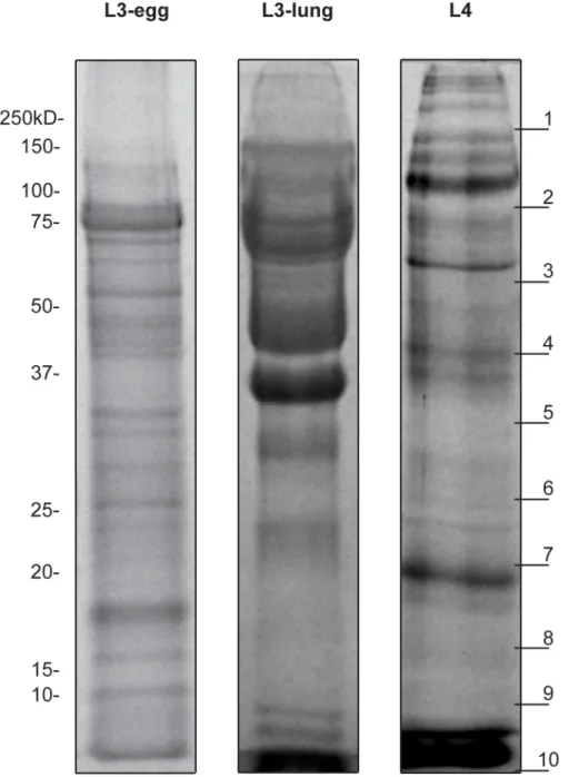
Figure 1. Protein profile of theA. suumES products.Protein profile of theA. suumL3-egg, L3-lung and L4 ES products displayed on a 12% SDS-PAGE stained with Coomassie blue. Each lane was loaded with 15mg of protein. Molecular weight markers are indicated to the left. The 10 gel slices used in the trypsin digests are indicated on the right.
Database searching and sequence analysis
Data were searched against an in-house Ascaris sequence database (18,542 protein entries), which is based on the recently publishedA. suum genome [23], using the search engine Mascot Daemon (v.2.3, Matrix Science, London, UK), allowing a maximum of one miscleavage. Carbamidomethyl (C) was specified as fixed modification and carbamidomethyl (N-term), deamidated (NQ) and oxidation (M) were considered as variable modifications for in-gel digest. For in solution digests, methylthio (C) was selected as the fixed modification, and deamidated (NQ) and oxidation (M) as variable modifications. An error-tolerant Mascot search was performed as well. The peptide tolerance and MS/MS tolerance were set to 0.35 Da and 0.45 Da, respectively. Only the most parsimonious group of protein identifications were reported from the identified proteins, and the identification threshold was set at p,0.01. For the proteins that were annotated based on only one peptide, the identification threshold was set at p,0.0001. An estimate of the relative abundance of the predicted proteins in the trypsin digestion was assessed using the Exponentially Modified Protein Abundance Index (emPAI) [28] together with the MS score, sequence coverage, detected peptides numbers. For redundant identifications, the emPAI value from the hit with the
highest score was considered. The Gene Ontology (GO) database was used for inferring the molecular function of individual proteins identified. The protein sequences were analysed for the presence of signal peptides and transmembrane regions with SignalP 3.0 and TMHMM 2.0 (http://www.cbs.dtu.dk/services/TMHMM/), respectively. The subcellular localization was predicted with SecretomeP 2.0. The sequences of the identified proteins were then used to BLAST search the A. suum genome to identify homologous sequences. This was done through the WormBase (http://www.wormbase.org/) (E-value threshold = 1E-16).
Amino acid sequences of selected eukaryotic glycosyl hydrolases listed in the CAZy database (http://www.cazy.org/) were downloaded and used for multiple alignment and consecutive phylogenetic analyses. These sequences included: Homo sapiens alpha acid glycosidase (AAG) (P10253), dual catalytic sucrase-isomaltase (SUIS) (P14410), maltase-glucoamylase (MGA) (O43451), alpha glucosidase AB (GANAB) (Q14697), alpha glucosidase C (GANC) (Q8TET4);Bos taurus AAG (Q9MYM4); Mus musculus AAG (P70699); Coturnix japonica AAG (O73626); Oryctolagus cuniculusSUIS (P07768);Suncus murinusSUIS (O62653); Rattus norvegicus SUIS (P23739); Sus scrofa GANAB (P79403); Drosophila melanogaster AAG-like (Q7KMM4) and Caenorhabditis
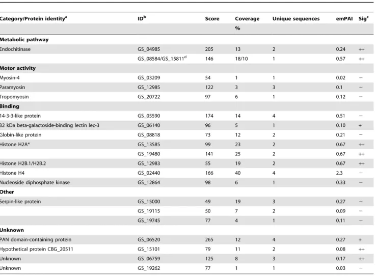
Table 1.Protein identifications inA. suumL3-egg ES products.
Category/Protein identitya IDb Score Coverage Unique sequences emPAI Sigc
%
Metabolic pathway
Endochitinase GS_04985 205 13 2 0.24 ++
GS_08584/GS_15811d 146 18/10 1 0.57
++
Motor activity
Myosin-4 GS_03209 54 1 1 0.02 2
Paramyosin GS_12985 122 3 3 0.1 2
Tropomyosin GS_20722 97 6 1 0.12 2
Binding
14-3-3-like protein GS_05590 174 14 4 0.51 2
32 kDa beta-galactoside-binding lectin lec-3 GS_06140 96 5 1 0.10 +
Globin-like protein GS_08818 73 12 2 0.21 2
Histone H2A* GS_13585 99 23 2 0.67 ++
GS_19480 141 25 2 0.67 ++
Histone H2B.1/H2B.2 GS_12983 55 19 2 0.67 ++
Histone H4 GS_02440 166 40 4 2.3 2
Nucleoside diphosphate kinase GS_12864 98 6 1 0.33 2
Other
Serpin-like protein GS_15000 49 19 3 0.27 2
GS_19115 50 7 2 0.09 2
GS_19745 77 4 1 0.11 2
Unknown
PAN domain-containing protein GS_06520 265 12 4 0.27 +
Hypothetical protein CBG_20511 GS_15101 79 11 2 0.08 ++
Unknown GS_06759 125 8 3 0.17 ++
Unknown GS_19262 77 1 1 0.03 2
aThe proteins identified were categorized by their molecular function according to information obtained from the Gene Ontology database.
bThe accession number inA. suumgenome database (available on WormBase, www.wormbase.org). cThe identified proteins were predicted to be either a classical secretory protein (
+), non-classical secretory protein (++) or not secreted (2) by secretion prediction using SignalP and SecretomeP.
Table 2.Protein identifications inA. suumL3-lung ES products.
Category/Protein identitya IDb Score Coverage Unique sequences emPAI Sigc
%
Metabolic pathway
Maltase-glucoamylase GS_07553 78 2 1 0.05 +
GS_15893 142 6 2 0.15 2
GS_16769 132 8 3 0.18 ++
GS_23879 171 4 4 0.08 ++
Neprilysin-1 GS_08219 66 1 1 0.02 +
Sucrase-isomaltase GS_01568 277 19 4 0.66 2
GS_02444 258 13 3 0.28 2
GS_05716 80 11 1 0.19 2
GS_08447 69 2 1 0.04 ++
GS_17323 129 3 1 0.05 2
GS_20796 121 2 1 0.04 2
GS_22047 151 6 3 0.12 ++
Structural
Cuticlin-1 GS_10816 63 3 1 0.09 2
Cuticle collagen 12 GS_16238 147 17 2 0.27 ++
Cuticle collagen 13 GS_12737 289 8 1 0.34 ++
Peptidyl-prolyl cis-trans isomerase B GS_15602 197 7 2 0.29 2
Binding
14-3-3-like protein GS_05590 73 3 1 0.11 2
C-type lectin GS_12842 102 49 2 1.70 2
Latent-transforming growth factor beta-binding protein 1 GS_21305 102 0 1 0.01 ++
Thyrotropin-releasing hormone-degrading ectoenzyme GS_02555 123 4 1 0.08 ++
Transmembrane cell adhesion receptor mua-3 GS_11192 253 2 6 0.06 ++
Other
Aspartic protease 6 GS_13572 239 9 2 0.25 +
Pepsin inhibitor Dit33 GS_22518 81 6 1 0.13 ++
Poly(U)-specific endoribonuclease GS_22743 101 4 1 0.05 +
Protein DAO-2 GS_24324 120 13 1 0.54 +
Serine protease GS_07735 78 2 1 0.05 +
Serpin-like protein GS_19115 303 14 2 0.29 2
Unknown
24 kDa protein of As22 GS_08591 219 17 2 0.36 +
DOMON domain-containing protein GS_00339 144 20 2 0.43 2
Excretory/secretory mucin MUC-5 GS_22776 529 56 1 4.62 ++
Heh-1 GS_20415 66 5 1 0.12 2
Transthyretin-like protein 5 GS_01881 85 9 1 0.25 +
Venom allergen 3 GS_10381 103 12 2 0.26 ++
von Willebrand factor domain-containing protein GS_02090 66 3 1 0.07 ++
Hypothetical protein LOAG_00319 GS_14306 72 14 1 0.30 2
Hypothetical protein LOAG_07538 GS_11367 109 5 1 0.12 2
Unknown GS_01811 72 14 1 0.29 2
Unknown GS_02698 83 11 1 0.33 2
Unknown GS_03310 89 4 1 0.14 +
Unknown GS_09456 119 20 2 0.56 +
Unknown GS_10718 171 34 2 0.92 +
Unknown GS_12589 133 11 1 0.58 +
elegans AAGR1-4. The protein sequences were subjected to MUSCLE alignment (http://www.ebi.ac.uk/Tools/msa/muscle/ ), and alignments verified and visually checked and edited, as required, in Jalview (http://www.jalview.org/). The program ClustalX 2.0.10 was used to generate phylogenetic tree following analysis using the neighbour-joining method (1000 replicates) [29]. Finally, the program WebLogo application (http://weblogo. threeplusone.com/create.cgi) was used to provide a graphical representation of the amino acid homology around the catalytic sites of some of the glycosyl hydrolases ofA. suumandC. elegans.
RNA extraction and quantitative real-time PCR (qPCR) Total RNAs from larvae and adult worm tissue samples were isolated using TRIzol (Invitrogen), followed by further purification with the RNeasy Mini kit (Qiagen), according to the manufactur-er’s instructions. An on-column DNase digestion was performed using the RNase-free DNase set (Qiagen) to remove any possible genomic DNA. The RNA concentrations were determined (NanoDrop ND-1000 spectrophotometer, NanoDrop Technolo-gies) and its quality was verified (Experion Automated Electro-phoresis System, Bio-Rad). For all samples, the RNA quality indicator (RQI) calculated (ExperionTM software, Bio-Rad) was 8.0, demonstrating high RNA integrity.
The qPCR analyses were performed as described previously [30]. Tubulin and glyceraldehyde-3-phosphate dehydrogenase (GAPDH) were selected as housekeeping genes. The primer sets used were designed by Primer3 software (http://frodo.wi.mit.edu/ primer3/) and are listed in Table S1.
Analysis of differential transcription
A transcriptome dataset was generated from the egg, the L3-liver, the L3-lung and the intestinal L4 stages as part of a previous study [23]. Briefly, following RNA-seq, all paired-end reads for each library constructed were aligned to the predictedA. suumgene set using TopHat. Levels of transcription (reads per kilobase per Million mapped reads (RPKM)) were calculated using Cufflinks [31]. To obtain the RPKM values for genes of interest, accession numbers from the A. suum genome were used to search the transcriptomic datasets.
Protein extraction and enzymatic assays
Protein extracts of larval stages or adult worm tissues were produced by grinding the frozen material to a fine powder in a liquid nitrogen-cooled pestle and mortar. The powder was sequentially subjected to a two-step process with reagents of increasing solubilising power [32]. For the water-soluble protein fraction, 4 ml of PBS, pH 7.4, were used to resuspend the powder for 2 h at 4uC by gentle ‘head-over-head’ mixing. The insoluble material was pelleted by centrifugation at 120,0006gfor 15 min
and the supernatant retained. For the water-insoluble protein fraction, the pellet was incubated at 22uC for 3 h using an extraction buffer consisting of 5 M urea (Sigma), 2 M thiourea (Sigma), 2% CHAPS (Sigma) and 2% SB3-10 (Sigma) in 40 mM Tris, pH 7.4. The supernatant was collected, as described for the water-soluble protein fraction. A general use cocktail of protease inhibitor (Sigma) was added to each extracts to avoid proteolytic degradation. Protein concentrations were measured with the Bradford reagent (Sigma), and proteins stored at280uC.
The glycosidase assays were conducted by incubating 5mg of protein extract with 30 mM of substrate at pH 6.5 for 40 min at 37uC. Reactions were quenched by the addition of 3 M Tris. The glucose was quantified using the Glucose Assay Kit (Sigma). The substrates used in the assays included maltose, lactose and sucrose. Each analysis was performed three times, and the results presented as the average of the three readings. For statistical analysis, the unpaired student t-test was used to test differences in activity between the different protein homogenates. The level of signifi-cance for analyses was set at P#0.05.
Results
Proteins profiles of the excretory/secretory material The protein profiles of the ES products from each of the three larval stages ofA. suum, displayed by SDS-PAGE and Coomassie staining, are shown in Figure 1. The analysis revealed a complex and distinct banding pattern for the ES of three individual stages. Most ES proteins from L3-egg were distributed between 10– 120 kDa, whereas those of L3-lung were mainly between 30 and 100 kDa, with a smear above 40 kDa. L4 ES represented a complicated profile, with major bands between 37 and 150 kDa, and some fainter bands in the 20–30 kDa range.
Protein identifications
Mascot searches of the MS/MS spectra for both the in-gel and in-solution approaches yielded 20, 45 and 58 protein identities within ES products of L3-egg, L3-lung and L4 stages, respectively. The full lists of proteins identified are provided in Tables 1 (L3-egg), 2 (L3-lung) and 3 (L4). Most ES proteins detected were inferred to be stage-specific [85% (n = 17) for L3-egg, 69% (n = 31) for L3-lung and 74% (n = 43) for L4], and 15 proteins identified in ES products were shared by at least two larval stages. The identities of proteins shared by all three stages are given in Figure 2. ES products from L3-lung and L4 shared 14 proteins, representing 31% and 24% of their sub-total, respectively, whereas the L3-egg shared only 2 and 3 proteins with L3-lung and L4, respectively. Finally, two proteins shared by all three ES samples included a 14-3-3-like protein and a serpin (Figure 2).
In silicoprediction of classical and non-classical secretion showed that 9 (45%), 25 (56%) and 42 (72%) of the identified proteins Table 2.Cont.
Category/Protein identitya IDb Score Coverage Unique sequences emPAI Sigc
%
Unknown GS_17230 198 11 2 0.49 2
Unknown L3E_00366 150 4 1 0.21 2
a
The proteins identified were categorized by their molecular function according to information obtained from the Gene Ontology database. bThe accession number in
A. suumgenome database (available on WormBase, www.wormbase.org). cThe identified proteins were predicted to be either a classical secretory protein (
+), non-classical secretory protein (++) or not secreted (2) by secretion prediction using SignalP and SecretomeP.
Table 3.Protein identifications inA. suumL4 ES products.
Category/Protein identitya IDb Score Coverage % Unique sequences emPAI Sigc
Metabolic pathway
Fructose-bisphosphate aldolase 1 GS_19276 189 14 3 0.28 2
Fumarate reductase GS_20429 78 2 1 0.07 2
Glutathione S-transferase 1 GS_16802 150 11 1 0.16 2
Maltase-glucoamylase GS_00984 65 5 1 0.13 ++
GS_07553 93 21 1 0.16 +
GS_15893 642 32 9 1.00 2
GS_18934 101 7 2 0.20 ++
GS_21210 66 15 1 0.43 ++
GS_23879 1143 16 20 0.46 ++
Neprilysin-1 GS_08219 1114 14 14 0.39 +
GS_10348 198 4 5 0.13 ++
GS_19140 331 6 6 0.14 ++
Phosphoenolpyruvate carboxykinase GTP GS_20378 63 4 1 0.08 2
Sucrase-isomaltase GS_05716 175 25 3 0.67 2
GS_08447 97 1 1 0.04 ++
GS_16354 95 5 1 0.08 ++
GS_19777 259 6 4 0.14 ++
Structural
Peptidyl-prolyl cis-trans isomerase 3 GS_07454 82 8 1 0.21 ++
Binding
14-3-3-like protein GS_05590 96 3 1 0.11 2
Aminopeptidase N GS_04166 106 4 3 0.13 ++
GS_05584 143 3 1 0.05 ++
GS_05746 696 14 12 0.29 +
C-type lectin protein 160 GS_02845 194 7 2 0.36 +
GS_04559 835 35 8 1.76 +
GS_12996 170 10 3 0.26 ++
Enolase GS_21295 87 3 1 0.08 2
GH family 25 lysozyme 2 GS_22190 441 39 5 3.27 ++
Nucleoside diphosphate kinase GS_12864 66 9 1 0.15 2
Phosphatidylethanolamine-binding protein GS_22941 103 16 2 0.19 ++
Thyrotropin-releasing hormone-degrading ectoenzyme GS_02555 404 26 7 0.66 ++
Zonadhesin GS_01761 771 22 9 1.00 ++
GS_11354 66 1 1 0.04 +
GS_11656 693 24 4 0.71 +
Other
Aspartic protease 6 GS_14901 340 15 2 0.17 +
GS_15316 753 24 5 0.90 +
GS_19445 919 26 5 1.24 +
Poly(U)-specific endoribonuclease GS_22743 638 16 7 0.56 +
Serpin-like protein GS_19115 395 18 3 0.29 2
Unknown
24 kDa protein [Anisakis simplex] GS_07900 68 16 1 0.41 ++
As14 GS_02102 217 25 1 0.26 +
Transthyretin-like protein 5 GS_21838 69 18 1 0.40 2
Transthyretin-like protein 46 GS_02516 119 18 2 0.45 +
Venom allergen 3 GS_10381 131 16 3 0.41 ++
from L3-egg, L3-lung and L4 ES products, respectively, were predicted to be either a classical or non-classical secreted protein (Tables 1–3).
All proteins identified were subsequently categorized based on their molecular function, according to information from the GO database. Assigned were: metabolic pathway, structural, motor Table 3.Cont.
Category/Protein identitya IDb Score Coverage % Unique sequences emPAI Sigc
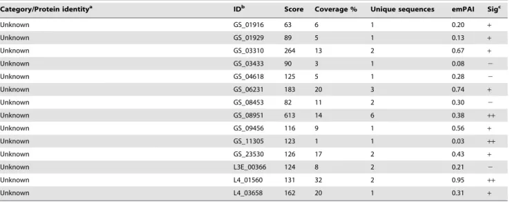
Unknown GS_01916 63 6 1 0.20 +
Unknown GS_01929 89 5 1 0.13 +
Unknown GS_03310 264 13 2 0.67 +
Unknown GS_03433 90 3 1 0.08 2
Unknown GS_04618 125 5 1 0.28 2
Unknown GS_06231 183 20 3 0.74 +
Unknown GS_08453 82 11 2 0.30 2
Unknown GS_08951 613 14 6 0.38 ++
Unknown GS_09456 116 9 1 0.56 +
Unknown GS_11305 123 1 1 0.03 ++
Unknown GS_23530 126 17 2 0.43 +
Unknown L3E_00366 124 8 2 0.21 2
Unknown L4_01560 131 32 2 0.95 ++
Unknown L4_03658 162 20 1 0.31 +
aThe proteins identified were categorized by their molecular function according to information obtained from the Gene Ontology database.
bThe accession number inA. suumgenome database (available on WormBase, www.wormbase.org). cThe identified proteins were predicted to be either a classical secretory protein (
+), non-classical secretory protein (++) or not secreted (2) by secretion prediction using SignalP and SecretomeP.
doi:10.1371/journal.pntd.0002467.t003
Figure 2. Venn diagram of similar proteins.Venn diagram showing the distribution of the number of proteins identified in ES products from L3-egg, L3-lung and L4 ofA. suum. The proteins identified are listed on the right.
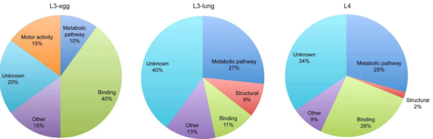
activity, binding, other functions and proteins of unknown function (Figure 3). From the entire annotated ES protein dataset, 24% (n = 38) of proteins did not have any known function or known homologues in other organisms. Comparison of the results obtained for the three larval stages indicated an increase in the number of proteins involved in metabolic pathways from the L3-egg stage to the L3-lung and L4 stage larvae, whereas only two endochitinase homologues were identified from the L3-egg. In contrast, motor activity proteins, including proteins such as myosin-4, paramyosin and tropomyosin, were unique to L3-egg. Finally, 9% of proteins identified in L3-lung ES products, including cuticlin-1, cuticle collagen 12 and 13, represented ‘structural’ proteins, whereas those belonging to this category were less represented in L3-egg (none) and L4 (2%). Of the 17 binding proteins identified 82% of them were ATP-, ion-, carbohydrate-and DNA-binding proteins.
Glycosyl hydrolases inAscaris suum
The most frequently identified proteins in ES products were glycosyl hydrolases belonging to family 31 (GH31). In total 16 GH31 proteins were identified in the ES products of L3-lung and L4 larvae with homology to maltase-glucoamylases and sucrase-isomaltases. Six and 5 GH31 proteins were identified in L3-lung and L4, respectively, and another 5 for both of these larval stages. In order to obtain more information on these proteins, we subsequently BLAST searched theA. suumgenome for additional members of this GH31 family. In total, 32 protein sequences were identified, all showing homology to GH31 proteins (Table 4). The length of the protein sequences ranged from 80 to 1772 amino acids (aa), suggesting that some of the sequences were not full length. Twenty of the predicted GH31 proteins were predicted as either secreted through a classical or non-classical pathway.
The GH31 protein sequences ($700 aa) representing Ascaris were aligned with those of homologous proteins from other species for subsequent phylogenetic analysis (Figure 4, panel A). The unrooted tree indicated clustering of the majority of the GH31 proteins of A. suum with acid-active GH31 enzymes (i.e. AAG, SUIS, MGA, AAGR1-2), whereas only one (i.e. GS_18807) clustered with neutral-active GH31 enzymes (i.e. GANAB and GANC). The results of a comparative analysis of the amino acid sequence homology around the catalytic site of 13A. suumGH31 proteins (codes GS_0471, GS_05082, GS_06701, GS_08447, GS_13054, GS_17123, GS_17323, GS_18807, GS_19777, GS_20796, GS_22047 and GS_23879) and the 4 GH31 proteins
present in C. elegans (AAGR1- AAGR 4) (Figure 4, panel B) indicated that the signature motifs around the catalytic nucleophile are largely conserved between these two nematode species.
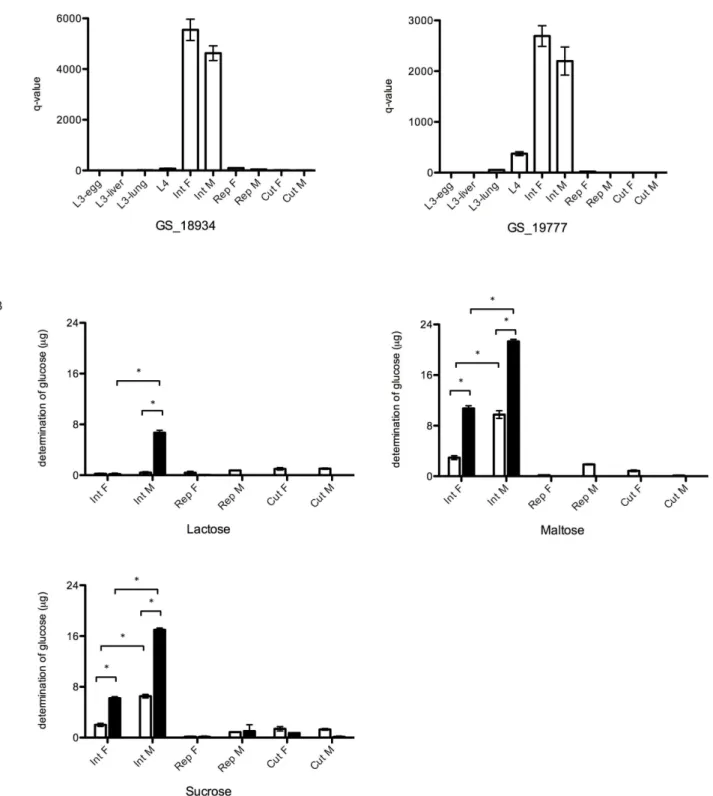
In the transcriptomic analysis, the RPKM values for all GH31 proteins identified here showed that most of them are transcrip-tionally upregulated in the late larval stages (L3-lung and L4) ofA. suum(Table 4). Based on the RPKM values, GH31 proteins with the highest transcription were GS_18934, GS_13054 and GS_19777, with RPKM values of.500 in L4. A qPCR analysis of genes encoding GH31 proteins (codes GS_18934 and GS_19777) was conducted to (1) verify the transcriptomic data and (2) to analyse their transcription profiles in different tissues of adult A. suum(Figure 5 panel A). Indeed, transcription levels of both genes were higher in L4 compared with other stages. In addition, the transcription linked to these GH31 was in the intestine of both female and male adults ofA. suum, whereas almost no transcription was detected in either the reproductive system or the cuticle of both sexes (Figure 5, panel A).
To confirm the intestinal location of the GH31 proteins, enzymatic assays were performed to measure glycolytic activity in protein homogenates from different adultA. suumtissues (Figure 5, panel B). Particularly maltose and sucrose were degraded following incubation with homogenates from the intestinal tracts of both adult male and female worms. The glycolytic activity measured was markedly higher in the water-insoluble protein fractions compared with the water-soluble fraction (P,0.05). In addition, the intestinal homogenates from males showed higher activity compared with females (P,0.05). The degradation of lactose was only observed after incubation with the water-insoluble protein fraction produced from the adult male intestines.
Discussion
The goal of this study was to identify the ES proteins produced and released by the larval stages ofA. suum in vitroand to infer the functions of these molecules during the migratory phase of the parasite through the body of the host animal. In total, 106 proteins were identified, of which 62% were predicted to either contain a signal peptide, suggesting secretion through a classical pathway, or predicted to be secreted viaa non-classical pathway. The other 38% of proteins lacked a detectable signal sequence. Although no changes were observed in the motility or physical appearance of the larvae during the in vitro culture, some atypical secreted proteins were detected. The highest number of ‘non-secreted’
Figure 3. Gene ontology.Gene Ontology terms relating to molecular function assigned to the proteins identified in ES products from egg, L3-lung and L4 ofA. suum.
proteins for L3-egg was 55% compared with 45% and 26% for L3-lung and L4, respectively. Some of these ‘atypical secreted’ proteins, including 14-3-3 and serpin, may include their secretion in extracellular vesicles as described for other helminths, such asC. elegans[33],Fasciola hepaticaandEchinostoma caproni[34]. However, the presence of some typical intracellular proteins in the ES material, such as histones, for example, suggests that there was some cellular damage in the larvae leading to leakage of intracellular proteins into the medium. The precise reason for this is unclear, but it is possible that the hatching procedure, and
the subsequent washing steps have a role. Moreover, keeping thein vitroculture as short as possible may help reducing the possibly invisible leakage of intracellular proteins into the medium. Therefore, in the future it would be interesting to analyse ES material that has been collected after only few hours ofin vitro culture.
Among the 106 ES proteins identified in this study, two (i.e. a serpin-like and a 14-3-3 protein) were released by all three larval stages investigated. Serpins are serine protease inhibitors with a wide spectrum of functions in numerous biological systems, such as blood coagulation, complement activation and inflammation [17,19,35,36]. Analysis of theA. suumgenome and transcriptomes showed that they contain 10 serpin-encoding genes [23], whereas 8 and 3 serpin genes have been identified in the genomes ofC. elegansandB. malayi[35], respectively. A number of studies have previously reported on the presence of serpins in nematode ES products and experimental evidence indicates that many of them can have an immune-evasive function [36]. Interestingly, earlier studies ofA. suumhave shown that the activities of host proteases, such as trypsin and chymotrypsin, were greatly decreased from the micro-environment of live worms with a functioning gastrointes-tinal system [37]. Subsequently, Martzenet al.[38,39] showed that inactive chymotrypsin complexes were formed in the muscle sarcolemma and in the epithelial surface of the gut of adultA. suum as well as in developing eggs and larvae of this nematode. In this way, the serine protease inhibitors may not only protect the worms from degradation in host digestive environment but might also mask the surface of developing larvae, permitting them to evade the host’s immune system as they migrate from the intestine to the liver and the lungs. Whether the serpins detected in the ES products from A. suum are involved in these processes is still unclear.
In addition to the serpin, a 14-3-3 protein was also detected in the ES material of all three larval stages. Such 14-3-3 proteins represent a family of relatively conserved regulatory proteins, which can bind a range of functionally diverse signaling proteins. InC. elegans, a 14-3-3 protein regulatesdaf-2/insulin-like signaling pathway, which is critical for regulating development, longevity, metabolism and stress resistance [40]. Although the 14-3-3 proteins have been isolated and characterized recently as molecules with a significant role in the parasite biology and immunology within the context of the host–parasite relationship [41–43], currently, little information is available on their actual role in parasites.
Further comparison of the protein composition of the larval ES proteins showed that more overlap existed between L3-lung and L4 compared with L3-egg. Glycosyl hydrolases belonging to family 31 (GH31) were particularly prominent in ES products from L3-lung and L4. The identification of 16 GH31 proteins is an intriguing outcome of this study, particularly since no other studies have reported the presence of such enzymes in the ES products from nematodes. An analysis of the A. suum genome and transcriptomes revealed 32 putative GH31 protein encoding genes/sequences. Although the exact number of GH31 protein genes inA. suumis less than 32, because of short or incomplete sequences in the current dataset, it is still clear that this gene family has undergone a large expansion compared with other nematode species. A preliminary analysis indicated the presence of only 4 GH31 protein genes in the genomes ofC. elegans,B. malayiandT. spiralis (results not shown). The results presented in the present study also indicated that most of the GH31 proteins were transcriptionally upregulated from the L4 larval stage onwards, with a peak in the adult stage ofAscaris, in particular in intestinal tissues. This finding was also confirmed by enzymatic assays, Table 4.List of glycosyl hydrolases identified in theA. suum
genome, their sequence length and their gene levels in different larval stages.
RPKM values
Gene IDa AAlength SigbL3-egg L3-liver L3-lung L4 ESmaterialc
GS_05082 735 2 0,2 0,02 0,03 0,02
GS_16769 587 ++ 0,06 13,45 0,2 1,14 +
GS_17323 600 + 0,33 141,38 11,02 9,61 +
GS_04731 1047 + 0,02 4,64 1,33 1,05
GS_10423 226 ++ 29,27 435,4 182,9 11,19
GS_00493 153 2 0,11 0,11 0,16 0,09
GS_01568 311 2 0,05 0,05 0,08 0,05 +
GS_02444 380 2 0,04 0,04 0,06 0,04 +
GS_08026 107 ++ 0,15 0,15 0,22 0,13
GS_08447 895 ++ 0,02 0,02 0,03 0,02 +
GS_16354 408 ++ 0,04 0,04 0,06 0,03 +
GS_23821 195 + 0,08 0,08 0,12 0,07
GS_24300 573 ++ 0,03 1,3 2,52 0,05
GS_07553 217 + 0,08 0,08 0,11 0,07 +
GS_15893 459 2 0,04 0,04 26,65 0,93 +
GS_22047 830 ++ 0,02 0,02 0,03 0,02 +
GS_00984 255 2 0,06 0,06 0,09 0,06 +
GS_20796 743 2 0,02 0,02 0,03 0,02 +
GS_21210 80 2 0,2 0,2 0,3 0,18 +
GS_18807 935 2 8,58 17,06 62,88 22,98
GS_06701 828 + 2,07 2,87 50,98 39,96
GS_18934 359 ++ 8,29 2,02 249,19 989,82 +
GS_05716 181 2 0,09 0,09 0,13 1,1 +
GS_13054 457 ++ 0,22 5,71 15,67 705,7
GS_17123 737 ++ 0,02 0,02 0,03 0,48
GS_21706 521 ++ 0,03 0,67 0,09 11,18
GS_23879 1772 ++ 0,61 0,39 9,98 56,22 +
GS_04250 479 2 1,65 9,45 4,42 38,01
GS_00096 460 ++ 0,04 0,11 0,05 97,35
GS_12078 373 2 0,04 0,04 0,06 3,05
GS_19777 966 ++ 1,07 24,87 159,14 1054,46+
GS_23076 995 + 0,02 0,02 2,49 17,89
aThe accession number inA. suumgenome database (available on WormBase, www.wormbase.org).
bThe identified proteins were predicted to be either a classical secretory protein
(+), non-classical secretory protein (++) or not secreted (2) by secretion prediction using SignalP and SecretomeP.
cGH31 proteins identified in the ES material are marked with an ‘
Figure 4. Phylogenetic tree and signature motifs of GH31 proteins.A. Unrooted phylogenetic tree of theA. suumGH31 proteins (with a minimum sequence length of 700 amino acids) and other selected eukaryotic GH31 protein following neighbour-joining analysis. The values at the branch nodes represent bootstrap values (maximum 1000). B. Comparative analysis of the amino acids around the catalytic nucleophile (Trp and Asp) of GH31 proteins for theC. elegansGH31 proteins AAGR1-4 and theA. suumGH31 proteins (GS_04731, GS_05082, GS_06701, GS_08447, GS_13054, GS_17123, GS_17323, GS_18807, GS_19777, GS_20796, GS_21706, GS_22047 and GS_23879).
doi:10.1371/journal.pntd.0002467.g004
Figure 5. qPCR and glucosidase hydrolytic activity analysis of GH31 proteins.A. A qPCR analysis for two GH31 proteins on cDNA produced from different larval stages and adult worm tissues. B. Comparison of the glucosidase hydrolytic activity in soluble (white bars) and water-insoluble extract (black bars) from different adult tissues. Results are shown as average+SD. Substrates used in the assays were lactose, maltose and sucrose. (Int F: female intestine; Int M: male intestine; Rep F: female reproductive system; Rep M: male reproductive system; Cut F: female cuticle; Cut M: male cuticle). (* P,0.05).
showing the highest glycosidase activity in intestinal protein extracts from adult worms. It has been suggested [44] thatAscaris takes most of its nutrients from the partially digested host food in the intestine. The present findings suggest that the degradation of complex carbohydrates forms an essential part of the energy metabolism of this parasite once it is established in the small intestine. The highest level of glycolytic activity was consistently found in the water insoluble protein fraction, suggesting that the enzymes are associated or directly bound to a cell membrane. In mammals, it has been shown that the sucrose-isomaltase complex is anchored to the small intestinal brush border through a highly hydrophobic segment in the N-terminal region of the isomaltase subunit [45,46]. However, none of the A. suum GH31 protein sequences were predicted to contain such a transmembranic region. Therefore, further research is needed to determine the exact cellular location of the GH31 proteins within the intestinal tissues ofA. suum. If the results would show that they are actually located on the intestinal surface, it would make them interesting drug and/or vaccine targets.
Apart from the shared proteins, most of the proteins identified were unique to a particular larval stage. Amongst the ES proteins identified in L3-egg, there were at least two different endochiti-nases. Chitinases are enzymes that catalyze the hydrolysis of beta-1, 4-N-acetyl-d-glucosamine linkages in chitin polymers. Studies of B. malayi (a filarioid nematode) showed that a chitinase was secreted during the exsheathment process of the microfilariae in the mosquito vector and from the eggshell during hatching of the larvae within the reproductive tract of the adult stage [47]. RNAi studies of Acanthocheilonema viteae (also a filarioid) showed that chitinase was also critical in the moulting process of the nematode [48]. Interestingly, Geng et al. [49] previously reported on the abundant secretion of a chitinase in the perivitelline fluid surrounding the infectiveA. suumlarva just prior to hatching from the egg. This chitinase is however different from that identified here. During the larval cultivation, many of the larvae need to lose the L2 cuticle, which is usually still present around the infective L3 larvae when they hatch from the egg. Therefore, it is possible that the chitinases identified herein are involved in the exsheathment process.
Analysis of ES products from L3-lung and L4-ES resulted in the identification of various proteins that have consistently been found in ES material of other parasites, such as the transthyretin-like proteins, C-type lectins and venom allergens [15,17,18,50]. The transthyretin-like proteins are one of the largest conserved nematode-specific protein families of which the function is still largely unclear. Recent data published by Wang et al. [51] on TTR-52, one of the 57 transthyretin-like proteins present inC. eleganssuggest that these proteins act extracellulary to mediate cell-cell interactions.
C-type lectins belong to a type of carbohydrate-binding protein family, known as lectins. These molecules are widely distributed throughout the animal kingdom and have a diverse range of
functions, including cell-cell adhesion, immune responses to pathogens and apoptosis [15,52]. Notably, C-type lectins were also particularly abundant in the secretions from T. canis and hookworms [50,53]. The recent report of the sequence similarity of C-type lectins fromA. suum to host to dendritic cell receptors suggests that the parasites may utilize lectins to bind to carbohydrate moieties on the surface of host cells to avoid pathogen recognition mechanisms in hosts [54]. The identification of several C-type lectins in the current study could indeed indicate that they play an important, yet undiscovered, role at the parasite-host interface.
Venom allergens belong to the SCP/TAPS protein family and are basically found in every species investigated so far. Despite the fact that the exact function of SCP/TAPS proteins remains unknown, various studies have shown that they are amongst the most abundant proteins expressed and secreted during the transition from the free-living to the parasitic life stages, suggesting an important role in the onset of parasitism [55]. Compared with the number of SCP/TAPS proteins identified in the ES material of some other species [20,56,57], it is surprising that only 2 were identified in the current study. However, this finding is concordant with previous evidence from genomic and transcriptomic datasets [23] indicating the presence of only 12 SCP/TAPS-encoding genes in the A. suum with relatively limited transcription levels (results not shown).
In conclusion, this study provides the first in-depth character-ization of the ES products from the larval stages ofA. suum, a crucial step in enhancing our knowledge and understanding of the biology of this parasite and its interactions with its mammalian host. The study provides a basis for further molecular investiga-tions aimed at exploring the biological role of the proteins identified and their potential as vaccine and/or therapeutic targets.
Supporting Information
Table S1 Nucleotide sequences of the primers used in the qRT-PCR assays.
(DOC)
Acknowledgments
The authors acknowledge the technical assistance of Mr. Dries Masure, Ms. Iris Peelaers and Dr. Jimmy Borloo of Department of Virology, Parasitology and Immunology, Faculty of Veterinary Medicine, Ghent University, Belgium.
Author Contributions
Conceived and designed the experiments: TW JV PG. Performed the experiments: TW KVS MD. Analyzed the data: TW KVS MD. Contributed reagents/materials/analysis tools: DD ARJ RBG. Wrote the paper: TW RBG PG.
References
1. de Silva NR, Brooker S, Hotez PJ, Montresor A, Engels D, et al. (2003) Soil-transmitted helminth infections: updating the global picture. Trends Parasitol 19: 547–551.
2. Bethony J, Brooker S, Albonico M, Geiger SM, Loukas A, et al. (2006) Soil-transmitted helminth infections: ascariasis, trichuriasis, and hookworm. Lancet 367: 1521–1532.
3. Dold C, Holland CV (2011)Ascarisand ascariasis. Microbes Infect 13: 632–637. 4. Barker KR (1993) The future of nematology: introduction. J Nematol 25: 365–
366.
5. Jungersen G, Eriksen L, Nansen P, Lind P, Rasmussen T, et al. (2001) Regional immune responses with stage-specific antigen recognition profiles develop in
lymph nodes of pigs followingAscaris suumlarval migration. Parasite Immunol 23: 185–194.
6. Bradley JE, Jackson JA (2004) Immunity, immunoregulation and the ecology of trichuriasis and ascariasis. Parasite Immunol 26: 429–441.
7. Masure D, Vlaminck J, Wang T, Chiers K, Van den Broeck W, et al. (2013) A Role for Eosinophils in the Intestinal Immunity against InfectiveAscaris suum
Larvae. PLoS Negl Trop Dis 7: e2138.
8. Lightowlers MW, Rickard MD (1988) Excretory-secretory products of helminth parasites: effects on host immune responses. Parasitology 96 Suppl: S123–166. 9. Blaxter ML, Page AP, Rudin W, Maizels RM (1992) Nematode surface coats:
10. Kennedy MW, Qureshi F (1986) Stage-specific secreted antigens of the parasitic larval stages of the nematodeAscaris. Immunology 58: 515–522.
11. Kennedy MW, Qureshi F, Fraser EM, Haswell-Elkins MR, Elkins DB, et al. (1989) Antigenic relationships between the surface-exposed, secreted and somatic materials of the nematode parasitesAscaris lumbricoides,Ascaris suum, andToxocara canis. Clin Exp Immunol 75: 493–500.
12. Kennedy MW, Qureshi F, Haswell-Elkins M, Elkins DB (1987) Homology and heterology between the secreted antigens of the parasitic larval stages ofAscaris lumbricoidesandAscaris suum. Clin Exp Immunol 67: 20–30.
13. Fetterer RH (1996) Growth and cuticular synthesis inAscaris suumlarvae during development from third to fourth stagein vitro. Vet Parasitol 65: 275–282. 14. Islam MK, Miyoshi T, Yokomizo Y, Tsuji N (2004) The proteome expression
patterns in adultAscaris suumunder exposure to aerobic/anaerobic environments analyzed by two-dimensional electrophoresis. Parasitol Res 93: 96–101. 15. Mulvenna J, Hamilton B, Nagaraj SH, Smyth D, Loukas A, et al. (2009)
Proteomics analysis of the excretory/secretory component of the blood-feeding stage of the hookworm,Ancylostoma caninum. Mol Cell Proteomics 8: 109–121. 16. Bennuru S, Semnani R, Meng Z, Ribeiro JM, Veenstra TD, et al. (2009)Brugia
malayi excreted/secreted proteins at the host/parasite interface: stage- and gender-specific proteomic profiling. PLoS Negl Trop Dis 3: e410.
17. Moreno Y, Geary TG (2008) Stage- and gender-specific proteomic analysis of
Brugia malayiexcretory-secretory products. PLoS Negl Trop Dis 2: e326. 18. Hewitson JP, Harcus YM, Curwen RS, Dowle AA, Atmadja AK, et al. (2008)
The secretome of the filarial parasite,Brugia malayi: proteomic profile of adult excretory-secretory products. Mol Biochem Parasitol 160: 8–21.
19. Yatsuda AP, Krijgsveld J, Cornelissen AW, Heck AJ, de Vries E (2003) Comprehensive analysis of the secreted proteins of the parasiteHaemonchus contortusreveals extensive sequence variation and differential immune recogni-tion. J Biol Chem 278: 16941–16951.
20. Nisbet AJ, Smith SK, Armstrong S, Meikle LI, Wildblood LA, et al. (2010)
Teladorsagia circumcincta: activation-associated secreted proteins in excretory/ secretory products of fourth stage larvae are targets of early IgA responses in infected sheep. Exp Parasitol 125: 329–337.
21. Craig H, Wastling JM, Knox DP (2006) A preliminary proteomic survey of thein vitroexcretory/secretory products of fourth-stage larval and adultTeladorsagia circumcincta. Parasitology 132: 535–543.
22. Robinson MW, Greig R, Beattie KA, Lamont DJ, Connolly B (2007) Comparative analysis of the excretory-secretory proteome of the muscle larva ofTrichinella pseudospiralisandTrichinella spiralis. Int J Parasitol 37: 139–148. 23. Jex AR, Liu S, Li B, Young ND, Hall RS, et al. (2011)Ascaris suumdraft genome.
Nature 479: 529–533.
24. Urban JF, Jr., Douvres FW (1981)In vitrodevelopment ofAscaris suumfrom third-to fourth-stage larvae and detection of metabolic antigens in multi-well culture systems. J Parasitol 67: 800–806.
25. Laemmli UK (1970) Cleavage of structural proteins during the assembly of the head of bacteriophage T4. Nature 227: 680–685.
26. Tilleman K, Van Beneden K, Dhondt A, Hoffman I, De Keyser F, et al. (2005) Chronically inflamed synovium from spondyloarthropathy and rheumatoid arthritis investigated by protein expression profiling followed by tandem mass spectrometry. Proteomics 5: 2247–2257.
27. Van Steendam K, De Ceuleneer M, Dhaenens M, Van Hoofstat D, Deforce D (2013) Mass spectrometry-based proteomics as a tool to identify biological matrices in forensic science. Int J Legal Med 127: 287–298.
28. Ishihama Y, Oda Y, Tabata T, Sato T, Nagasu T, et al. (2005) Exponentially modified protein abundance index (emPAI) for estimation of absolute protein amount in proteomics by the number of sequenced peptides per protein. Mol Cell Proteomics 4: 1265–1272.
29. Thompson JD, Gibson TJ, Plewniak F, Jeanmougin F, Higgins DG (1997) The CLUSTAL_X windows interface: flexible strategies for multiple sequence alignment aided by quality analysis tools. Nucleic Acids Res 25: 4876–4882. 30. Dreesen L, Rinaldi M, Chiers K, Li R, Geurden T, et al. (2012) Microarray
analysis of the intestinal host response inGiardia duodenalisassemblage E infected calves. PLoS One 7: e40985.
31. Mortazavi A, Williams BA, McCue K, Schaeffer L, Wold B (2008) Mapping and quantifying mammalian transcriptomes by RNA-Seq. Nat Methods 5: 621–628. 32. Braschi S, Wilson RA (2006) Proteins exposed at the adult schistosome surface
revealed by biotinylation. Mol Cell Proteomics 5: 347–356.
33. Liegeois S, Benedetto A, Michaux G, Belliard G, Labouesse M (2007) Genes required for osmoregulation and apical secretion in Caenorhabditis elegans. Genetics 175: 709–724.
34. Marcilla A, Trelis M, Cortes A, Sotillo J, Cantalapiedra F, et al. (2012) Extracellular vesicles from parasitic helminths contain specific excretory/ secretory proteins and are internalized in intestinal host cells. PLoS One 7: e45974.
35. Zang X, Maizels RM (2001) Serine proteinase inhibitors from nematodes and the arms race between host and pathogen. Trends Biochem Sci 26: 191–197. 36. Molehin AJ, Gobert GN, McManus DP (2012) Serine protease inhibitors of
parasitic helminths. Parasitology 139: 681–695.
37. Juhasz S, Nemeth I (1979) Proteolytic enzymes and enzyme inhibitors inAscaris suum. IV. Estimation of molecular weights of chymotrypsin inhibitors and an intestinal protease by gel chromatography. Acta Vet Acad Sci Hung 27: 217– 224.
38. Martzen MR, Geise GL, Hogan BJ, Peanasky RJ (1985)Ascaris suum: localization by immunochemical and fluorescent probes of host proteases and parasite proteinase inhibitors in cross-sections. Exp Parasitol 60: 139–149.
39. Martzen MR, Geise GL, Peanasky RJ (1986)Ascaris suum: immunoperoxidase and fluorescent probe analysis of host proteases and parasite proteinase inhibitors in developing eggs and second stage larvae. Exp Parasitol 61: 138– 145.
40. Li J, Tewari M, Vidal M, Lee SS (2007) The 14-3-3 protein FTT-2 regulates DAF-16 inCaenorhabditis elegans. Dev Biol 301: 82–91.
41. McGonigle S, Loschiavo M, Pearce EJ (2002) 14-3-3 proteins inSchistosoma mansoni; identification of a second epsilon isoform. Int J Parasitol 32: 685–693. 42. Siles-Lucas M, Merli M, Mackenstedt U, Gottstein B (2003) TheEchinococcus
multilocularis14-3-3 protein protects mice against primary but not secondary alveolar echinococcosis. Vaccine 21: 431–439.
43. Siles-Lucas M, Merli M, Gottstein B (2008) 14-3-3 proteins inEchinococcus: their role and potential as protective antigens. Exp Parasitol 119: 516–523. 44. Hall A, Hewitt G, Tuffrey V, de Silva N (2008) A review and meta-analysis of
the impact of intestinal worms on child growth and nutrition. Matern Child Nutr 4 Suppl 1: 118–236.
45. Semenza G, Brunner J, Wacker H (1983) Biosynthesis and assembly of the largest and major intrinsic polypeptide of the small intestinal brush borders. Ciba Found Symp 95: 92–112.
46. Semenza G (1986) Anchoring and biosynthesis of stalked brush border membrane proteins: glycosidases and peptidases of enterocytes and renal tubuli. Annu Rev Cell Biol 2: 255–313.
47. Wu Y, Preston G, Bianco AE (2008) Chitinase is stored and secreted from the inner body of microfilariae and has a role in exsheathment in the parasitic nematodeBrugia malayi. Mol Biochem Parasitol 161: 55–62.
48. Tachu B, Pillai S, Lucius R, Pogonka T (2008) Essential role of chitinase in the development of the filarial nematodeAcanthocheilonema viteae. Infect Immun 76: 221–228.
49. Geng J, Plenefisch J, Komuniecki PR, Komuniecki R (2002) Secretion of a novel developmentally regulated chitinase (family 19 glycosyl hydrolase) into the perivitelline fluid of the parasitic nematode,Ascaris suum. Mol Biochem Parasitol 124: 11–21.
50. Loukas A, Mullin NP, Tetteh KK, Moens L, Maizels RM (1999) A novel C-type lectin secreted by a tissue-dwelling parasitic nematode. Curr Biol 9: 825–828. 51. Wang X, Li W, Zhao D, Liu B, Shi Y, et al. (2010) Caenorhabditis elegans
transthyretin-like protein TTR-52 mediates recognition of apoptotic cells by the CED-1 phagocyte receptor. Nat Cell Biol 12: 655–664.
52. McGreal EP, Martinez-Pomares L, Gordon S (2004) Divergent roles for C-type lectins expressed by cells of the innate immune system. Mol Immunol 41: 1109– 1121.
53. Loukas A, Doedens A, Hintz M, Maizels RM (2000) Identification of a new C-type lectin, TES-70, secreted by infective larvae ofToxocara canis, which binds to host ligands. Parasitology 121 Pt 5: 545–554.
54. Yoshida A, Nagayasu E, Horii Y, Maruyama H (2012) A novel C-type lectin identified by EST analysis in tissue migratory larvae ofAscaris suum. Parasitol Res 110: 1583–1586.
55. Hewitson JP, Grainger JR, Maizels RM (2009) Helminth immunoregulation: the role of parasite secreted proteins in modulating host immunity. Mol Biochem Parasitol 167: 1–11.