Evaluation of an alternative technique to optimize direct
bonding of orthodontic brackets to temporary crowns
Francilena Maria Campos Santos Dias1, Célia Regina Maio Pinzan-Vercelino2, Rudys Rodolfo de Jesus Tavares3,
Júlio de Araújo Gurgel4, Fausto Silva Bramante5, Melissa Nogueira Proença Fialho6
Objective: To compare shear bond strength of different direct bonding techniques of orthodontic brackets to acrylic resin surfaces. Methods: The sample comprised 64 discs of chemically activated acrylic resin (CAAR) randomly divided into four groups: discs in group 1 were bonded by means of light-cured composite resin (conventional adhesive); discs in group 2 had surfaces roughened with a diamond bur followed by conventional direct bonding by means of light-cured composite resin; discs in group 3 were bonded by means of CAAR (alternative adhesive); and discs in group 4 had surfaces roughened with a diamond bur followed by direct bonding by means of CAAR. Shear bond strength values were determined after 24 hours by means of a universal testing machine at a speed of 0.5 mm/min, and compared by analysis of variance followed by post-hoc Tukey test. Adhesive remnant index (ARI) was measured and compared among groups by means of Kruskal-Wallis and Dunn tests. Results: Groups 3 and 4 had significantly greater shear bond strength values in comparison to groups 1 and 2. Groups 3 and 4 yielded similar results. Group 2 showed better results when compared to group 1. In ARI analyses, groups 1 and 2 predominantly exhibited a score equal to 0, whereas groups 3 and 4 predominantly exhibited a score equal to 3. Conclusions: Direct bonding of brackets to acrylic resin surfaces using CAAR yielded better results than light-cured composite resin. Sur-face preparation with diamond bur only increased shear bond strength in group 2.
Keywords:Orthodontic brackets. Dental bonding. Shear bond strength. Temporary dental restoration.
How to cite this article: Dias FMCS, Pinzan-Vercelino CRM, Tavares RRJ, Gurgel JA, Bramante FS, Fialho MNP. Evaluation of an alternative technique to optimize direct bonding of orthodontic brackets to temporary crowns. Dental Press J Orthod. 2015 July-Aug;20(4):57-62.
DOI: http://dx.doi.org/10.1590/2176-9451.20.4.057-062.oar
Submitted: August 06, 2014 - Revised and accepted: November 25, 2014
Contact address: Francilena Maria Campos Santos Dias
Rua Duque Bacelar, casa 20, quadra 22 - Quintas do Calhau. São Luís/MA, Brazil E-mail: [email protected]
DOI: http://dx.doi.org/10.1590/2176-9451.20.4.057-062.oar
1 MSc in Orthodontics, Universidade Ceuma, São Luís, Maranhão, Brazil.
2 Assistant professor, Universidade Ceuma, Department of Orthodontics, São
Luís, Maranhão, Brazil.
3 PhD in Oral Rehabilitation, Universidade de São Paulo (USP), School of
Dentistry, Bauru, São Paulo, Brazil.
4 PhD in Orthodontics, Universidade Ceuma, São Luís, Maranhão, Brazil.
5 Associate Professor, Master’s Program in Odontology, Universidade Ceuma,
São Luís, Maranhão, Brazil.
6 MSc in Orthodontics, Universidade Ceuma, São Luís, Maranhão, Brazil.
» The authors report no commercial, proprietary or financial interest in the products or companies described in this article.
Objetivo: comparar a resistência ao cisalhamento de diferentes técnicas para colagem direta de braquetes ortodônticos em superfície de resina acrílica. Métodos: sessenta e quatro (64) discos de resina acrílica ativada quimicamente (RAAQ) foram divididos aleatoriamente em quatro grupos: Grupo 1 = colagem com resina composta fotopolimerizável (adesivo convencional); Grupo 2 = superfície abrasionada com broca diamantada e colagem com resina composta fotopolimeri-zável; Grupo 3 = colagem com RAAQ (adesivo alternativo); Grupo 4 = superfície abrasionada com broca diamantada e colagem com RAAQ. A resistência ao cisalhamento foi avaliada 24h após a colagem, utilizando-se uma máquina universal de ensaios, operando a uma velocidade de 0,5mm/min, e comparada por meio da análise de variância, seguida pelo teste post-hoc de Tukey. O índice de adesivo remanescente (IAR) foi mensurado e comparado entre os grupos por meio dos testes Kruskal-Wallis e Dunn. Resultados: os Grupos 3 e 4 demonstraram resistência ao cisalhamento signi-ficativamente maior do que os Grupos 1 e 2. Os Grupos 3 e 4 apresentaram resultados similares. O Grupo 2 apresentou melhores resultados do que o Grupo 1. Na análise do IAR, observou-se predominância do escore 0 para os Grupos 1 e 2 e do escore 3 para os Grupos 3 e 4. Conclusões: a colagem de braquetes em superfície de resina acrílica utilizando-se a RAAQ apresentou melhores resultados do que a colagem com resina composta fotopolimerizável. O processo de abrasão da superfície com broca diamantada apenas aumentou a resistência ao cisalhamento no Grupo 2.
INTRODUCTION
In recent decades, there has been a growing con-cern for esthetics, in addition to increased life expec-tancy of individuals. As a result, the number of adult patients seeking orthodontic treatment has signiicantly increased.1 Patients seek orthodontists for personal
rea-sons, including esthetic or functional improvements of occlusion, or because other dentists refer them to have dental movements carried in order to aid diferent re-storative procedures.
In many cases, adult patients use deinitive or tem-porary crowns.2 Temporary crowns are commonly used
to protect tissues and render dental position stable be-fore deinitive crown manufacture.3 Additionally, they
also contribute to reestablish both esthetics and function during rehabilitation treatment.
Whenever a patient has temporary crowns, deinitive restoration before orthodontic treatment is not recom-mended due to occlusal changes resulting from dental movement. In these cases, the orthodontist must bond or band the accessories on the surface of temporary material. Banding is recommended for posterior teeth; however, in the anterior region, direct bonding of ac-cessories is used due to the poor esthetic aspect of bands. During orthodontic movement of healthy teeth, bracket bonding on tooth enamel is part of orthodontic routine. However, bonding brackets on acrylic surfaces of temporary crowns is critical and presents high bonding failure rates. Frequent rebonding procedures hinders the advance of mechanotherapy, thereby con-tributing to increase treatment time, costs and chair-side time.4 Therefore, these procedures are undesirable
for both orthodontist and patient.
Among the diferent types of material available to manufacture temporary crowns, chemically activated acrylic resin (CAAR) is the most commonly used5 due
to being inexpensive, easily manipulated and allowing repair, adjustment and relining, all of which may prove necessary during treatment. Additionally, this resin is resistant to oral function, including mastication; sup-ports orthodontic forces and does not damage dental crown surface during debonding.
Despite the advantages of using CAAR as a temporary restorative material, its surface has lower bond strength values in comparison to what is clinically acceptable, when testing orthodontic accessories bonding.5 Various
studies have compared diferent types of material used to
manufacture temporary crowns,6-10 diferent methods of
preparing the provisional material surface before direct bracket bonding5,11,12 and diferent adhesives.7 However,
studies assessing the use of CAAR as a material for direct bonding of orthodontic brackets are scarce.
Composite resin is a common adhesive used for di-rect bracket bonding regardless of the surface (tooth or restoration). However, this type of material yields results lower than what is clinically acceptable when brackets are bonded to provisional acrylic resin material.5 Thus,
it has been speculated that the use of an alternative ad-hesive could optimize the results. Therefore, the objec-tive of this study was to test the following null hypoth-esis: there is no diference in shear bond strength values of diferent direct bonding techniques (varying adhesive material and surface treatment) of orthodontic brackets to acrylic resin surfaces.
MATERIAL AND METHODS
Sample size calculation was carried out by means of the statistical program SAS, version 9.1.3. (SAS Insti-tute Inc., Cary, USA). The following parameters were adopted: shear bond strength value of 5.3 ± 3.3 MPa;6
signiicance level set at 5%; power of test of 80%; and efect size equal to 1. A sample size (n) of 12 specimens in each group was then determined. As a precaution, n = 16 was adopted.
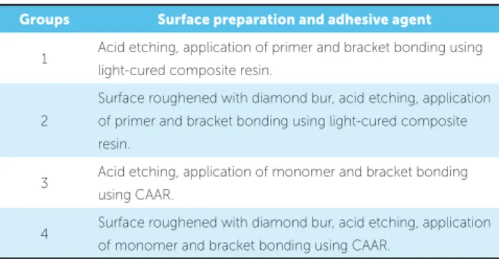
Sixty-four CAAR Duralay discs (Dental Mfg. Co, Worth, USA) were prepared using rigid polyvinyl chlo-ride (PVC) cylinders as matrix. Resin was prepared fol-lowing the manufacturer’s instructions (volume ratio of 3 : 1). Ater manipulation, the material was poured into the PVC rings until 3-mm thickness was reached. The remaining space in the PVC ring was illed with colorless CAAR also prepared according to the manu-facturer’s instructions (volume ratio of 2.5 : 1). To ho-mogenize the bonding surfaces, CAAR surfaces were inished and polished with silicon carbide sandpaper in decreasing order of roughness (400 and 600). Specimens were then randomly divided into four groups (Table 1).
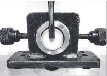
Figure 1 - Surface preparation with a diamond bur. Figure 2 - Specimen ready to be tested.
30 seconds and drying with oil-free air spray for 15 sec-onds. A thin coat of Transbond XT primer (3M Uni-tek, Monrovia, Calif., USA ) was applied, followed by drying with a brief air spray and light-curing for 20 seconds. Stainless steel 14.79-mm2 brackets (Standard
Edgewise maxillary central incisors, Morelli Ortodon-tia, Sorocaba, SP, Brazil) were bonded to the surface prepared with Transbond XT light-cured composite resin (3M Unitek , Monrovia, Calif). Brackets remained in the manufacturer’s package until immediately before bonding and were handled with bonding tweezers to avoid contamination of the bonding base.
Brackets were positioned at the center of specimens and then irmly pressed during 5 seconds, so as to ob-tain a ine layer of bonding material.13 Excess
mate-rial was carefully removed by means of an exploratory probe. Light curing was performed with a light inten-sity of 450 mW/cm2 measured by a radiometer. The
duration of light incidence was 20 seconds, 10 seconds on each side (mesial and distal), following the manufac-turer’s instructions.
In group 2 (conventional adhesive with surface treat-ment), surface was initially roughened with a cylindrical diamond bur, medium granulation (PM 82. Vortex; São Paulo, Brazil). The bur was positioned parallel to the sur-face of the specimen with a rotation speed of 4,000 rpm (Fig 1). Brushing movements were made with the bur over the specimens, using a device for standardization. Subsequently, prophylaxis, acid etching and direct brack-et bonding were performed as described for group 1.
In group 3 (alternative adhesive without surface treatment), after prophylaxis, 37% gel phosphoric
acid was applied for 30 seconds, followed by copious rinsing for 30 seconds and drying with oil-free air spray for 15 seconds. Afterwards, CAAR monomer was applied with the aid of a brush, and direct bracket bonding was performed with Duralay resin applied to the base of brackets with a brush, according to the powder/liquid technique; subsequently, brackets were positioned. After excess material removal, self-curing of CAAR was carried out.
Group 4 (alternative adhesive with surface treat-ment) was subjected to the same roughening procedure described for group 2. The direct bonding procedure was carried out as described for group 3.
Ater the bonding procedure, specimens were im-mediately stored in distilled water, in a bacteriologi-cal incubator at 37 ± 1 oC. Tests were performed ater
24 hours.14 Shear bond strength was assessed by means
of a universal testing machine, with a 50-kg load cell, operating at a speed of 0.5 mm/min15 (Fig 2).
A single previously calibrated operator performed all bonding procedures. Laboratory tests were conducted by another operator who was blind with respect to the technique used for bonding.
Ater bracket debonding, the surfaces were analyzed by two examiners using a magnifying glass under 5 x magniication (Illuminated Magniier, Fujian, China), so as to determine the adhesive remnant index (ARI). Scores recommended by Artur and Bergland were used.16 They range from 0 to 3, as follows: 0 = no
enamel, including impression of the bracket mesh. Although they were developed to assess the enamel surface, in the present study, these scores were applied to assess acrylic resin surface.17,18
Shapiro-Wilk test conirmed that data followed nor-mal distribution. Analysis of variance (ANOVA) and post-hoc Tukey test were used to compare groups. Data regarding ARI were analyzed by means of Kruskal-Wallis and Dunn tests for multiple comparisons. Sig-niicance level was set at p < 0.05. Statistical analyses were performed with SAS sotware version 9.1.3 (SAS Institute Inc., Cary, USA).
RESULTS
Results revealed that groups 3 and 4, in which brackets were bonded by means of CAAR, had signiicantly greater shear bond strength values than groups 1 and 2, in which brackets were bond-ed with light-curbond-ed composite resin. Groups 3 and 4 yielded similar results (Table 2).
The surface prepared with diamond bur yielded bet-ter bonding only for the group bonded with light-cured composite resin (Table 2).
Regarding ARI, groups 1 and 2 predominantly exhibited a score equal to 0, whereas groups 3 and 4 predominantly exhibited a score equal to 3. Multiple comparisons demonstrated that groups 1 and 2 and groups 3 and 4 were similar. However, ARI differed significantly when comparing groups 1 and 2 with groups 3 and 4 (Table 3).
DISCUSSION
Direct bracket bonding to temporary material must be of good quality to support orthodontic forces applied during dental movement, as well as masticatory forces. The present study was developed to test an alternative technique using CAAR as adhesive to improve shear bond strength in these cases. Results suggest that the null hypothesis was rejected because signiicant difer-ences were observed among the techniques analyzed.
The methods used in this study were based on the literature about bonding of artiicial acrylic resin teeth to the base of complete dentures9 and surfaces of
tempo-rary material bonded by means of light-cured compos-ite resin6,7,11,12 due to shortage of studies on this subject.
Before the direct bonding procedure was carried out, specimens had their surfaces treated with 37%
phosphoric acid. Phosphoric acid is a bactericidal agent that increases the energy of dental enamel surface by removing non-reactive hydroxyapatite crystals and the acquired pellicle, thereby transforming the surface into a highly porous tissue.19 Although the acid does not alter
the original topography of acrylic resin, this substance was used to promote cleaning of debris generated during the preparation of the bonding surfaces.7,12 A previous
study conducted by Thean, Chew and Goh20 reported
that removal of contaminants, such as saliva and mate-rial residues, was more important than the mechanical preparation of surfaces. Bonding failure can occur if the surface is contaminated before the bonding procedures.
In the present study, groups 3 and 4 yielded the best shear bond strength values. The results obtained
Table 1 - Description of groups.
Groups Surface preparation and adhesive agent
1 Acid etching, application of primer and bracket bonding using
light-cured composite resin.
2
Surface roughened with diamond bur, acid etching, application of primer and bracket bonding using light-cured composite
resin.
3 Acid etching, application of monomer and bracket bonding
using CAAR.
4 Surface roughened with diamond bur, acid etching, application
of monomer and bracket bonding using CAAR.
Table 2 - Means, standard deviation (SD), minimum and maximum values in MPa and comparison between groups.
*Different letter suggest statistically significant difference (Tukey test).
Groups Mean ± SD* Min. value Max. value
Group 1 1.38 ± 0.40a 0.82 2.14
Group 2 4.37 ± 1.14b 2.55 6.61
Group 3 12.19 ± 1.58c 9.7 15.2
Group 4 12.41 ± 1.96c 10.19 16.22
Table 3 - ARI scores and comparison between groups.
*Different letter suggest statistically significant difference.
Groups Scores Median*
0 1 2 3
Group 1 16 0 0 0 0a
Group 2 13 1 0 2 0a
Group 3 0 0 2 14 3b
for the alternative technique using CAAR as adhesive showed values that could be considered as clinically acceptable.21 Studies demonstrating satisfactory
bond-ing forces between orthodontic brackets and tempo-rary crowns are scarce in the literature. Various studies assessing direct bonding of brackets onto temporary material have reported bond strength values below what is clinically acceptable.11,12
Shear bond strength values for groups 3 and 4 were greater than those obtained by Chay et al,5 Maryanchik
et al,6 Blakey and Mah11 and Masioli et al.12 The main
diference between our study and the aforementioned ones was the adhesive used to bond brackets to provi-sional crowns. However, when compared to group 1, the values reported by certain authors5,11 were greater.
This result is most likely related to the use of diferent procedures in the preparation of the tested surfaces.
In groups 3 and 4, it is believed that moistening the test surface with monomer provided an additional mea-sure to improve efectiveness of the acrylic resin/acrylic resin chemical interaction.10
Despite the high values obtained for groups 3 and 4, it is important to emphasize that in vivo and in vitro stud-ies comparing bond strength demonstrate that the val-ues obtained in vivo are signiicantly lower than those obtained in vitro.22
Orthodontists commonly use diamond burs to pre-pare bonding surfaces on provisional crowns.5 As in a
previous study,7 surface preparation with burs was
per-formed following a systematic protocol conducted by a single operator. Minor diferences in the surfaces, if present, most likely did not afect the results obtained. Masioli et al12 assessed roughness of surfaces prepared
with burs and demonstrated reasonable uniformity with a variation coeicient below 30%.
When the groups were compared in terms of sur-face treatment, results revealed that shear bond strength values difered signiicantly between groups 1 and 2, with group 2 exhibiting higher values than group 1. It is speculated that this result occurred because, ater curing, the composite resin requires macromechanical retentions to become attached to other material. Com-posite resin does not chemically react with the acrylic resin of temporary crowns, which gives support to the better bonding values yielded by roughened surfaces compared with surfaces without preparation.12 It is
further speculated that the tertiary amines present in
CAAR composition can inhibit adequate polymeriza-tion of light-cured composite resins, thereby impairing bonding between diferent types of material. Inhibi-tion could also explain the lower bond strength values observed in groups 1 and 2 compared with the values observed in groups 3 and 4. It is emphasized that the strength values obtained for groups 1 and 2 were lower than what is considered as clinically acceptable.21
Groups 3 and 4, bonded with CAAR, exhibited similar strength values. It is speculated that this result occurred because the test surface had a short aging time, and the mechanical properties of the material were not altered ater bonding.5
The predominant ARI score for groups 1 and 2 was 0. This result indicates that when light-cured com-posite resin was used as adhesive, there was no efec-tive bond between the bracket and the tested surface. In groups 3 and 4, in which CAAR was used as bonding material, the predominant ARI score was 3, which can be explained by the fact that CAAR underwent chemi-cal interaction with a new material of the same compo-sition ater being moistened with the monomer.10 These
indings suggest that groups 3 and 4 exhibited adequate bonding between the specimen and CAAR used as bonding material.
Results suggest that when there is a need to bond accessories to acrylic resin surfaces, the orthodontist can efectively use CAAR as adhesive and monomer before the procedure.
CONCLUSIONS
Direct bonding of orthodontic brackets to acrylic resin surfaces using CAAR was efective in increasing shear bond strength.
Surface preparation by means of a diamond bur in-creased shear bond strength only in the group bonded with light-cured composite resin; however, the values obtained for this group were lower than what is clini-cally acceptable.
Author contributions
1. Pabari S, Moles DR, Cunningham SJ. Assessment of motivation and psychological characteristics of adult orthodontic patients. Am J Orthod Dentofacial Orthop. 2011;140(6):e263-72.
2. Wassell RW, Steele JG, Welsh G. Considerations when planning occlusal rehabilitation: a review of the literature. Int Dent J. 1998;48(6):571-81. 3. Yannikakis SA, Zissis AJ, Polyzois GL, Caroni C. Color stability of provisional resin
restorative materials. J Prosthet Dent. 1998;80(5):533-9.
4. Pasquale A, Weinstein M, Borislow AJ, Braitman LE. In-vivo prospective comparison of bond failure rates of 2 self-etching primer/adhesive systems. Am J Orthod Dentofacial Orthop. 2007;132(5):671-4.
5. Chay SH, Wong SL, Mohamed N, Chia A, Yap AUJ. Efects of surface treatment and aging on the bond strength of orthodontic brackets to provisional materials. Am J Orthod Dentofacial Orthop. 2007;132(5):577.e7-11.
6. Maryanchik I, Brendlinger EJ, Fallis DW, Vandewalle KS. Shear bond strength of orthodontic brackets bonded to various aesthetic pontic materials. Am J Orthod Dentofacial Orthop. 2010;137(5):684-9.
7. Rambhia S, Heshmati R, Dhuru V, Lacopino A. Shear bond strength of orthodontic brackets bonded to provisional crown materials utilizing two diferent adhesives. Angle Orthod. 2009;79(4):784-9.
8. Cardash HS, Applebaum B, Baharav H, Liberman R. Efect of retention grooves on tooth-denture base bond. J Prosthet Dent. 1990;64(4):492-6.
9. Chung RWC, Stanford JW, Serio A. Properties of self-curing denture base resins. J Adv Prosthodont. 2011;3(3):136-9.
10. Cunningham JL, Benington IC. An investigation of variables that may afect the bond between plastic teeth and denture base resin. J Dent. 1999;27(2):129-35. 11. Blakey R, Mah J. Efects of surface conditioning on the shear bond strength of
orthodontic brackets bonded to temporary polycarbonate crowns. Am J Orthod Dentofacial Orthop. 2010;138(1):72-8.
12. Masioli DLC, Almeida MAO, Masioli MA, Almeida JRM. Assessment of the efect of diferent surface treatments on the bond strength of brackets bonded to acrylic resin. Dental Press J Orthod. 2011;16(1): 37-47.
REFERENCES
13. Machado CT, Borges BCD, Araujo GJR, Santos AJS, Dametto FR, Pinheiro FHSL. Inluence of adhesion promoters and curing-light sources on the shear bond strength of orthodontic brackets. Indian J Dent Res. 2012;23(6):747-52. 14. Endo T, Ozoe R, Shinkai K, Aoyagi M, Kurokawa H, Katoh Y, et al. Shear bond
strength of brackets rebonded with a luoride-releasing and recharging adhesive system. Angle Orthod. 2009;79(3):564-70.
15. Isber H, Ambrosio AR, Carvalho PEG, Valle-Corotti KM, Siqueira DF. Comparative in vitro study of the shear bond strength of brackets bonded with restorative and orthodontic resins. Braz Oral Res. 2011;25(1):49-55.
16. Artun J, Bergland S. Clinical trials with crystal growth conditioning as an alternative to acid-etch enamel pretreatment. Am J Orthod. 1984;85(4):333-40. 17. Northup RG, Berzins DW, Bradley TG, Schuckit W. Shear bond strength
comparison between two orthodontic adhesives and self-ligating and conventional brackets. Angle Orthod. 2007;77(4):701-6.
18. Bishara SE, Soliman MM, Oonsombat C, Lafoon JF, Ajlouni R. The efect of variation in mesh-base design on the shear bond strength of orthodontic brackets. Angle Orthod. 2004;74(3):400-4.
19. Vieira TI, Valença AMG, Santiago BM, Gondim BLC. Antimicrobial activity of phosphoric acid associated or not associated with 2% chlorhexidine over dental bioilm bacteria. Int J Dent. 2011;10:143-7.
20. Thean HP, Chew CL, Goh KI. Shear bond strength of denture teeth to base: a comparative study. Quintessence Int. 1996;27(6):425-8.
21. Reynolds PR. A review of direct orthodontic bonding. Br J Orthod. 1975;2:171-8. 22. Hajrassie MKA, Khier SE. In-vivo and in-vitro comparison of bond strengths of