Effe ct o f m e dro xypro ge ste ro ne ace tate
o n thyro tro pin se cre tio n in adult and
o ld fe m ale rats
1Laboratório de Fisiologia Endócrina, Instituto de Biofísica Carlos Chagas Filho,
Universidade Federal do Rio de Janeiro, Rio de Janeiro, RJ, Brasil
2Departamento de Ciências Fisiológicas, Universidade do Estado do Rio de Janeiro,
Rio de Janeiro, RJ, Brasil R.M. Moreira1, P.P. Borges2,
P.C. Lisboa1, F.H. Curty1,
E.G. Moura2 and
C.C. Pazos-Moura1
Abstract
Steroid hormones have been implicated in the modulation of TSH secretion; however, there are few and controversial data regarding the effect of progesterone (Pg) on TSH secretion. Medroxyprogesterone acetate (MPA) is a synthetic a-hydroxyprogesterone analog that has been extensively employed in therapeutics for its Pg-like actions, but that also has some glucocorticoid and androgen activity. Both hormones have been shown to interfere with TSH secretion. The objective of the present study was to investigate the effects of MPA or Pg administration to ovariectomized (OVX) rats on in vivo and in vitro TSH release and pituitary TSH content. The treatment of adult OVX rats with MPA (0.25 mg/100 g body weight, sc, daily for 9 days) induced a significant (P<0.05) increase in the pituitary TSH content, which was not observed when the same treatment was used with a 10 times higher MPA dose or with Pg doses similar to those of MPA. Serum TSH was similar for all groups. MPA administered to OVX rats at the lower dose also had a stimulatory effect on the in vitro basal and TRH-induced TSH release. The in vitro basal and TRH-stimulated TSH release was not significantly affected by Pg treatment. Conversely, MPA had no effect on old OVX rats. However, in these old rats, ovariectomy alone significantly reduced (P<0.05) basal and TRH-stimulated TSH release in vitro, as well as pituitary TSH content. The results suggest that in adult, but not in old OVX rats, MPA but not Pg has a stimulatory effect on TSH stores and on the response to TRH in vitro.
Co rre spo nde nce C.C. Pazos-Moura
Laboratório de Fisiologia Endócrina Instituto de Biofísica Carlos
Chagas Filho, CCS, Bloco G, UFRJ 21949-900 Rio de Janeiro, RJ Brasil
Fax: + 55-21-280-8193 E-mail: cpazosm@ biof.ufrj.br
Research supported by CNPq,
FINEP, FUJB and FAPERJ.
Received O ctober 5, 1999 Accepted June 21, 2000
Ke y wo rds
·Medroxyprogesterone acetate
·Progesterone ·Thyrotropin ·Aging ·O variectomy ·Thyrotropin-releasing
hormone
Intro ductio n
Although thyroid hormones and TRH are the main regulators of serum TSH, steroid hormones have been shown to modulate TSH secretion. Previous reports indicated that, physiologically, estrogen has a stimulatory effect on TRH-stimulated TSH release (1,2). Progesterone (Pg) has been reported to have no effect (3) or to stimulate (4) TSH secre-tion, although there are very few reports on this subject. Testosterone seems to be a
receptors; however, because of its structural homology with other steroids, MPA exhibits limited binding to androgen and glucocorti-coid receptors, and therefore also has andro-gen- and glucocorticoid-like activities (10-13), Therefore, since medroxyprogesterone might activate steroid receptors that have been implicated in the regulation of TSH secretion, we investigated whether medroxy-progesterone modifies the in vivo and in vitro TSH release from the glands of
ovari-ectomized (OVX) adult rats. We also inves-tigated if aging interferes with TSH responses to MPA. Furthermore, since there is little and controversial information regarding Pg modulation of TSH secretion, we also evalu-ated the effects of Pg on TSH release and on the response to TRH in vitro.
Mate rial and Me tho ds
Female rats were bred in our animal fa-cilities and housed under controlled condi-tions of temperature (24 ± 1o
C) and light (12-h lig(12-ht starting at 7 a.m.).
Exp erim ent I: Effe ct o f MPA and Pg tre atm e nt o f adult O VX rats o n se rum and pituitary TSH
Adult rats showed a regular 4-5-day es-trous cycle monitored by vaginal cytology at least for two weeks before starting the experi-ments. Groups of rats weighing 160-175 g were ovariectomized and one group subjected to surgical stress (sham operated - normal) was used as control. The OVX groups were in-jected subcutaneously (sc) with 0.25 or 2.5 mg/100 g body weight of MPA (Depo-Provera, Rhodia Pharma, São Paulo, SP, Brazil), daily for 9 days. One group of OVX rats received vehicle (saline) instead of MPA and was used as control. The normal group was treated with vehicle (saline). The estrous cycles of sham-operated rats were monitored by daily morn-ing vaginal smears. In another set of experi-ments, OVX rats received 0.25 or 2.5 mg Pg/ 100 g body weight, sc, daily for 9 days. In all
experiments, rats were sacrificed 24 h after the last injection and three weeks after ovariec-tomy. After sacrifice by decapitation, blood was collected from the trunk and serum was obtained and stored at -20oC until the time for TSH assay. The anterior pituitaries were dis-sected out and homogenized in 500 µl of cold phosphosaline buffer, pH 7.6, and stored at -20o
C for TSH determination.
Exp erim ent II: Effe ct o f MPA and Pg tre atme nt o f adult O VX rats o n basal and
TRH-stim ulate d TSH re le ase in vitro
Rats were treated as described above with MPA or Pg (Sigma Chemical Co., St. Louis, MO, USA) administered sc at the dose of 0.25 mg 100 g body weight-1 day-1 for 9 days. On the day of sacrifice the pituitaries were quickly dissected out, the anterior pituitary was separated from the posterior pituitary and transected with a longitudinal midline cut. Each anterior hemipituitary was imme-diately transferred to a tube containing 1 ml of Krebs-Ringer bicarbonate medium, pH 7.4, and incubated at 37oC in an atmosphere of 95% O2/5% CO2 in a Dubnoff metabolic shaker. After a 30-min pre-incubation pe-riod, the medium was removed and the hemi-pituitaries were resuspended in 1 ml of fresh medium. At the end of 1-h incubation, an aliquot was removed for measurement of basal TSH and TRH (Sigma) was added to a final concentration of 50 nM in all groups. The incubation was continued for 30 min to determine the TSH released in response to TRH. Data are reported as DTSH, calculated by the difference in medium TSH after TRH and basal TSH. Each hemipituitary was ho-mogenized in phosphate-buffered saline, pH 7.6, for measurement of intrapituitary TSH.
Exp erim ent III: Effe ct o f MPA tre atme nt o f o ld O VX rats o n basal and TRH-stimulate d TSH
re le ase in vitro
estrous cyclicity, as evaluated by vaginal smears, were OVX or sham operated (normal) and then treated sc with MPA (0.25 mg 100 g body weight-1
day-1
for 9 days) or saline fol-lowing the same protocol as described before. The in vitro experiment was performed fol-lowing the same procedures as described pre-viously.
Radio im m uno assay
TSH concentrations in serum, homoge-nates and incubation medium were deter-mined by radioimmunoassay using reagents supplied by the National Institute of Diabe-tes, Digestive and Kidney Diseases and ex-pressed in terms of the preparation reference provided (RP2). Serum Pg was measured using a commercial kit (Coat-a-Count, Diag-nostic Products Co., Los Angeles, CA, USA). For TSH, the within-assay variation was 7.9%, the coefficient of variation between assays was 6.7% and the minimum assay sensitivity was 0.52 ng/ml.
Statistical analysis
Data are reported as mean ± SEM. One-way analysis of variance (ANOVA) followed by the Newman-Keuls multiple comparison test was applied to the data, with the level of significance set at P<0.05.
Re sults
Exp erim ent I: Effe ct o f MPA and Pg tre atm e nt o f adult O VX rats o n se rum and pituitary TSH
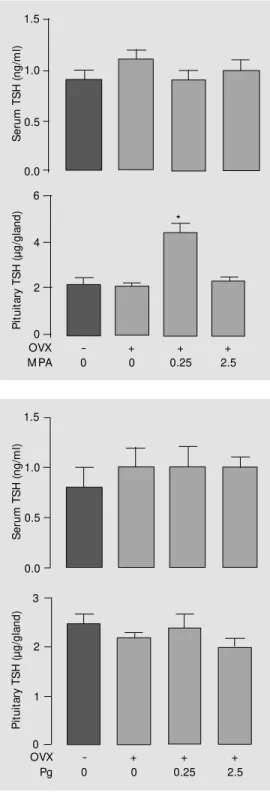
As shown in Figure 1, TSH content was increased approximately 2-fold in pituitaries of OVX rats treated sc with 0.25 mg MPA 100 g body weight-1
day-1
for 9 days, com-pared to both OVX control and normal sham-operated rats. However, the OVX group treated with a 10 times higher dose of MPA (2.5 mg/100 g body weight) showed a TSH content similar to that of the normal and
S
e
ru
m
T
S
H
(
n
g
/m
l)
1.5
1.0
0.5
0.0
-0
+ 0
+ 0.25
+ 2.5 OVX
Pg
P
it
u
it
a
ry
T
S
H
(
µ
g
/g
la
n
d
)
3
2
1
0
Figure 2 - Serum and pituitary TSH of young adult (3 months old) sham-operated or ovariecto-mized (OVX) rats treated w ith progesterone (Pg, 0.25 or 2.5 mg/100 g body w eight) or sa-line, sc, for 9 days before sacri-f ice. Values are report ed as mean ± SEM ; number of ani-mals per group: 6-11.
OVX groups. MPA did not modify serum TSH at either dose used (Figure 1). Proges-terone treatment, regardless of the dose em-ployed, did not change pituitary or serum TSH (Figure 2). Serum Pg concentration of the OVX group treated with 0.25 mg Pg was
Figure 1 - Serum and pituitary TSH of young adult (3 months old) sham-operated or ovariecto-mized (OVX) rats treated w ith medroxyprogesterone acetate (M PA, 0.25 or 2.5 mg/100 g body w eight) or saline, sc, for 9 days before sacrifice. Values are report ed as m ean ± SEM . * P<0.05 compared to all other groups (ANOVA f ollow ed by New man-Keuls test). Number of animals per group: 10-16.
S
e
ru
m
T
S
H
(
n
g
/m
l)
1.5
1.0
0.5
0.0
P
it
u
it
a
ry
T
S
H
(
µ
g
/g
la
n
d
)
6
4
2
0 -0
+ 0
+ 0.25
+ 2.5 OVX
M PA
Figure 3 - In vitro basal (panel A) and TRH-stimulated (panel B) TSH release from hemipituitaries of young adult (3 months old) sham-operated or ovariectomized (OVX) rats treated w ith medroxyprogesterone acetate (M PA, 0.25 mg/100 g body w eight, sc, for 9 days) or saline. After incubation, pituitary TSH content w as measured (panel C). Number of hemipituitaries per group: 6-8; * P<0.05 vs normal and OVX; * * P<0.05 vs
normal and OVX + M PA (ANOVA follow ed by New -man-Keuls test). DTSH values w ere calculated as the difference betw een medium TSH and basal TSH after TRH.
Figure 4 - In vitro basal (panel A) and TRH-stimulated (panel B) TSH release from hemipituitaries of young adult (3 months old) sham-operated or ovariectomized (OVX) rats treated w ith progesterone (Pg, 0.25 mg/100 g body w eight, sc, for 9 days) or saline. Number of hemipituitaries per group: 9-13. DTSH values w ere cal-culated as the difference betw een medium TSH and basal TSH after TRH.
B a s a l T S H ( n g /m l) 75 50 25 0 D T S H a ft e r T R H ( n g /m l) 75 50 25 0 T S H ( µ g /h e m ip it u it a ry ) 1.00 0.75 0.50 0.00 0.25 1234567890 1234567890 1234567890 1234567890 1234567890 1234567890 1234567890 1234567890 1234567890 1234567890 1234567890 1234567890 1234567890 1234567890 1234567890 1234567890 1234567890 1234567890 1234567890 1234567890 1234567890 1234567890 1234567890 1234567890 123456789 123456789 123456789 123456789 123456789 123456789 123456789 123456789 123456789 123456789 123456789 123456789 A * -0 + 0 + 0.25 OVX M PA B -0 + 0 + 0.25 OVX M PA 123456789 123456789 123456789 123456789 123456789 123456789 123456789 123456789 123456789 123456789 123456789 123456789 123456789 123456789 123456789 123456789 123456789 1234567890 1234567890 1234567890 1234567890 1234567890 1234567890 1234567890 * * 1234567890 1234567890 1234567890 1234567890 1234567890 1234567890 1234567890 1234567890 1234567890 1234567890 1234567890 1234567890 1234567890 1234567890 1234567890 1234567890 123456789 123456789 123456789 123456789 123456789 123456789 123456789 123456789 123456789 123456789 123456789 123456789 123456789 123456789 123456789 123456789 123456789 123456789 123456789 123456789 123456789 123456789 C -0 + 0 + 0.25 OVX M PA
similar to that of normal rats at different phases of the estrous cycle, while serum levels were significantly higher than normal when 2.5 mg Pg was used (proestrus: 10.9 ± 1.2, estrus: 9.6 ± 1.0, diestrus I: 12.0 ± 1.1, diestrus II: 14.3 ± 2.6, OVX + 0.25 mg Pg: 7.9 ± 2.1, OVX + 2.5 mg Pg: 50.1 ± 14.2 ng/ ml).
Experiment II: Effect of MPA and Pg treatment of young adult OVX rats on the
basal and TRH-stimulated TSH release in vitro
The data from the in vitro study with MPA and Pg treatment of young adult rats
are summarized in Figures 3 and 4, respec-tively. In young adult rats, OVX alone did not change significantly the basal TSH re-lease from isolated hemipituitaries. How-ever, the OVX group receiving MPA (0.25 mg/100 g body weight for 9 days) showed an increase (P<0.05) in basal TSH release com-pared to normal and OVX saline-treated groups (Figure 3, panel A). The TRH-stimu-lated TSH release from the glands of OVX saline-injected rats was lower (P<0.05) than that of the normal group (Figure 3, panel B). However, the treatment of OVX rats with MPA induced an increment in the TRH re-sponse of the OVX group to levels compa-rable to those of the normal group. The TSH content of the incubated glands did not differ significantly among groups (Figure 3, panel C). Treatment of OVX rats with Pg had no significant effect on basal or TRH-stimulat-ed TSH secretion in vitro (Figure 4). How-ever, as observed in experiment I, treatment with MPA or Pg did not significantly change serum TSH (data not shown).
Exp erim ent III: Effe ct o f MPA tre atme nt o f o ld O VX rats o n basal and TRH-stimulate d TSH
re le ase in vitro
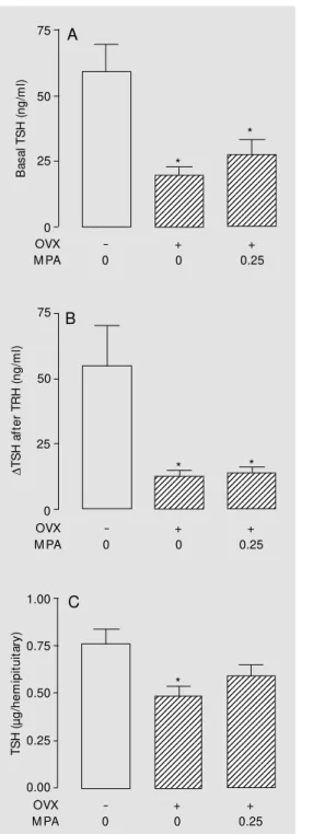
These results are presented in Figure 5. In old rats, basal and TRH-stimulated TSH release from glands of saline- or MPA-treated OVX groups were significantly (P<0.05) re-duced compared to the sham-operated old group. Pituitary TSH content (P<0.05) of the saline-treated OVX old rats was also signif-icantly lower than that of sham-operated old rats. Serum TSH did not differ significantly among the old groups (sham = 0.49 ± 0.08, OVX = 0.75 ± 0.07, OVX + MPA = 0.76 ± 0.1 ng/ml).
D iscussio n
In this study we demonstrate that MPA treatment increased the pituitary TSH con-tent of adult OVX rats treated sc for 9 days.
Figure 5 - In vitro basal (panel A) and TRH-stimulated (panel B) TSH release from hemipituitaries of 23-month-old sham-operated or ovariectomized (OVX) rats treated w ith medroxyprogesterone acetate (M PA, 0.25 mg/ 100 g body w eight, sc, for 9 days) or saline. After incubation, pituitary TSH content w as measured (panel C). Number of hemipituitaries per group: 8-9; * P<0.05
vs normal (ANOVA follow ed by New man-Keuls test).
DTSH values w ere calculated as the difference be-tw een medium TSH and basal TSH after TRH.
123456789 123456789 123456789 123456789 123456789 123456789 123456789 123456789 123456789 123456789 123456789 123456789 123456789 123456789 123456789 123456789 123456789 123456789
1234567890 1234567890 1234567890 1234567890 1234567890 1234567890 1234567890 1234567890 1234567890 1234567890 1234567890 1234567890 1234567890 1234567890 1234567890 1234567890 1234567890 1234567890 1234567890 1234567890 1234567890 1234567890
T
S
H
(
µ
g
/h
e
m
ip
it
u
it
a
ry
)
1.00
0.75
0.50
0.00
-0
+ 0
+ 0.25 OVX
M PA 0.25
C
*
1234567890 1234567890 1234567890 1234567890 1234567890 1234567890 1234567890
1234567890 1234567890 1234567890 1234567890 1234567890 1234567890 1234567890
D
T
S
H
a
ft
e
r
T
R
H
(
n
g
/m
l)
75
50
25
0
-0
+ 0
+ 0.25 OVX
M PA
B
* *
B
a
s
a
l
T
S
H
(
n
g
/m
l)
75
50
25
0
1234567890 1234567890 1234567890 1234567890 1234567890 1234567890 1234567890 1234567890 1234567890 1234567890
1234567890 1234567890 1234567890 1234567890 1234567890 1234567890 1234567890 1234567890 1234567890 1234567890 1234567890 1234567890 1234567890 1234567890
-0
+ 0
+ 0.25 OVX
M PA
A
*
This stimulatory effect depended on the dose used, being observed only at the lower dose (0.25 mg/100 g body weight). The in vitro assay also showed a stimulatory effect of MPA treatment on basal and TRH-induced TSH release from the pituitaries of adult OVX rats. We (14) and others (3,15,16) had shown that the decreased TSH response to TRH of OVX rats was restored by estrogen treatment. We also had reported before (5) a stimulatory effect of testosterone given chronically to castrated male rats on in vitro basal and TRH-stimulated TSH release. Here, we found that a synthetic progestin pro-moted the same effect.
However, the mechanism by which MPA exerts its effect on TSH is uncertain. It is possible that the effect of MPA was not through the activation of Pg receptors since there was no effect of the same treatment with Pg on TSH content or on basal and TRH-stimulated TSH secretion in vitro (Fig-ures 2 and 4). Although early reports (3) did not show any effect of Pg on TSH release in response to TRH injected into OVX rats, Tsai et al. (4) recently showed that a 3-day treatment of rats with Pg led to an increased TSH and prolactin response to TRH in vitro. Additionally, these investigators demon-strated a stimulatory effect of Pg added to the incubation medium of isolated pituitaries from OVX rats, which was associated with a parallel increase in cAMP production. These results suggest that Pg might act directly on the pituitary gland, modulating TRH actions on TSH and prolactin secretion. However, in the present study, Pg did not change pituitary TSH content or in vitro TSH secretion. The disagreement on the results might be related to the use of different protocols, especially the duration of treatment.
However, there are other points to be considered. The half-lives of MPA and Pg are different. MPA injected into muscle has a long-acting activity because it stays in the circulation for months, due to the slow re-lease from the injection site (17). However,
in the present study, we used subcutaneous administration which probably favored rapid absorption since the clearance of this aque-ous solution should be rapid (17). Neverthe-less, the elimination half-life of Pg is very short (5 min) compared to that of MPA (24 h) (18), and therefore it is possible that the concentration of Pg achieved in the circula-tion during the experiment, although much higher than the physiological values in rats receiving the higher dose, was not sufficient to activate Pg receptors at the same intensity as observed with the lower dose of MPA. In any case, the effects observed seem not to be dose dependent, since they were not ob-served with the higher dose of MPA.
Another important consideration con-cerns the status of Pg receptors in OVX rats. Pg receptors exert a dual control of estrogen and Pg, which act together to regulate the cellular concentration of Pg receptors. Es-trogen increases the Pg receptor mRNA lev-els and protein synthesis (19) in most tis-sues, although a recent report (20) showed a depressive effect of estrogen on Pg receptor content in rat uterine epithelial cells. On the other hand, Pg induces a decrease in the Pg receptor protein concentration and mRNA levels (21) and consequently reduces the effects of the hormone after prolonged treat-ment, although this is not true for all tissues, with the reverse effect also having been re-ported (22). In the pituitary gland, the regu-lation of Pg receptors seems to show simi-larities to as well as differences from that of the uterus. Recently (23), it was demon-strated that ovariectomy reduced mRNA lev-els for both isoforms, A and B, of the Pg receptor in the pituitary gland, and that estro-gen treatment increased the levels of these mRNAs, but Pg had no effect. Therefore, in our experiments, Pg receptors may have been reduced at the thyrotrope level in rats receiv-ing MPA or Pg, although this hypothesis needs experimental confirmation.
ac-tivity (24,25) and in addition it had been demonstrated that MPA interacts with cyto-sol androgen receptors in the rat hypothala-mus and anterior pituitary (26). The present report, together with our previous observa-tion that testosterone can also stimulate ba-sal and TRH-induced TSH secretion (5), raises the possibility that the stimulatory effect of MPA on TSH might be mediated trough androgen receptors. In any case, the mechanism underlying the effects of MPA on TSH secretion needs to be further inves-tigated.
Since serum TSH was not changed by MPA treatment we suggest that in vivo coun-terregulatory mechanisms maintained the ba-sal TSH secretion in the normal range. How-ever, the decreased TSH storage might im-pair the compensatory rise of TSH neces-sary, for example, to correct primary hypo-thyroidism.
However, contrary to the effect observed in young adult rats, the same MPA dose had no influence on the in vitro basal or TRH-stimulated TSH release from pituitaries of old OVX rats or on their pituitary TSH con-tent. These results suggest that aging is asso-ciated with alterations in the pituitary re-sponsiveness to the steroid. There have been reports of decreased estrogen receptors in the brain and pituitary of aged female
ro-dents (27,28). The significant decrease in the in vitro TSH release and intrapituitary TSH content caused by ovariectomy alone in old rats suggests that the ovary plays an important role in the regulation of the TSH response to TRH, even in old rats that lost their estrous cyclicity (29,30). This leads us to speculate that ovarian factors other than estrogen and Pg might be important, directly or indirectly, in the maintenance of TSH stores and TSH response to TRH in old rats. It is known that stroma cells continue to produce androgens after the end of folliculo-genesis and testosterone has been reported to have stimulatory effects on TSH secretion (3,30). Chen and Walfish (31) showed a lower TSH response to TRH administered to old OVX rats compared to the adult OVX group, although the TRH response was not affected in intact old rats.
In summary, chronic treatment of OVX rats with MPA, but not Pg, increased TSH storage and the response to TRH in vitro, an effect that was not observed in aged OVX rats.
Ackno wle dgm e nts
We are grateful to Ms. Norma de Araújo Faria, Mr. Advaldo Bezerra and Mr. Wagner Bezerra for excellent technical assistance.
Re fe re nce s
1. Chen HJ & Walfish PG (1978). Effects of estradiol benzoate on thyroid-pituitary function in female rats. Endocrinology, 103: 1023-1030.
2. Kimura N, Arai K, Sahara Y, Suzuki H & Kimura N (1994). Estradiol transcription-ally and posttranscriptiontranscription-ally up-regulates thyrotropin-releasing hormone receptor messenger ribonucleic acid in the rat pitu-itary cells. Endocrinology,134: 432-440. 3. Watanobe H & Takebe K (1987). Role of
postnatal gonadal function in the determi-nation of thyrotropin (TSH) releasing hor-m one-induced TSH response in adult male and female rats. Endocrinology, 120:
1711-1718.
4. Tsai Sc, Lu Cc, Lau Cp, Hw ang Gs, Lee Hy, Chen Sl, Huang Sw , Shih Hc, Chen YH, Chiao YC, Wang SW & Wang PS (1996). Progesterone stimulates in vitro
release of prolactin and thyrotropin involv-ing cAM P production in rat pituitary. Chi-nese Journal of Physiology, 39: 245-251. 5. Borges PP, Curty FH, Pazos-M oura CC & M oura EG (1998). Effect of testosterone propionate treatment on thyrotropin se-cretion of young and old rats in vitro. Life Sciences, 62: 2035-2043.
6. Brabant G, Ocran K, Ranft U, von zur M uhlen A & Hesch RD (1989).
Physiologi-cal regulation of thyrotropin. Biochimie, 71: 293-301.
7. Lee PA (1981). M edroxyprogesterone therapy for sexual precocity in girls. A-merican Journal of Diseases of Children, 135: 443-445.
8. Wheeler M D & Styne DM (1991). Drug treatment in precocious puberty. Drug, 41: 717-728.
10. Pridjian G, Schmit V & Schreiber J (1987). M edroxyprogesterone acetate: receptor binding and correlated effects on steroido-genesis in rat granulosa cells. Journal of Steroid Biochemistry, 26: 313-319. 11. Poulin R, Baker D, Poirier D & Labrie F
(1989). Androgen and glucocorticoid re-ceptor-mediated inhibition of cell prolif-eration by medroxyprogesterone acetate in ZR-75-1 human breast cancer cells.
Breast Cancer Research and Treatment, 13: 161-172.
12. M athew s JH, Abrams CA & M onshim A (1970). Pituitary-adrenal function in the patients receiving medroxyprogesterone acetate for true precocious puberty. Jour-nal of Clinical Endocrinology and M etabo-lism, 30: 653-658.
13. Sadeghi-Nejad A, Kaplan SL & Grumbach M M (1971). The effect of medroxypro-gesterone acetate on adrenocortical func-tion in children w ith precocious puberty.
Journal of Pediatrics, 78: 616-624. 14. M oreira RM , Lisboa PC, Curty FH &
Pazos-M oura CC (1997). Dose-dependent effects of 17-ß estradiol on pituitary thy-rotropin content and secretion in vitro.
Brazilian Journal of M edical and Biological Research, 30: 1129-1134.
15. Fisher JS & D’Angelo SA (1978). Stimula-tory and inhibiStimula-tory action of estradiol on TSH secretion. Endocrinology, 88: 687-691.
16. Castro-Vazquez A, Ojeda SR, Krulich L & M cCann SM (1981). TSH release during the estrous cycle of the rat: Variations in responsiveness to cervicovaginal stimula-tion. Neuroendocrinology, 33: 193-198. 17. Cornette JC, Kirton KT & Duncan GW
(1971). M easurement of medroxyproges-terone acetate (Provera) by radioimmu-noassay. Journal of Clinical Endocrinology, 33: 459-466.
18. Williams CL & Stancel GM (1996). Estro-gens and progestins. In: Goodman and Gilman’s The Pharmacological Basis of Therapeutics. 9th edn. M cGraw -Hill, New York.
19. Nardulli AM , Greene GL, O’M alley BW & Katzenellenbogen BS (1988). Regulation of progesterone receptor messenger ri-bonucleic acid and protein levels in M CF-7 cells by estradiol: analysis of estrogen’s effect on progesterone receptor synthe-sis and degradation. Endocrinology, 122: 935-944.
20. Parczyk K, M adjno R, M ichna H, Nishino Y & Schneider M R (1997). Progesterone re-ceptor repression by estrogens in rat uter-ine epithelial cells. Journal of Steroid Bio-chemistry and M olecular Biology, 63: 304-316.
21. Nardulli AM & Kat zenellenbogen BS (1988). Progesterone receptor regulation in T 47D human breast cancer cells: analy-sis by density labeling of progesterone receptor synthesis and degradation and their modulation by progestin. Endocri-nology, 122: 1532-1540.
22. M artel D, M oner M N, Roche D, De Feo VJ & Psychoyos A (1989). Hormonal de-pendence of the metrial gland: further studies on oestradiol and progesterone receptor levels in the rat. Journal of Endo-crinology, 120: 465-472.
23. Szabo M , Kilen SM , Nho SJ & Schw artz NB (2000). Progesterone receptor A and B messenger ribonucleic acid levels in the anterior pituitary of rats are regulated by estrogen. Biology of Reproduction, 62: 95-102.
24. Couture P, Thériault C, Simard J & Labrie F (1993). Androgen receptor-mediated stimulation of 17ß-hydroxysteroid dehy-drogenase activity by dihydrotestosterone and medroxyprogesterone acetate in
ZR-75-1 human breast cancer cells. Endocri-nology, 132: 179-185.
25. Bentel JM , Birrell SN, Pickering M A, Holds DJ, Horsfall DJ & Tilley WD (1999). Androgen receptor agonist activity of the synthetic progestin, medroxyprogester-one acetate, in human breast cancer cells.
M olecular and Cellular Endocrinology, 154: 11-20.
26. Perez-Palacios G, Chavez B, Vilchis F, Escobar N, Larrea F & Perez AE (1983). Interaction of medroxyprogesterone ace-tate w ith cytosol androgen receptors in the rat hypothalamus and pituitary. Jour-nal of Steroid Biochemistry, 19: 1729-1735.
27. Haji M , Kato KI, Naw ata H & Ibayashi H (1981). Age-related changes in the con-centrations of cytosol receptors for sex steroid hormones in the hypothalamus and pituitary gland of the rat. Brain Re-search, 204: 373-386.
28. Steiger RN, Huang HH, Chamberlain DS & M utes J (1980). Changes in control of gonadotropin secretion in the transition period betw een regular cycles and con-stant estrus in ageing female rats. Biol-ogy of Reproduction, 22: 595-603. 29. Nass TE, La Polt PS, Judd HL & Lu JHK
(1984). Alterations in ovarian steroid and gonadotropin secretion preceding the ces-sation of regular oestrus cycles in ageing female rats. Journal of Endocrinology, 100: 43-50.
30. Christianson D, Roti E, Vagenakis AG & Braverman LE (1981). The sex related dif-ference in serum thyrotropin concentra-tion is androgen mediated. Endocrinology, 108: 529-535.