*e-mail: [email protected]
Effect of Solvents on the Morphological Characterization of Enteric Nanoparticles
Danay Rosa Dupeyrón Martella, Jacques Rieumont Brionesb*,
Mayra González Hurtadoc, Alicia del Real Lópezd, Víctor Manuel Castaño Menesesd
a
Instituto de Ciencia y Tecnología de Materiales, Universidad de la Habana, Cuba
b
Departamento de Química-Física, Facultad de Química, Universidad de la Habana,
Ciudad de la Habana, Cuba
c
Centro de Ingeniería e Investigaciones Químicas,
Ciudad de la Habana, Cuba
d
Centro de Física Aplicada y Tecnología de Avanzada,
Universidad Nacional Autónoma de México
A.P. 1-1010 Santiago de Querétaro, Querétaro 7600 México
Received: March 18, 2009; Revised: September 24, 2009
Size and external and internal morphologies of nanoparticles and microparticles are very important on the design of drug devices for controlled release. Random enteric copolymers such as poly (methacrylic acid-co-ethyl acrylate) and poly (methacrylic acid-co-macid-co-ethyl methacrylate) were used to produce nanoparticles, which contain a model drug and could be employed as drug carriers for proteins. The solvent effect on re-dispersion of such nanoparticles was studied by Scanning Electron Microscopy (SEM) and revealed not only differences in size, but also several shapes, depending on the chemical nature of the polymer matrix and the non-solvent used. Acrylate containing copolymers in acidic aqueous dispersions lead to spheroidal particles. However for the copolymer containing methyl methacrylate, spheroidal particles collapsed in a “grenade” type morphology and besides some cubic structures are also formed.
Dynamic Light Scattering (DLS) studies of the re-dispersed nanoparticles showed the strong tendency to form agglomerates not only in acidic water but also in hexane and the presence of bimodal size distributions.
Keywords: nanoparticles morphology, enteric copolymers, solvent effect, agglomerates
1. Introduction
Drug delivery is one of the most actives areas in today’s nanotechnology and biotechnology. Indeed, from the pharmacologi-cal standpoint, an increasing number of proteinic drugs present some sort of drawbacks, in particular for oral formulations, due to their instability in the gastro intestinal tract1-4. In this regard, many attempts to face this challenge have been reported recently4,5 and one of them is by using an enteric copolymer6. The very concept of preparing small particles as carriers of core material trapped within a polymeric material dates back, at least, to the 1930s7. Ever since, a number of methods for preparing carrier microparticles have been developed, some based exclusively on physical or chemical phenomena, whereas others combine both physical and chemical phenomena. Recently, the use of nanoparticles has become increasingly important because their size and their surface properties offer many advantages, such as improved solubility, targetability and adhesion to tissue8-11. Control-ling the size is also important because only certain particle size can be used for a specific use and release characteristics greatly depend on size and morphology of the particles involved.
There are numerous factors that can influence the morphology obtained with a specific drug/polymer combination, among them: the nature of the drug, the nature of the polymer, the amount of drug in the nanoparticles and the nature and amount of the emulsifier used in the aqueous phase. Amorphous polymers generally give particles with a smooth surface whereas semi-crystalline polymers did not12. The encapsulation process can also play its role in determining the external and internal morphology of particles. A process of double emulsion (water-oil-water) can induce multivesicular particles as
has been reported from transmission electron microscopy studies13,14 and also the stability of the emulsion can induce morphological changes15,16.
The present work deals with the morphological characterization of nanoparticles obtained by using three different matrixes, two types of enteric copolymer and a blend of one of them with poly(ethylene glycol) (PEG). The enteric matrix was selected, according to a pre-vious work6 using a model drug such as the bovine serum albumin (BSA) in order to protect protein from the lower pH of the stomach and enzymes, whereas the blend with PEG was used to enhance further the protection of the protein encapsulated6,17.
The main purpose of the present paper was to analyze by SEM the particle size and morphology changes induced by solvents on re-dispersion of nanoparticles and their diameter distribution by dynamic light scattering.
2. Experimental
2.1. Materials and procedures
• Bovineserumalbumin(BSA)(MW66kD)wassuppliedby Sigma Chemical;
• Kollicoat® MAE 100 P: Anionic copolymer based on meth-acrylic acid and ethyl acrylate (1:1), Tg: 110 °C from BASF; • Eudragit® L-100: Anionic copolymer of methacrylic acid and methylmethacrylate(1:1),MW:135.000,Tg>150°C,sup -plied by Röhm Pharma Polymers;
2.3. Characterization techniques
i) Scanning Electron Microscopy (SEM)
Samples obtained by the encapsulation procedure were ultra-sonically re-dispersed for 10 minutes and dried and coated by sputtering with a layer of gold, of approximately 20 nm. An EMS 550 Sputter coater was used. Samples were observed in a Scanning Electron Microscope (JEOL-JSM -6060LV). One hundred particles were random taken for each sample for evaluating the average diameter.
ii) Dynamic Light-Scattering (DLS).
Prior to use, all the solvents used were filtrated with 0.2 µm filters to eliminate dust and the sample holder was cleaned with distilled water followed by acetone, to prevent contamination. For each sample, 2 mg of particles were suspended in 20 mL of solvent and filtrated. Filters of 2 µm Sartorious AG PTFE and Advantec MFS Nylon were used for organic and acid viscosity 375-480 mPas, n20
D 1,472;
• Methanol, Hexane,and Xylene (reagent grade), supplied by MERCK;
• 0,2µmFilters(SartoriousAGPETFandAdvantecMFSNy -lon); and
• UltraTurraxhomogenizer(ModelT-25IKA).
2.2. Encapsulation procedure
An aqueous solution (2 mL), containing BSA (20 mg) and PEG (40 mg), was added to a copolymer solution in methanol (20 mL, 1%) and the mixture was homogenized at 9500 rpm/min, by using anUltraTurraxhomogenizer(ModelT-25IKA).Then,thedisper -sion obtained in this first step, was emulsified over a buffer solution (120 mL, pH = 2) containing Tween 80 at 9500 rpm/min. More details are described in a previous work6. Table 1 contains a description of all the samples prepared and the encapsulation parameters.
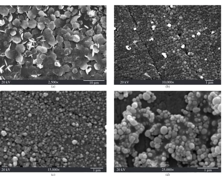
Figure 1. Scanning electron micrographs at different magnifications of sample B6: a) 2500× b) 10000× c) 15000× and d) 25000×. Table 1. Experimental conditions for BSA encapsulation with the different matrixes.
Sample Polymer Matrix Surfactant amount (mg) Stirring rate (rpm/min)
B6 Kollicoat® MAE 100 P 20 9500
B2- PEG Kollicoat® MAE 100 P + PEG 20 9500
Nanoparticles behaviour with solvents will be discussed sepa-rately in the following items:
3.1. Copolymer: poly (methacrylic acid-co-ethyl acrylate)
In the case of sample B6 when re-dispersed in pure water, where particles can swell and even dissolve, a leaf-like morphology was ob-served (Figure 1a). However, when the same sample is re-suspended in an acidic aqueous medium where the copolymer is insoluble, the particles show spheroidal shapes (Figures 1b, 1c and 1d); surfaces are rough and agglomeration is present to some extent.
One feature of these enteric random copolymers is that contain polymethacrylic acid in their structure and the common drying proc-esses do not exclude completely the water, due to its hydrophilic nature. Thus, it is unavoidable that traces of water remain inside the particle or near its surface and seem to act as plasticizers, contribut-ing to agglomeration.
3.2. Blend of the copolymer poly (methacrylic acid-co-ethyl
acrylate) and poly (ethylene glycol)
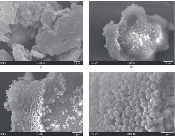
For sample B2-PEG re- dispersed in xylene, a better solvent for the acrylate units than for the methacrylic acid ones, a layered com-pact structure is shown (Figure 2a). However, re-suspension of this sample in hexane, a non-solvent for this copolymer, results in perfect spheres with smooth surfaces (Figures 2c and 2d). This interesting aqueous solutions respectively. The samples were maintained
in an ultrasonic bath for 10 minutes. The scattering cells (10 mL cylindrical vials) were immersed in a large-diameter thermostated bath of index-matching liquid (transdecalin). DLS measurements were performed in a Dynamic Light Scattering Brookhaven Instrument machine with a Model BI900 correla-tor. The results were analyzed by using the Non Negative Least Square (NNLS) and Contin methods18,19,20.
3. Results and Discussion
For comparison purposes all samples were obtained using identi-cal procedures and amounts as is shown in Table 1. The control of particle size and shape depends mainly on parameters such as the amount of surfactant, the stirring rate and the chemical nature of the polymer matrix. Thus, surfactant and stirring rate were the same for all the experiments.
All the samples were obtained as described in the Experimental Part. By this procedure, the enteric copolymer closes their conforma-tions to a more compact one and the protein is trapped by the polymer coating. Nanoparticles obtained were redispersed in some solvents such water at pH = 2 and hexane where the enteric copolymers are insoluble, and xylene that is a selective solvent for the hydrophobic units.
result may be due to the extremely high insolubility of the copolymer in hexane, which leads to a more compact material. PEG is a semi-crystalline polymer and the poly (methacrylic acid-co-ethyl acrylate) is amorphous. Amorphous polymers, as was said in the Introduction, give as a rule particles with smooth surfaces. Thus, possibly PEG is forming the core of particles.
Nevertheless, agglomeration is not ruled out completely as to be expected in hexane and some clusters, such as those shown in Figure 2b, are present. This material is a blend and differences on the rate of shrinkage of the two components could be producing such result.
3.3. Copolymer: poly (methacrylic acid-co-methyl
methacrylate)
Sample B1 L100 shows interesting unique features. The suspen-sion in acidic solution shows two types of morphologies (Figures 3a and 3d), namely spheroidal and cubic. These morphologies are produced simultaneously possibly due to the fact that the amount of surfactant for this copolymer or its hydrophilic-lypophilic bal-ance, is not enough to stabilize all the spheres that can be formed. Furthermore, this copolymer possesses a higher glass temperature since it contains methyl methacrylate in its composition. Thus, this copolymer is stiffer in comparison to the other samples prepared with
an ethyl acrylate-containing copolymer. It is well known that acrylates have lower glass temperatures than their methacrylates homologues and both are amorphous21,22. Those factors could make possible the formation of non-spheroidal structures such as the cubes observed, that can also agglomerate, forming separated groups (Figure 3c). Furthermore, a higher magnification micrograph of the spheroidal entities shows that they in fact are not “true” spheres, but the result of the compactation of smaller spheres, that produce a morphology resembling a “grenade” (see Figure 3b).
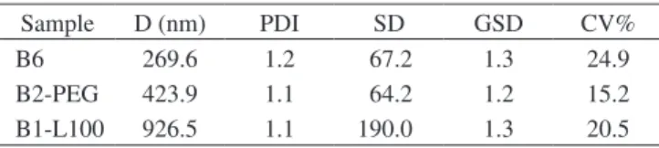
Those morphological changes are accompanied by changes in the particle diameters. Table 2 indicates that, under the same experimental nanoprecipitation conditions (amount of surfactant, stirring rates, acidic water); the composition of the enteric matrix practically determines not only the morphology but also the size of the particles. As a matter of fact the poly methacrylic acid-co-methyl methacrylate gave particles with greater diameters and more complex morphologies.
3.4. Size distribution
Table 2. Results obtained by SEM (D: average diameter; PDI: polydisper-sion index; SD: standard deviation; GSD: geometric standard deviation; CV: variance coefficient).
Sample D (nm) PDI SD GSD CV%
B6 269.6 1.2 67.2 1.3 24.9
B2-PEG 423.9 1.1 64.2 1.2 15.2
B1-L100 926.5 1.1 190.0 1.3 20.5
Table 3. Results obtained by Dynamic Light Scattering.
Sample NNLS
Diameter (µm)
Deviation Contin
Diameter (µm)
Deviation
B6 2.9 0.01 2.8 0.37
B2-PEG 6.9 0.04 5.2 0.44
B1-L100 1.9 0.026 2.2 0.40
process nanoparticles are formed separately and in a second step become agglomerated.
4. Conclusions
SEM micrographs revealed how the specific polymeric matrix influences the morphology and size of nanoparticles obtained under the very same experimental conditions when are re-dispersed in dif-ferents solvents. In the particular case of the copolymer with methyl methacrylate that posseses the highest Tg value, two different shapes were observed, one spherical that resembles a grenade and the other one cubic. These morphologies are simultaneously produced under the same experiment. The dual character of these enteric materials (hydrophilic-hydrophobic) and their difference in physical properties seem to rule the complex behaviour observed.
It was also demonstrated by DLS the strong tendency of these copolymer nanoparticles to agglomerate and increase agglomerates sizes with re-dispersion, leading even to bimodal diameter distribu-tions, more significantly when PEG is used in the matrix.
Acknowledgements
The authors are grateful to the Red de Macrouniversidades for inancialsupport.WealsothankDra.SusanaVargasandDr.Rogelio Rodríguez for their kind help with the DLS experiments and theory. The copolymers were a courtesy of Röhm Pharma Polymer.
References
1. ShenWC.Oralpeptideandproteindelivery:unfulilledpromises?Drug Discovery Today. 2003; 8(14):607-608.
2. NakamuraK,MurrayR,JosephJ,PeppasN,MorishitaMandLowmanA.
Oral insulin delivery using P(MMA-g-EG) hydrogels effects on network morphology on insulin delivery characteristics. Journal of Controlled Release. 2004; 95(3):589-599.
3. Caliceti P and Veronese FM. Phamacokinetic and biodistribution properties on poly (ethylene glycol) protein conjugates. Advanced Drug Delivery Reviews. 2003; 55(10):1261-1277.
4. Whelan J. Beyond pegylation. Drug Discovery Today. 2005; 10(5):301-306.
5. Carino G, Jacob J and Mathiowitz E. Nanosphere based oral insulin delivery. Journal of Controlled Release. 2000; 65(1-2):261-269.
6. Dupeyrón D, González M, Sáez V, Ramón J and Rieumont J. Nano-encapsulation of protein using an enteric polymer as carrier. IEE Proceedings Nanobiotechnology. 2005; 152(5):165-168.
7. Benitas S. Microencapsulation: methods and industrial applications. New York: Marcel Dekker Inc.; 1996
8. Donini C, Robinson DN, Colombo P, Giordano F and Peppas NA. Preparation of poly (methacrylic acid-g-poly (ethylene glycol) nanospheres from methacrylic monomers for pharmaceutical applications. International Journal of Pharmaceutics. 2002; 245(1-2):83-91.
9. KawashimaY. Preface nanoparticulate systems for improved drug
delivery. Advanced Drug Delivery Reviews. 2001; 47(1):1-2.
10. Gonzalez M, Galano A, Rieumont J, Lopez T, Dupeyron D and Albaran L. Drug-Matrix interactions in nanostructured materials containing Acetyl
SalicylicAcidUsinganEntericPolymerasaCoating.Journal of Physical Chemistry C. 2008; 112(51):20222-20226.
11. Gonzalez M, Rieumont J, Dupeyron D, Perdomo I, Abdón L and Castaño VM. Nanoencapsulation of acetyl salicylic acid within enteric polymer nanopartilcles.Review on Advanced Matererial Science. 2008; 17:71-75.
12. Martin M, Miguens FC, Rieumont J and Sanchez R. Tailoring of the external and internal morphology of poly-3-hydroxy butyrate microparticles. Colloid Surface. 2000; B17(2):111-116.
Figure 4. Results of Dynamic light Scattering of Sample B6. (NNLS)
Figure 5. Results of Dynamic light Scattering of Sample B2 PEG. (NNLS)
Figure 6. Results of Dynamic light Scattering of Sample B1L100. (NNLS)
Figure 4, 5 and 6 show the presence of two well-separated size groups, one corresponding to smaller particles and the other one to agglomer-ated clusters that are in much greater amounts than the first one. For the sample B2-PEG re-dispersed in hexane a bimodal distribution is even more noticeable as shown in Figure 5.
17. Ferrero C, Bravo I and Jiménez MR. Drug release kinetics and fronts movement studies from methyl methacrylate (MMA) copolymer matrix tablets: effect of copolymer type and matrix porosity. Journal of Controlled Release. 2003; 92(1-2):69-82.
18. Giacomelli C, Le-Men L, Borsali R, Lai-Hee-Him J, Brisson A, Armes SP and Lewis AL. Phosphorylcholine-based pH-responsive diblock copolymer micelles as drug delivery vehicles: light scattering, electron microscopy, and fluorescence experiments. Biomacromolecules. 2006; 7(3):817-828.
19. Provencher SW. Contin: a general purpose constrained regularization
programm for inverting noisy linear algebraic and integral equations. Computer Physics Communications. 1982; 27:229-242.
20. StockRSandRayWH.Interpretationofphoton-correlationspectroscopy
data: a comparison of analysis methods. Journal of Polymer Science. Part B: Polymer Physics. 1985; 23(7):1393-1398.
21. Luskin L and Myers RJ. Acrylic Ester Polymers. In: Mark HF. (Ed.). Encyclopedia of Polymer Science and Technology. 1 ed. New York: John
Wiley&Sons;1964.p.246-328.(vol.1).
22. Brandrup I and Immergut EH. Polymer Handbook. New York: Wiley InterScience; 1989.
13. Eligio T, Rieumont J and Sanchez R. Characterization of chemically modified poly (3-hydroxyalcanoates) and their performance as matrix for hormone release. Die Angewandte Makromolekulare Chemie. 1999; 270:69:70.
14. Schugens CH, Laruelle N, Nihant R, Grandphils C, Jérôme R and Teyssie P. Effect of the emulsion stability on the morphology and porosity of semicrystalline poly-L-lactide microparticles prepared by w/o/w double emulsion-evaporation. Journal of Controlled Release. 1994; 32(2):161-176
15. Nihant N, Schugens CH, Grandphils C, Jérôme R and Teyssie P. Polylactide microparticles prepared by double emulsion/evaporation tecnique. 1. Effect of primary emulsion stability. Pharmaceutical Research. 1994; 11(10):1479-1484