Int. braz j urol. vol.29 número5
Texto
Imagem
Documentos relacionados
For the diagnosis, it is necessary to use imaging methods during the investigation, especially computed tomography and magnetic resonance imaging, both with emphasis on
Key words: cone beam computed tomography, keratocystic odontogenic tumor, magnetic resonance imaging, odontogenic keratocyst, oral diagnosis.. T he keratocystic odontogenic tumor
However, the appearance of this cyst on cone beam computed tomography (CBCT) and magnetic resonance imaging (MRI) have received relatively little attention.. CBCT and
In the present study, the authors describe several non-dysfunctional conditions affecting the temporomandibular joints through computed tomography and magnetic resonance
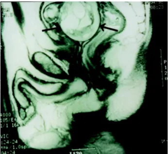
The present article presents information on the prostate gland anatomy, the tumor aspect at magnetic resonance imaging, specific signs of extracapsular extension and seminal
Amongst the available imaging methods (Doppler ultrasonography, computed tomography, magnetic resonance imaging and digital subtraction angiography), angiography is considered the
The diagnosis of mature cystic ter- atoma by computed tomography (CT) and magnetic resonance imaging (MRI) is fairly straightforward as such modalities are more fat sensitive.. At
With the development of imaging studies, the enterogra- phy, either by computed tomography or magnetic resonance, is replacing intestinal transit and enteroclysis procedures in
