Introduction
Pulmonary sequestration (PS) is a rare congenital malformation characterized by a mass of nonfunctioning lung tissue that does not communicate with the tracheobronchial tree and is vascularized by an anomalous systemic artery. It is composed of cystic embryonic tissue and contains disorganized non-aerated alveoli, as well as bronchi, cartilage, and respiratory epithelium.(1)
In 75% of cases of PS, the blood supply is derived from the thoracic or abdominal aorta, and venous drainage is via the systemic veins or via the pulmonary vein. Accidental transection of an anomalous systemic artery can cause massive hemorrhage with fatal consequences, and it is of
paramount importance that anomalous vessels be identified in the preoperative period.(2)
The objective of the present study was to report an intraoperative finding of PS following hemorrhage resulting from the transection of an anomalous pulmonary vessel during resection of a carcinoid tumor.
Case report
A 39-year-old female patient presented with a history of recurrent pneumonia, together with sporadic episodes of productive cough and fever. In the two preceding years, the patient had had four episodes of moderate volume
Carcinoid tumor and pulmonary sequestration*
Tumor carcinoide e sequestro pulmonar
Fernando Luiz Westphal, Luís Carlos de Lima, José Corrêa Lima Netto, Maria do Socorro Lucena Cardoso, Márcia dos Santos da Silva,
Danielle Cristine Westphal
Abstract
Pulmonary sequestration is defined as a mass of lung tissue separated from the tracheobronchial tree and irrigated by an anomalous systemic artery. It is rarely seen in conjunction with lung neoplasms. We report the case of a 39-year-old female patient diagnosed with a carcinoid tumor, located in the intermediate bronchus and accompanied by bronchiectasis in the right lower lobe. The patient underwent thoracotomy for the resection of the affected area. During surgery, she presented with significant hemorrhage resulting from the transection of the anomalous artery that irrigated an intralobar pulmonary sequestration, which was located in right lower lobe and had not been identified in pre-operative examinations.
Keywords: Bronchopulmonary sequestration; Hemorrhage; Carcinoid tumor.
Resumo
O sequestro pulmonar é definido como uma massa de tecido pulmonar separada da árvore traqueobrônquica e irrigada por uma artéria sistêmica anômala. Sua associação com neoplasias pulmonares é rara. Relatamos o caso de uma paciente de 39 anos com o diagnóstico de tumor carcinoide localizado no brônquio intermediário, associado a alterações caracterizadas como bronquiectasias em lobo inferior direito. A paciente foi submetida à toracotomia para ressecção da área acometida e, durante a cirurgia, apresentou hemorragia importante decorrente da transecção da artéria anômala que nutria o sequestro pulmonar intralobar localizado em lobo inferior direito, não identificado nos exames pré-operatórios.
Descritores: Sequestro broncopulmonar; Hemorragia; Tumor Carcinoide.
* Study carried out in the Department of Clinical Surgery, Federal University of Amazonas School of Medicine, Manaus, Brazil. Correspondence to: Fernando Luiz Westphal. Hospital Universitário Getúlio Vargas, Coordenação de Ensino e Pesquisa. Avenida Aripuanã, 4, Praça 14 de Janeiro, CEP 69020-170, Manaus, AM, Brasil.
Tel. 55 92 234-6334. E-mail: [email protected] Financial support: None.
malformation, PS, congenital lobar emphysema, bronchogenic cysts, and pulmonary arteriovenous malformations. The reported annual incidence of these malformations ranges from 30 to 42 cases per 100,000 population, PS accounting for 0.15-6.45% of all cases.(3,4)
Depending on its pleural covering, PS is classically divided into intralobar and extralobar. An intralobar PS (ILPS) share the pleural covering with the rest of the lung, whereas an extralobar PS (ELPS) is completely covered by its own visceral pleura.(5) Although the extralobar form is well defined as a congenital abnormality, the intralobar form has a controversial pathogenesis, with some evidence indicating that, in many cases, it is an acquired disease.(6,7)
Accounting for approximately 75% of all cases of PS, ILPS is more common in the lower lobes and on the left, involving the posterior basal segment.(5,8) Its arterial supply is nearly always derived from the aorta or from one of its branches, typically one of a large diameter. Venous drainage is via the pulmonary veins into the left atrium, creating a left-to-left shunt. In a minority of cases, drainage is via the inferior vena cava or via the azygos system.(9) In the case presented here, the PS was located in the right lower lobe and the blood supply came from an anomalous branch derived from the thoracic aorta.
Cases of ILPS typically occur in adolescents and young adults with a history of recurrent infections of the respiratory tract, hemoptysis, and dyspnea. Some ILPS patients develop cardiac symptoms, which are a consequence of the left-hemoptysis. In the preoperative investigation, a
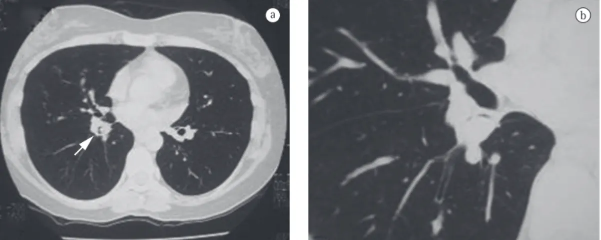
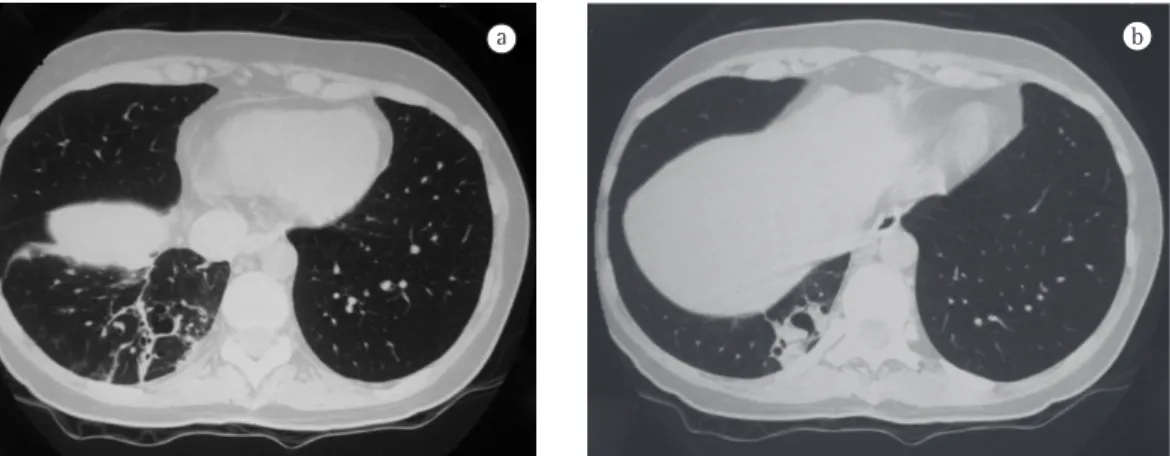
CT scan of the chest revealed a lesion in the intermediate bronchus (Figure 1), as well as cystic bronchiectasis in the right lower lobe (Figure 2). Fiberoptic bronchoscopy confirmed the tomographic finding of a wine-colored, exophytic endobronchial lesion at the root of the intermediate bronchus, and biopsy of the lesion revealed a typical carcinoid tumor.
Clinical reasoning based on those findings suggested bronchiectasis resulting from chronic bronchial obstruction. The patient underwent right posterolateral thoracotomy with bilobectomy (middle and lower lobes). During the intraoperative period, sectioning of the pulmonary ligament resulted in a hemorrhagic process originating from an anomalous arterial vessel that had irrigated the parenchyma of the PS (Figure 3). Hemostasis was achieved by ligation of the vessel. Histopathology results were consistent with the finding of PS. The postoperative course was satisfactory, without any complications, and the patient was discharged on postoperative day 5.
Discussion
Despite being defined as distinct entities, lung malformations constitute a spectrum of abnormalities, with quite similar clinical presentations, arising from flaws in the development of the primitive intestine and its differentiation into a respiratory system during the embryonic period.(2) Chief among the most common malformations are cystic adenomatoid
a b
cyanosis, and feeding difficulties are common. More rarely, recurrent episodes of respiratory tract infection or gastrointestinal symptoms can occur.(2,11) Cases of ELPS can be accompanied by pulmonary, cardiac, and vertebral malformations, as well as by malformations of the chest wall and gastrointestinal tract, being most commonly accompanied by diaphragmatic hernia.(12)
On chest X-rays, PS can be initially identified as a mass of homogeneous opacity. Diagnostic confirmation is made through chest CT, magnetic resonance imaging, or arteriography. Chest CT is more useful in detecting abnormalities in the lung parenchyma and only in some cases can it identify PS anomalous irrigation. Arteriography is the best test for diagnostic confirmation of PS, because it detects the anomalous artery irrigating the PS with precision. More recently, reconstruction with multichannel CT scanners to-left shunt and of the high flow diverted to
the anomalous vessel.(10) The recurrent episodes of infection cause fibrosis and visceral pleural thickening. The lung parenchyma shows areas of consolidation and fibrosis containing multiple cystic formations, which can be mistaken for bronchiectasis,(5) as in the case reported here.
In general, an ELPS is located in the posterior costodiaphragmatic recess, between the lower lobe and the left hemidiaphragm. More rarely, it can occur in the mediastinum or in the abdominal region. An ELPS is typically irrigated by an artery originating directly from the aorta, and, in 80% of cases, venous drainage is systemic, occurring via the azygos-hemiazygos system or via the superior vena cava, which creates a left-to-right shunt.(2,5) Unlike in cases of ILPS, clinical manifestations appear in the first six months of life in cases of ELPS. Neonatal asphyxia, dyspnea,
a b
Figure 2 - In A, CT scan of the chest revealing cystic areas permeated by fibrotic tissue in the right lower lobe. In B, cystic areas in the posterior segment of the right lower lobe.
a b
complex, and, if unsuspected, it might appear only as an intraoperative finding following potentially fatal complications.
References
1. Salmons S. Pulmonary sequestration. Neonatal Netw. 1995;14(6):69-73.
2. Felker RE, Tonkin IL. Imaging of pulmonary sequestration. AJR Am J Roentgenol. 1990;154(2):241-9.
3. Ferreira HP, Fischer GB, Felicetti JC, Camargo Jde J, Andrade CF. Surgical treatment of congenital lung malformations in pediatric patients. J Bras Pneumol. 2010;36(2):175-80.
4. Van Raemdonck D, De Boeck K, Devlieger H, Demedts M, Moerman P, Coosemans W, et al. Pulmonary sequestration: a comparison between pediatric and adult patients. Eur J Cardiothorac Surg. 2001;19(4):388-95. 5. Abbey P, Das CJ, Pangtey GS, Seith A, Dutta R, Kumar
A. Imaging in bronchopulmonary sequestration. J Med Imaging Radiat Oncol. 2009;53(1):22-31.
6. Clements BS, Warner JO. Pulmonary sequestration and related congenital bronchopulmonary-vascular malformations: nomenclature and classification based on anatomical and embryological considerations. Thorax. 1987;42(6):401-8.
7. Stocker JT, Malczak HT. A study of pulmonary ligament arteries. Relationship to intralobar pulmonary sequestration. Chest. 1984;86(4):611-5.
8. Halkic N, Cuénoud PF, Corthésy ME, Ksontini R, Boumghar M. Pulmonary sequestration: a review of 26 cases. Eur J Cardiothorac Surg. 1998;14(2):127-33. 9. Nacif MS, Lima Filho HS, Mello RA, Jauregui GF, Miranda
BJ, Caramel JM, et al. Seqüestro broncopulmonar intralobar: relato de caso. Radiol Bras. 2005;38(1):65-7. 10. Pêgo-Fernandes PM, Freire CH, Jatene FB, Beyruti R,
Suso FV, Oliveira SA. Seqüestro pulmonar: uma série de nove casos operados. J Pneumol. 2002;28(4):175-9. 11. Mezzacappa MA, Bianchi MO, Furtado PL, Sbragia
Neto L, Alvares BR. Derrame pleural no recém-nascido: uma manifestação incomum de seqüestro pulmonar extralobar. Rev Paul Pediatria. 2006;24(1):85-9. 12. Sousa A, Costa J, Silva LJ. Sequestro pulmonar com
diagnóstico pré-natal. Caso clínico. Acta Pediatr Port. 2007;38(3):117-9.
13. Pugliese JG, Bártholo TP, Santos HT, Saito EH, Costa CH, Rufino R. Usefulness of chest CT in the diagnosis of pulmonary sequestration. J Bras Pneumol. 2010;36(2):260-4.
14. Wan IY, Lee TW, Sihoe AD, Ng CS, Yim AP. Video-assisted thoracic surgery lobectomy for pulmonary sequestration. Ann Thorac Surg. 2002;73(2):639-40. 15. Okamoto T, Masuya D, Nakashima T, Ishikawa S,
Yamamoto Y, Huang CL, et al. Successful treatment for lung cancer associated with pulmonary sequestration. Ann Thorac Surg. 2005;80(6):2344-6.
16. Pinchot SN, Holen K, Sippel RS, Chen H. Carcinoid tumors. Oncologist. 2008;13(12):1255-69.
and venous contrast has allowed definitive diagnosis because of its enhanced capacity to depict communication between the anomalous artery and the PS.(10,13)
The treatment for PS is surgical resection of the sequestered lobe or segment by thoracotomy or even by video-assisted thoracoscopy. In both cases, the success of the procedure depends on adequate knowledge of the vascular anatomy of the PS and on early ligation of the artery irrigating the PS, because, as previously stated, accidental transection of that artery can lead to massive hemorrhage, with fatal consequences. (4,14) In the case reported here, there was
significant hemorrhage, but it was contained by mass clamping and subsequent ligation with continuous sutures.
There have been few reports of PS accompanied by lung cancer, and there has been only one case in which the histological subtype was carcinoid tumor.(15) In a report published in 1985, the PS and the carcinoid tumor were located in the left lower lobe.(16) In the case presented here, the PS and the carcinoid tumor were located in different lobes, but both were located in the right lung.
A carcinoid tumor is a rare type of neuroendocrine tumor, derived from enterochromaffin cells, and occurs primarily in the gastrointestinal tract. When a carcinoid tumor occurs in the bronchopulmonary system, it can manifest as recurrent episodes of pneumonia, cough, hemoptysis, and chest pain. (16) In the case reported here, the major symptom
experienced by the patient was hemoptysis, and there was a history of recurrent pneumonia. These two characteristics are common to PS and to carcinoid tumors, a fact that made the preoperative diagnosis of PS difficult, because the symptoms of the patient were attributed to the neoplasm and to chronic obstruction.
About the authors
Fernando Luiz Westphal
Coordinator of the Teaching and Research Center. Getúlio Vargas University Hospital, Federal University of Amazonas School of Medicine, Manaus, Brazil.
Luís Carlos de Lima
Physician-in-Chief. Department of Thoracic Surgery, Getúlio Vargas University Hospital, Federal University of Amazonas School of Medicine, Manaus, Brazil.
José Corrêa Lima Netto
Attending Physician. Department of Thoracic Surgery, Getúlio Vargas University Hospital, Federal University of Amazonas School of Medicine, Manaus, Brazil.
Maria do Socorro Lucena Cardoso
Professor of Pulmonology. Federal University of Amazonas School of Medicine, Manaus, Brazil.
Márcia dos Santos da Silva
Physician. Getúlio Vargas University Hospital, Federal University of Amazonas School of Medicine, Manaus, Brazil.
Danielle Cristine Westphal