Brain white matter lesions correlated to
newborns death and lethality
Fatores correlacionados ao óbito e à
letalidade hospitalar em neonatos com lesão
da substância branca cerebral
Nayara Argollo 1
Ines Lessa 2
Suely Ribeiro 3
Katiusha C. Abreu 4
Juliana M.S.Pinto 5
Raquel P. Faria 6
Tatiana G. Telles 7
Gabriel Santos 8
1,4-8Laboratório de Pesquisa em Neurociências. Curso de Psicologia. Faculdade Ruy Barbosa. Rua Theodomiro Batista, 422. Rio Vermelho. Salvador, BA, Brasil.
E-mail: [email protected]
2Curso de Pós-Graduação em Medicina e Saúde. Faculdade de Medicina. Universidade Federal da Bahia. Salvador, BA, Brasil. 3Unidade de Terapia Intensiva Neonatal. Hospital Santo Amaro. Salvador, BA, Brasil.
Abstract
Objectives: to describe hospital lethality rates and factors correlated to death in neonates with brain white matter lesions.
Methods: a retrospective study was performed from January 1994 to December 2001. Neonates with white brain matter lesions were divided into survival and death groups and their medical files reviewed through the single blind method to determine evolu-tion. Death certificates provided the cause of death. The groups were compared through correlation coef-ficients. Hospital lethality rate was calculated.
Results: ninety three cases of white brain matter lesions and seven deaths were determined. Hospital lethality rate was of 8.2.% (95%CI: 2.4-14.0) inde-pendently from lesion occurrence time, and of 10.3% (95%CI: 3.3-17.3) for deaths occurred during prenatal and perinatal periods. Death was correlated to: Apgar score, non-cephalic presentation, gesta-tional age, hyperglicemia, hypercalcemia, convulsion, respiratory insufficiency and atelectasy.
Conclusions: hospital lethality was of 10.3% generating the following hypothesis: perinatal asphyxia must be the principal direct and indirect etiologic factor (aggravating the expression of prema-turity and infection diseases), of prenatal and peri-natal mortality among newborns with white brain matter lesions; and <7 Apgar score in the 5thminute
associated to brain white matter lesions, are markers for perinatal asphyxia diagnosis.
Key words Brain diseases, Cerebral infarction, Apgar score, Infant mortality
Resumo
Objetivos: descrever a taxa de letalidade hospi-talar e fatores correlacionados com o óbito em crianças com lesão da substância branca cerebral (LSB).
Métodos: estudo retrospectivo realizado de janeiro de 1994 a dezembro de 2001. Os neonatos com LSB foram divididos em sobreviventes ou óbito, e seus prontuários revisados de forma cega para a evolução. Dos atestados de óbito, a causa de morte. Os grupos foram comparados por coeficientes de correlação. Calculada a taxa de letalidade hospitalar.
Resultados: foram encontrados 93 casos de LSB e sete óbitos. A taxa de letalidade hospitalar foi de 8,2%, (IC95%: 2,4-14,0), independentemente da época de instalação da lesão, e de 10,3% (IC95%: 3,3-17,3) para aqueles de ocorrência pré/perinatal. O óbito correlacionou-se com: escore de Apgar, apre-sentação não-cefálica, idade gestacional, hiper-glicemia, hipercalcemia, convulsão, insuficiência respiratória e atelectasia.
Conclusões: a letalidade hospitalar foi de 10,3% e as seguintes hipóteses foram geradas: a asfixia perinatal deve ser o principal fator etiológico, direto e indireto (agravando a expressão das doenças da prematuridade e da infecção), da mortalidade pré/perinatal entre neonatos com LSB; e o escore de Apgar do 5o minuto <7, associado à LSB, são
marcadores para o diagnóstico de asfixia perinatal.
ventions in high risk pregnancies in addition to alert Neonatologists to anticipate and introduce preven-tive and curapreven-tive methods for these risk factors.
The objectives of this article are to determine hospital lethality rates in children with white brain matter lesions and generate hypothesis on the risk factors of death caused by this pathology.
Methods
A retrospective, correlated study, performed in the main reference hospital for high risk pregnancies among health insured and private patients. The annual number of deliveries (~ 2900) is high and the maternity wards hold the largest number in neonatal intensive care units (ICU) in the State of Bahia in Brazil. Cranial ultrasonography is systematically performed in all newborns admitted in the neonatal ICU in the first three days, in the seventh, fourteenth and twenty first day following birth or any clinical event. All cranial ultrasound exams are performed by a specialized neonatologist with ample experi-ence in neonates.
Through cranial ultrasound exams reports, neonates with signs of brain white matter lesions were identified, with or without intraventricular hemorrhage, hydrocephaly or ventriculomegaly, born between January 1994 and December, 2001. The medical files of this group of neonates were reviewed by a single blind method to the death outcome and data related to pregnancy, delivery, neonatal period, clinical events and complementary exams were registered by the researchers KA, RF and GS. Medical files were entered for: a) informa-tion related to pregnancy, serology and reason for early pregnancy interruption or Cesarean section, written in the anamneses, systematically performed by the Neonatologist to the Obstetrician in the delivery room; b) gestational age based on the last menstruation date and menstruation absence, Capurro score; c) Apgar score in the 1st and 5th
minutes; d) birthweight; e) diagnosis, problems and intercurrences during hospital stay; f) complemen-tary exams results, analyzed based on the normal variation for the age group; and g) discharge report data and death certificates written by another researcher (NA). A total of 51 variables were listed (day of birth, sex, maternal age, type of delivery, type of presentation, gemelarity, number of twins,
Introduction
Perinatal deaths account for 56.8% of all infant deaths in Brazil.1 and birthweight is the higher
predictor of morbidity and mortality in the first year of life2particularly, in the neonatal period: lower
birthweight results in higher perinatal morbidity and mortality.
Intrauterine growth retardation and prematurity are the two main causes of low birthweight (<2500 g). In Brazil 50% of the newborns with low birth-weight are premature3and among these, the risk for
child mortality is 2.5 times higher than in newborns that are small in relation to gestational age and 13 times higher than in newborns with adequate birth-weight.3,4
Premature neonates'deaths are secondary to immature physiology with "perinatal death causes" playing a prevalent role. The association of physio-logical immaturity, ischemic hypoxia events (peri-natal asphyxia, apnea with hypoxia, bradicardy, hyaline membrane condition, and cardiac diseases) and intrauterine infections are important causes for premature neonate mortality and etiological factors of the principal brain lesion in neonates: white matter lesion.
The two predominant types of white brain matter lesions are - hemorrhagic preventricular infarction and periventricular leukomalatia - can be determined through cranial ultrasonography through echoden-sity visualization. Periventricular leukomalatia and periventricular hemorrhagic infarction are neuropathological diagnosis, while echodensity and echolucency are descriptive ultrasonographic terms for these lesions. Abnormalities patterns of USc are heterogeneous and many times difficult to distin-guish between the two types of lesions. Therefore, the term "prematurity white matter lesion" is prefer-able when describing ultrasonographic abnormalities of this region in premature neonates.5
group. The survival group should be perceived as a specific neonate group, in which postnatal injury did not compromise neonates'survival, therefore, this sub-group within the survival group was removed from the analysis to enable comparison between prenatal and perinatal injuries. Thus, the group resulting in death (the "death" group comprised seven cases and the ones who survived ("survival group"), 61 cases.
Two hospital lethality rates were calculated (confidence interval at 95%) for neonates with brain white matter lesion: the denominator represented for all pathology cases noted in the study period, and afterwards, excluded the postnatal injury ones and the transferred ones; the numerator in both was the number of deaths caused by the lesion of the brain white substance itself in the same period. Medians with standard deviations and medians with variations were obtained for all quantitative variables. To assess similarity the subgroups were compared among themselves through non-parametric statistical tests: Mann-Whitney, for quantitative variables and Chi-Square (Fisher) for qualitative variables. Correlations between groups were accomplished by the Spearman (r) (quantitative variables) ETA (ε)
(nominal variables x quantitative variables) and PHI (ö) (between dichotomic variables) methods. All variables with the correlation value of p<0.05 were ponderated through Binary Logistic Regression (forward stepwise) to determine independent predic-tors. The value p<0.05 was considered significant.
The project was approved by the Research Ethics Committee of the Maternity Hospital Climério de Oliveira, Post-Graduate Course in Medicine and Health of the Federal University of Bahia.
Results
During the period studied 93 (3.46%) cases of white brain matter lesion were determined among the 2688 cases admitted in the Neonatal ICU, 8 (8.6% of the total of brain white matter lesion) were transferred and excluded. Of the total of 85,7 evolved to death, with a hospital lethality rate of 8.2% (95%CI: 2.4-14.0) independently from the time the lesion occurred. Nevertheless, hospital lethality conside-ring the prenatal and perinatal brain white matter lesion was of 10.3% (95%CI: 3.3-17.3). Of the 25 excluded cases (8 transferred and 17 with postnatal lesion) the median mothers'age (26.59 years old), of gestational age (30.79 weeks), birthweight (1360.87 g) and the low birthweight proportion (92%) and female sex (60%) showed no significant statistical suspected by the Obstetrician, fetusremoval
diffi-culty, other gestation complications, Apgar score in the 1st, 5thand 10thday, birthweight, hypoglicemia,
hyperglicemia, jaundice, hyponatremia, hyperna-tremia, prematurity of osteopaenia, hypopotasemia, other metabolites disorder, asphyxia diagnosis by the Neonatologist, apnea, infection, convulsion, retino-pathy, Ck-mb dosage, hospitalization time, death, death cause, other complications of the neonatal period, USc descriptive alteration, lesion time, anatomic and pathological exam of the placenta.
Variables were transformed into dichotomic (yes/no or present/absence). Birthweight was classi-fied into: <500 g; 501 g-999 g; 1000 g-1499 g; 1500 g-2000 g; 2001-2499 g; and >2500 g. Prematurity degree into: <32 weeks, 32 weeks to 33 weeks and six days; 34 weeks to 36 weeks and six days. Neonates between 37 weeks to 41 weeks and six days were considered at term neonates.
Classification for white matter lesion: increased echodensity (in the periventricular regions); and complication: echolucency and/or echodensity com-plicated with ventriculomegaly (and/or colpoce-phaly) and/or hydrocephaly (with or without intra-ventricular hemorrhage). This classification avoids anatomic and pathogenic terms of periventricular leukomalatia and periventricular hemorrhagic infarc-tion pathologies which are difficult to differentiate in terms of USc,5severeness is rated according to
brain mass loss, increased echodensity (edema without signs of brain tissues loss), echolucency (focal secondary necrosis to leukomalatia or periven-tricular hemorrhagic infarction). Both could evolve to complications such as atrophy (ventriculomegaly, indirect signal of brain tissue loss) or white matter compression (hydrocephaly, hemorrhage). To be placed into specific groups the worse result of a series of ultrasound performed on the neonate was determined. The time of lesion occurrence was divided into: antenatal, when periventricular echolu-cency was immediately present a few hours following birth; and postnatal when ultrasound alter-ations appeared following a known clinical event.6
Cases were broken into two groups: the one resulting in death and the survival group. Eight neonates that had been transferred from another hospital or any other hospital due to incomplete in-formation on pregnancy and delivery were excluded. Malformation syndromes, congenital infections or alterations of the screening test extended to meta-bolic innate errors.
Tabela 1
Gestational and demographic characteristics of survival and death sub-groups.
Characteristics Death Group(n=7) Survival Group(n=61) p value
Maternal age (years), x + dp Median 31.2 + 6.83 28.14 + 6.06 0.211 3
(variation) 33 (20 a 37) 1 29 (15-41) 2
Gestational age (weeks), x + dp Median 32.27 + 5.52 31.57 + 3.94 0.777 3
(variation) 34.30 (25-38.1) 31.000 (24-40)
Female sex (%) 4 (57.14) 32 (52.46) 0.100 4
Low birthweight 7 (100.0) 53 (86.9) 0.587 4
Hospitalization (days), x + dp Median 23 + 16.21 43.87 + 36.71 0.027 3
(variation) 23 (3 a 44) 49.98 (4–225)
1) n=5; 2) n=56; 3) Mann Whitney test; 4) Chi-Square test (Fisher).
Tabela 2
Time of appearance and type of lesion of the brain white matter.
Group 1 Type of brain white matter lesion Time of insult Total (%)
Prenatal (%) Perinatal (%)
Survivors Increased echodensity 1 (1.64) 38 (62.30) 39 (63.90)
(n=61) Aggravated 4 (6.56) 18 (29.51) 22 (36.10)
Total 5 (8.20) 56 (91.80) 61 (100.0)
Death Increased echodensity 0 (0.0) 4 (57.14) 4 (57.14)
(n=7) Aggravated 1 (14.29) 2 (28.57) 3 (42.85)
Total (%) 1 (14.29) 6 (85.71) 7 (100.0)
1 - Chi-Square test (Fisher): Group x Type of brain white matter lesion p= 0,702; Group x Time of insult
p= 0.493.
Tabela 3
Correlation between gestational complications/ patterns and neonate death.
Death x characteristics / pathology Correlation Coefficient PHI (ö) Correlation pvalue
Oligohydramnios -0.176 Weak 0.147
Polihydramnios -0.059 Weak 0.627
Eclampsy -0.041 Weak 0.733
Pre-eclampsy -0.149 Weak 0.220
two in the "death" groups). In the "death" group, a neonate had no Apgar score registered and in two of them birthweight had not been registered.
The type of predominant alteration found in the ultrasound exam was echodensity and the time of appearance concentrated in the perinatal period and not statistically different between the two groups (Table 2).
Correlation analysis between the two groups (Table 3) did not show significance for illnesses of the gestational history (oligohydramnios, polihy-dramnios, eclampsy, pre-eclampsy, maternal hyper-tension, infection of the urinary tract in pregnant women, gemeparity, and other complications).
As for the events occurring during delivery the difference from the non-excluded group (Chi-Square
test for sex and low birthweight with p= 0.544 and 0.721 respectively and Mann-Whitney with p = 0.283 and 0.264 for maternal age and gestational age respectively).
Table 1 shows that the ones who died were similar to the ones who survived as related to maternal age, gestational age, low birthweight and female sex predominance (p>0.05). The number of hospitalization time was lower than in the group evolving to death (p=0.027). Death occurred up to 60 days in the hospital in seven cases, while among the surviving cases, 95% were hospitalized up to 90 days. Maternal age was not found in the medical files in seven of the cases (five in the "survival" and
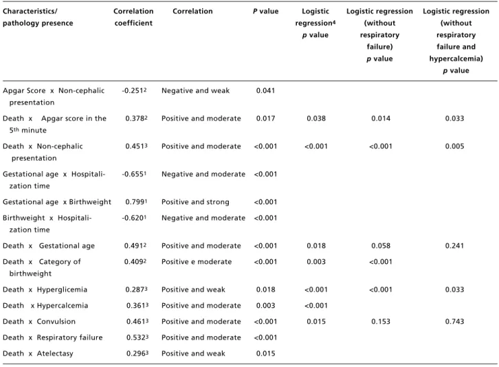
Tabela 4
Perinatal and delivery characteristics/complications with significant statistical correlation between death and survival groups.
Characteristics/ Correlation Correlation P value Logistic Logistic regression Logistic regression
pathology presence coefficient regression4 (without (without
pvalue respiratory respiratory
failure) failure and
p value hypercalcemia)
p value
Apgar Score x Non-cephalic -0.2512 Negative and weak 0.041 presentation
Death x Apgar score in the 0.3782 Positive and moderate 0.017 0.038 0.014 0.033
5thminute
Death x Non-cephalic 0.4513 Positive and moderate <0.001 <0.001 <0.001 0.005 presentation
Gestational age x Hospitali- -0.6551 Negative and moderate <0.001 zation time
Gestational age x Birthweight 0.7991 Positive and strong <0.001
Birthweight x Hospitali- -0.6201 Negative and moderate <0.001 zation time
Death x Gestational age 0.4912 Positive and moderate <0.001 0.018 0.058 0.241
Death x Category of 0.4092 Positive e moderate <0.001 0.003 <0.001 birthweight
Death x Hyperglicemia 0.2873 Positive and weak 0.018 <0.001 <0.001 0.033
Death x Hypercalcemia 0.3613 Positive and moderate 0.003 <0.001
Death x Convulsion 0.4613 Positive and moderate <0.001 0.015 0.153 0.743
Death x Respiratory failure 0.5323 Positive and moderate <0.001
Death x Atelectasy 0.2963 Positive and weak 0.015
1) Spearman correlation coefficient (r); 2) ETA Correlation Coefficient ( ε); 3) PHI (ö) Correlation Coefficient; 4) Logistic Regression
Discussion
Neonatal brain white matter lesion is a severe pathology that could lead to neurological conditions and death. This research is a pioneer study for brain white matter lesion and there are no literature data that could be compared with the current one. Nevertheless, lethality rates between 8.2% and 10.3% are considered high, demonstrating the value and severeness of these lesions in neonates.
Of the seven death cases, 42.9% had echodensity and/or complicated echolucency in contrast with 36.1% of the survivors, suggesting that evolution to brain white matter lesion is an alert signal for a higher death probability, even if this finding has not shown statistical significance, possibly because of the small number of deaths noted. But the difference of 6.8% noted among the survivors and deaths by complicated lesions should be considered clinically significant. Death did not occur among brain white matter lesion cases after the postnatal period. Theoretically, in late occurrences or following cli-nical insult, brain white matter lesion could be less severe because of continuous medical monitoring of the neonate, and more maturity spontaneously protecting from an unfavorable evolution.
Birthweight and gestational age correlated nega-tively with hospitalization time. Median hospitaliza-tion time was significantly inferior among the cases evolving to death (median of 23 days x 50 days in the survival group), with all deaths occurring up to 44 days. This is suggestive of: postnatal premature maturing protects from death and/or the death causes occurred early in the antenatal or perinatal period.
In relation to the first hypothesis, deaths were positively correlated with birthweight, as expected and amply demonstrated in the international7-9and
national literature.10,11Nevertheless, 85.3% of low
birthweight neonates survived being equivalent to the proportions of extremely low birthweight and one death occurred among the one between 2000 to 2499 g, a range of lower mortality risk. Other factors should have influenced evolution towards death in addition to birthweight, but in the evaluations or early neonates'history, no information of the maternal or gestational history were significantly correlated to the deaths. On the other hand, in the delivery history, Apgar score showed a positive and moderate correlation with the group evolving to death. Although 57% of the neonates in this group Apgar Score in the 5th minute was lower in the
"death" group (6.17 versus8.15) with positive corre-lation coefficient of 0.378 (moderate) (Table 4). In this group there were more deliveries with non-cephalic presentation (71.43% versus 13.12%) with moderate correlation (ö=0.451). These differences were statistically significant in contrast with the lack of statistical significance of the differences between: asphyxia reports, fetal suffering, placenta abruptio, premature membrane rupture, ascending infection signs, eclampsy or pre-eclampsy, difficulty of fetus
removal or tocotraumatism.
Correlation was negative for gestational age and hospitalization time(r=-0.655-moderate): the lower the gestational age the longer the hospitalization time and was positive (r=0.491-moderate) with death (Table 4). Birthweight assessment percategory has demonstrated that two cases of birthweight equal or under 500 grams evolved to death, with 71.5% (five cases) of death within the birthweight range of up to 999 grams, in contrast with 21.3% (13 cases) of the survivals within the same range. All deaths occurred among the low birthweight group while among the survivors, nine were over 2500 g. Birthweight had a positive correlation with gesta-tional age (r=0.799-strong) and with death ((r=0.409-moderate) and negative with hospitaliza-tion time (-0.620-moderate (Table 4).
Positive correlations in the death cases were also detected for hyperglicemia, hypercalcemia, convul-sion, respiratory insufficiency, atelectasy (Table 4) with convulsion being the more significant condi-tion. Infection, sepsis, septic chock, enterocolitis, pulmonary hypertension, coagulation disorders and severe malformations had no significant correlation with white cerebral matter lesion evolving to death.
Following binary logistic regression analysis, all variables remained statistically significant and in regression, after exclusion of respiratory insuffi-ciency (step 1) and hypercalcemia (step 2), the atelectasy and hyperglicemia variables did not remain statistically significant (Table 4).
group.
Hyaline membrane condition is the specific pathology of prematurity, secondary to low surfac-tant production. The more premature the neonate the more severe the condition. The occurrence of asphyxia and infectious diseases aggravate the condition and respiratory failure is its most severe expression. On the other hand, respiratory failure aggravates clinical and neurological conditions of the neonate who suffered from asphyxia and can in itself cause postnatal anoxia. Atelectasy is commonly associated to pneumonia and respiratory failure in premature newborns with severe hyaline membranes condition. Early respiratory disconfort in premature newborns is caused by the hyaline membrane disease leading to respiratory failure.
The careful review of the medical files with the survey of 53 variables and the one by one correlation of these variables with the evolution to death are among the positive aspects of the study. Never-theless, some aspects need to be justified: a) cases assessment occurred within an eight year period, but there was a concentration of seven deaths between 1996 and 1998. Technological and medical progress in Obstetrics and Neonatology are rapidly occurring, and many medical procedures are being changed15 -17and that could possibly favor death toll reduction;
b) the presence of absence of choriomnionitis is the second etiologic factor of brain white matter lesion in addition to asphyxia,18,19 and in some studies
determined as being the cause10,20and in others as
being a protection factor21for perinatal mortality.
Ascending infection is diagnosed by the anatomic pathological study of the placenta. This exam cannot be assessed in the study because it was not available in 45 cases. Nevertheless ascending infection cli-nical suspicion did not significantly correlate with death c) the study group size was based in only one hospital. Authors chose to use secondary data for convenience due to the lack of USc information availability in other maternity hospitals or inappro-priate exam performance (inapproinappro-priate transducer, low resolution device) and d) descriptive study based on medical files information. Because this was a pioneer study, it had the objective to survey data and generate hypothesis. Therefore, an option was made for a convenience sample.
Conclusions
Hospital lethality for brain white matter lesion starting during prenatal and perinatal phases was estimated in 10.3% and the following hypothesis A "normal" premature baby can receive a low
Apgar score solely because of immaturity therefore, this score cannot be used to diagnose "asphyxia" in this population making a clinical based diagnoses difficult.12The assumption of asphyxia was based on
gestational and delivery history. Nevertheless, this study did not find significant alterations in the group with fatal evolution, consequently many asphyxiated premature with a low Apgar score did not receive this clinical diagnosis.
Only in one case, perinatal asphyxia diagnosis was registered in the death certificate probably based encephalopathy and multiple organ failure. There's suspicion of non-diagnosed asphyxia in the remaining cases. Nelson and Ellenberg13through the
multicenter study "The National Collaborative Peri-natal Project" followed up 54.000 pregnant women and their children up to seven years old and confir-med that children with birthweight under 2500 g, low Apgar score in the first and fifth minutes, were associated with 95.7% death risk and a high rate of severe neurological disorders in the survivors. On the other hand, many premature newborns have long term neurological disorders without prior diagnosis of perinatal risk factors; therefore prematurity itself is considered as the cause. It is possible that many mild and moderate ischemic hypoxic insults are not being diagnosed because of the non-use of the Apgar score, as a predictor tool for future disorders.
Still in the delivery history, non-cephalic presen-tation correlated significantly with death. In this type of presentation delivery is more difficult and may lead to perinatal asphyxia, nevertheless, neonates with intrauterine suffering do not have a good move-ment pattern and are unable to get to the cephalic position. In both situation asphyxia could be related to the non-cephalic presentation and in this study non-cephalic presentation significantly correlated with the Apgar score.
Postnatal factors analysis pointed towards a moderate and positive correlation with death in cases of: hyperglicemia, convulsion, respiratory failure and atelectasy. In general metabolic disorders are frequent in ill premature newborns and have multiple etiologies. Possibly, hyperglicemia and hypercal-cemia are coadjutants and could signal the terminal phase of these patients.
Neonatal convulsion is strongly associated to pe-rinatal asphyxia, with ischemic hypoxic encephalo-pathy the principal cause of this event in the neonatal period.14Positive correlation between convulsion
9. Wilcox AL, Skjaerven R. Birth weight and perinatal mortality: the effect of gestational age. Am J Public Health. 1992; 82: 378-82.
10. Miura E, Fiori F. Mortalidade perinatal e neonatal no Hospital de Clínicas de Porto Alegre. Rev Assoc Med Bras. 1997; 43: 35-9.
11. Lansky S, França E, Leal MC. Mortes perinatais evitáveis em Belo Horizonte, Minas Gerais, Brasil, 1999. Cad Saúde Pública. 2002; 18: 1389-400.
12. Apgar V, Holaday DA, James LS. Evaluation of the newborn infant - second report. JAMA. 1958; 168: 1985-8.
13. Nelson KB, Ellenberg JH. Apgar scores as predictors of chronic neurologic disability. Pediatrics. 1981; 68: 36-44.
14. Tharp Br. Neonatal seizures and syndromes. Epilepsia. 2002; 43 (Suppl 3): 2-10.
15. LeFlore JD, Salhb WA, Broyles S, Engles WD. Association of antenatal and postnatal dexamethasone exposure with outcome in extremely low weight neonates. Pediatrics. 2003; 110: 275-9.
16. Ingemar I, Lamont RF. An update on the controversies of tocolytic therapy for the prevention of preterm birth. Acta Obstet Gynecol Scand. 2003; 82: 1-9.
17. Meneguel JF, Guinsburg R, Myoshi MH, Peres CA, Russo RH, Kopelman BI, Camano L. Antenatal treatment with corticosteroids for preterm neonates: impact on the inci-dence of respiratory distress syndrome and intra-hospital mortality. São Paulo Med J. 2003; 12: 45-52.
18. Dammann O, Kuban KCK, Leviton A. Perinatal infection, fetal inflammatory response, white matter damage, and cognitive limitations in children born preterm. Mental Retard Dev Disabil Res Rev. 2002; 8: 46-50.
19. Rezaie P, Dean A. Periventricular leucomalácia, inflamma-tion and white matter lesions within the developing nervous system. Neuropathology. 2002; 22: 106-32.
20. Jason JM. Infectious disease-related deaths of low birth References
1. Victora CG, Barros FC. Infant mortaliy due to perinatal causes in Brazil: trends, regional patterns and possible interventions. São Paulo Med J. 2001; 119: 33-42.
2. Wise PH. The anatomy of a disparity in infant mortality. Ann Rev Public Health. 2003; 24: 241-62.
3. Barros FC, Hurtly SRA, Victora CG, Kirkwood MA, Vaughan JP. Comparisons of the causes and consequences of prematurity and intrauterine growth retardation: a longi-tudinal study in Soutern Brazil. Pediatrics. 1992; 90: 238-44.
4. Gray RH, Ferraz EM, Amorin MS, Melo LF. Levels and determinants of early neonatal mortality in Natal, Northeastern Brazil: results of a surveillance and case-control study. Int J Epidemiol. 1991; 20 :467-73.
5. Kuban KC, Allred EN, Dammann O, Pagano M, Leviton A, Share J, Abiri M, Di Salvo D, Doubilet P, Kairam R, Kazam E, Kirpekar M, Rosenfeld DL, Sanocka UM, Schonfeld SM. Developmental epidemiology network. Topography of cerebral white-matter disease of prematurity studied prospectively in 1607 very-low-birthweight infants. J Child Neurol. 2001; 16: 401-8.
6. Kumazaki K, Nakayama M, Sumida Y, Ozono K, Mushiake S, Suehara N, Wada Y, Fujimura M. Placental features in preterm infants with periventricular leucomalácia. Pediatrics. 2002; 109: 650-5.
7. Richardson DK, Phibbs CS, Gray JE, McMormick MC, Workman-Daniels K, Goldmann DA. Birth weight and illness severity: independent predictors of neonatal mortality. Pediatrics. 1993; 91: 969-75.
8. Larroque B, Marret S, Ancel PY, Arnaud C, Marpeu L, Super Nant K, Pierrat V, Rozé JC, Matis J, Cambonie G, Burguet A, Andre M, Kaminski M, Bréar G, The EPIPAGE Study Group. White matter damage and intraventricular hemorrhage in very preterm infants. J Pediatr. 2003; 143: 477-83.
Acknowledgements
To CAPES for the doctorate scholarship and sand-wich scholarship for training in the Southern Illinois University for the principal author. To the Academic Administration of the Psychology Course of the Ruy Barbosa Faculty for the financial, material and personal support, to the research of the Neurosciences Laboratory, Dr. Rosana Pelegrinni for her valuable discussion of neonatal aspects of the study. Dr. Kimberly Espy, Neuropsychological Laboratory, Southern Illinois University for the statistical and librariam support.