R E V B R A S R E U M A T O L . 2 0 1 4 ;5 4 ( 3 ): 2 3 7 – 2 4 0
www.reumatologia.com.br
REVISTA BRASILEIRA DE
REUMATOLOGIA
Case report
First report of mild Brazilian spotted fever associated to
arthritis
Virgínia Lucia Nazario Bonoldi
a, Roberta Gonçalves Marangoni
a, Giancarla Gauditano
a,
Jonas Moraes-Filho
b, Marcelo Bahia Labruna
b, Natalino Hajime Yoshinari
a,*
a Department of Rheumatology, Faculty of Medicine, Universidade de São Paulo, São Paulo, SP, Brazil b Faculty of Veterinary Medicine, Universidade de São Paulo, São Paulo, SP, Brazil
a r t i c l e i n f o
Article history:
Received on 15 February 2012 Accepted on 18 February 2013
Keywords:
Brazilian spotted fever Mild rickettsiosis Arthritis
Infectious arthritis
a b s t r a c t
We describe the irst Brazilian case of mild Rickettsiosis, complicated by knee monoarthri-tis, in young adult bitten by a tick on his left leg in Camburi zone, located in São Sebastião municipality, southern coastal region of the State of São Paulo, in the Atlantic rainforest region, Brazil. The patient developed inoculation eschar at the tick bite site associated with enlarged lymph nodes in the left groin, fever, polyarthralgia, headache and macular rash. Twenty days after tick bite episode, he displayed monoarthritis in his right knee. The diag-nosis of mild Rickettsiosis was established by sequential immunological analysis in serum and synovial luid, using the indirect immunoluorescence (IF) assay for antibodies reactive with Rickettsia parkeri and Rickettsia rickettsii. The mild Rickettsiosis is an emerging zoonosis, that must be investigated by physicians, including rheumatologists, in patients that pre-sent macular rash, fever and eventually arthritis, after visiting the southern coastal Atlan-tic rainforest region in Brazil.
© 2014 Sociedade Brasileira de Reumatologia. Published by Elsevier Editora Ltda. All rights reserved.
* Corresponding author.
E-mail: [email protected] (N.H. Yoshinari).
0482-5004/$ - see front matter. © 2014 Sociedade Brasileira de Reumatologia. Published by Elsevier Editora Ltda. All rights reserved. http://dx.doi.org/10.1016/j.rbre.2013.02.002
Primeiro caso de branda maculosa brasileira branda associada à artrite
Palavras-chave:
Febre maculosa brasileira Riquetsiose branda Artrite
Artrite infecciosa
r e s u m o
Descrevemos o primeiro caso brasileiro de Riquetsiose branda, agravada por monoartrite em joelho, em adulto jovem picado por carrapato na perna esquerda na região de Camburi, lo-calizada no município de São Sebastião, sul da região costeira do estado de São Paulo, Mata Atlântica, Brasil. O paciente apresentou uma escara de inoculação no local da picada do car-rapato, associada ao aumento ganglionar em virilha esquerda, febre, poliartralgia, cefaleia e erupção macular. Vinte dias após o episódio da picada de carrapato, o paciente apresen-tou monoartrite em joelho direito. O diagnóstico de Riquetsiose branda foi estabelecido pela análise imunológica sequencial em amostras de soro e líquido sinovial, tendo sido empregada a técnica de imunoluorescência (IF) indireta para anticorpos reativos contra Rickettsia parkeri
238
R E V B R A S R E U M A T O L . 2 0 1 4 ;5 4 ( 3 ): 2 3 7 – 2 4 0pelos médicos, incluindo reumatologistas, em pacientes que apresentem erupção macular, febre e, eventualmente, artrite, após visita ao sul da região costeira da Mata Atlântica no Brasil.
© 2014 Sociedade Brasileira de Reumatologia. Publicado por Elsevier Editora Ltda. Todos os direitos reservados.
Introduction
Spotted fever caused by Rickettsia rickettsii is a serious zoono-sis transmitted by ticks described in Americas, including in Brazil.1 The disease can lead to death if not diagnosed and
treated at the onset of clinical symptoms.
Laboratory diagnosis is based on seroconversion of con-secutive samples from acute and convalescent phases of the disease using R rickettsii antigen. The clinical manifestations of BSF begin about a week after the tick bite with fever, head-ache, abdominal pain and maculopapular rash. The disease can progress to respiratory and renal complications, coagu-lation disorders and encephalitis. Due to the severity of the illness, antibiotic treatment should be started quickly despite laboratory analysis.1,2
Paddock et al. identiied a new tick-borne Rickettsia of the spotted fever group that causes disease. Patients exhibited characteristic skin ulceration at the tick bite site (eschar), fol-lowed by maculopapular rash, fever, headache, myalgia and arthralgia. However, the clinical picture is milder due to the absence of coagulopathy.3 The etiologic agent was identiied
as Rickettsia parkeri, which has a lot of genetic similarity with
R. conorii, R. africae and R. sibirica, species that cause similar clinical symptoms in the Mediterranean region.4
In Brazil, the etiologic agent of the mild rickettsiosis was identiied in the skin lesion biopsy (eschar) of two patients. The molecular studies demonstrated that this agent is geneti-cally similar to R. parkeri, R. africae and R. sibirica.5,6 The present
study reports the discovery of the third case of mild rickettsi-osis in Brazil, and importantly, for the irst time, related to oc-currence of monoarthritis following this rickettsial infection. In Brazil, all three cases were described in the Atlantic Forest, ecological complex with the occurrence of the Amblyomma ovale tick infected with the etiologic agent.7
Case report
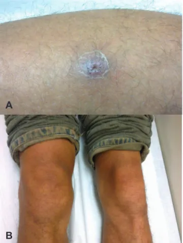
In February 2011, a 30-year-old man was bitten by a tick on his left leg while walking on an ecological trail within Atlantic rainforest area in Camburi, São Sebastião city, southern coastal region of the State of São Paulo. After 7 days, he presented ery-thematous skin lesions with central ulceration (eschar) at the tick bite site (Fig. 1a). After 10 days of the tick bite, he devel-oped fever not measured, polyarthralgia (hands, elbows, wrists, ankles), myalgia, neck pain, headache, nausea, enlarged lymph nodes on the left groin and chills, followed by generalized rash on the trunk and limbs. The patient sought medical care and, based on suspicion of rickettsial disease, the physician pre-scribed doxycycline even before the laboratorial tests. After 10 days of treatment, general symptoms had improved, but the doxycycline was continued due to the emergence of right knee monoarthritis (Fig. 1b).
In the fourth week of therapy, prednisone (10 mg/day) was prescribed associated with doxycycline due to the persistence of arthritis. After 7 days of therapy, the arthritis worsened with physical exercise and the prednisone was replaced by sulfasalazine (1g/ 12/12h). After 3 months of therapy with sul-fasalazine, the right knee monoarthritis remitted. Two samples of blood and synovial luid were collected (March 1st, 2011, and
March 15th, 2011); the irst, after 10 days of antibiotic therapy,
the other, 15 days after this. Additional laboratory tests showed alanine aminotransferase, alkaline phosphatase, aspartate aminotransferase, creatinine phosphokinase, gamma glutamyl transferase, lactic dehydrogenase, glucose, hemogram with platelets counting, protein electrophoresis, urea, creatinine, sodium, potassium and anti-streptolisin O levels within refer-ence ranges. Tests for hepatitis B and C, cytomegalovirus, HIV 1 and 2, syphilis, Brazilian borreliosis and bartonellosis were negative. IgE and C-Reactive Protein were increased, but levels became normal with treatment. Two serum samples revealed
Fig. 1 – (A) Inoculation eschar on the left leg of a patient with mild Brazilian spotted fever and acute knee monoarthritis. (B) Acute monoarthritis in right knee of a patient with mild Brazilian spotted fever, after a tick bite episode occurred in the Atlantic rainforest, São Sebastião, São Paulo State, Brazil.
A
239
R E V B R A S R E U M A T O L . 2 0 1 4 ;5 4 ( 3 ): 2 3 7 – 2 4 0
presence of anti-nuclear antibodies (ANA), exhibiting a dense ine speckled pattern on HEp-2 cells analysis (Table 1).
Paired samples of acute-phase and convalescent-phase of blood sera and synovial luids were evaluated under the same conditions by IF assay for antibodies to R. parkeri and R. rickett-sia. The samples were tested with a goat anti-human immuno-globulin (Ig) G for the irst and second serum and synovial luid samples or a goat anti-human IgM luorescein isothiocyanate conjugate (Sigma Diagnostics, St. Louis, MO, USA) for samples of acute-phase and convalescent-phase of synovial luids and a single convalescent-phase sample of serum.
In Table 1, we can see that the titles of the IgM to R. parkeri
and to R. rickettsii in synovial luid showed increased antibody reactivity between the irst and second samples. Titers of IgG to
R. parkeri and R. rickettsii were always high in synovial luid. The IgG titles to rickettsial antigens were high in both sequential samples of blood serum, but the IgG to R. parkeri showed an increase in antibody title in blood serum samples.
Discussion and conclusions
We described the third Brazilian case of mild rickettsiosis, at this time complicated with knee monoarthritis, which appeared nearly twenty days after tick bite episode. Like the previously de-scribed two cases of mild rickettsiosis,5,6 the patient caught the
disease walking on ecological trail, within Atlantic rain Forest. The speciic serologic diagnostic tests for rickettsiosis, per-formed in serum and synovial luid samples at acute and con-valescent phases of disease conirmed the diagnosis. Moreover, laboratorial investigations for other infectious diseases were negative, including the possibility of co-infection with Lyme like,8 which is transmitted by ticks and causes arthritis.
Anti-nuclear antibodies positivity was not correlated with clinical indings, and therefore, interpreted as an isolated phenomenon
or related to rickettsial infection. The increased level of IgE was understood as mast cell-dependent allergic response to the tick bite. This hypothesis is plausible, since the IgE levels decreased as the disease improved.
In patients with Mediterranean Spotted Fever disease, caused by R. conorii, arthritis in large joints with joint effusion in the hips, knees and ankles is described.9 Sundy et al.
report-ed knee acute monoarthritis in patient with RMSF.10 Ding et
al. reported polyarthritis in the wrists, metacarpal phalangeal and proximal interphalangeal joints in patient who acquired the illness in travel to Africa, where the disease is caused by R. parkeri or R. conorii.11
In the present case, the pathogenesis of knee monoarthritis following mild rickettsiosis infection is uncertain. The joint effu-sion can result of direct articular infection by rickettsial micro-organisms (infectious arthritis) or can relect synovial inlam-mation triggered by immune complexes depositions (reactive arthritis). The identiication of rickettsial infection by molecular procedures in the synovial luid and eschar biopsy was incon-clusive, because PCR was done lately when patient was treated with doxycycline. We believe that arthritis viewed in this patient is of reactive origin, since arthritis appeared late, nearly 20 days from bacteria inoculation. Additionally, this period of time was enough to produce speciic antibodies to rickettsial components. Outbreak of arthritis in the opposite leg of tick bite and uprising of antinuclear antibodies are further evidences to suggest occur-rence of immunological inlammatory arthritis.
In Rickettsia infection, the IgM and IgG serum levels increase by the second week of illness, IgM antibodies wane after 3 or 4 months and IgG titers persist for 7 or 8 months.12 We noted
that despite the existence of cross-reactivity between rickett-sial species,2 the IgG titer to R. parkeri increased, at least, 1-fold
higher in the serum, although it was a low increase in title, which did not occur to R. rickettsii. We can see an increase in R. parkeri IgM titer of paired synovial luid specimens taken early and later in the disease course, unlike in R. rickettsii, suggest-ing more IgM speciicity to R. parkeri. In addition, the R. parkeri
rickettsiosis can be associated with the presence of an eschar at the site of tick bite.3
High titers of IgM antibodies to rickettsial antigens in syno-vial luid conirmed the etiology of knee acute arthritis.
We conclude that Brazilian spotted fever is not a single dis-ease, because at least two pathogenic species of Rickettsia are present in Brazil causing similar symptoms. The mild form of the disease is reported in the region that attends the Atlantic Forest and, as described in this current case report, may be as-sociated to arthritis as a complication of systemic disease.
Conlicts of interest
The authors declare no conlicts of interest.
R E F E R E N C E S
1. Labruna MB. Ecology of rickettsia in South America. Ann N Y Acad Sci. 2009 May;1166:156-66.
2. Paddock CD, Finley RW, Wright CS, Robinson HN, Schrodt BJ, Lane CC et al. Rickettsia parkeri rickettsiosis and its clinical
Table 1 – Sequential Immunoluorescent (IF*) assays (IgM and IgG) to detect antibodies against R. parkeri
and R. rickettsii in serum and synovial luid samples.
C-Reactive protein, IgE and antinuclear antibodies (ANA**) in serum samples of a patient with mild Brazilian spotted fever and acute knee monoarthritis in Brazil, 2011.
Serum Synovial luid March 1 March 15 March 1 March 15
IF IgG R. parkeri
1/1024 1/2048 1/2048 1/2048
IF IgM R. parkeri
- - 1/256 1/512
IF IgG R. rickettsii
1/1024 1/1024 1/2048 1/2048
IF IgM R. rickettsii
- - 1/128 1/256
IgE (IU/ml) 1240 783 - -PCR (mg/L) 1,5 0,5 - -ANA > 1/320 > 1/320 - -*IF Cut off = 1/64
240
R E V B R A S R E U M A T O L . 2 0 1 4 ;5 4 ( 3 ): 2 3 7 – 2 4 0distinction from Rocky Mountain spotted fever. Clin Infect Dis. 2008;47:1188-96.
3. Paddock CD, Sumner JW, Comer JA, Zaki SR, Goldsmith CS, Goddard J et al. Rickettsia parkeri: a newly recognized cause of spotted fever rickettsiosis in the United States. Clin Infect Dis. 2004 Mar 15;38:805-11.
4. Goddard J. Historical and recent evidence for close
relationships among Rickettsia parkeri, R. conorii, R. africae, and
R. sibirica: implications for rickettsial taxonomy. J Vector Ecol. 2009 Dez;34:238-42.
5. Spolidorio MG, Labruna MB, Mantovani E, Brandao PE, Richtzenhain LJ, Yoshinari NH. Novel spotted fever group rickettsiosis, Brazil. Emerg Infect Dis. 2010 Mar;16:521-3. 6. Silva N, Eremeeva ME, Rozental T, Ribeiro GS, Paddock CD,
Ramos EA, Favacho AR, Reis MG, Dasch GA, de Lemos ER, Ko AI. Eschar-associated spotted fever rickettsiosis, Bahia, Brazil. Emerg Infect Dis. 2011 Fev;17:275-8.
7. Sabatini GS, Pinter A, Nieri-Bastos FA, Marcili A, Labruna MB. Survey of ticks (Acari: Ixodidae) and their rickettsia in an
Atlantic rain forest reserve in the State of São Paulo, Brazil. J Med Entomol. 2010 Set;47:913-6.
8. Yoshinari NH, Mantovani E, Bonoldi VLN, Marangoni RG, Gauditano G. Doença de Lyme-símile brasileira ou Síndrome Baggio-Yoshinari: Zoonose exótica e emergente brasileira transmitida por carrapatos. Rev Assoc Med Bras. 2010;56:363-9.
9. Pedro-Botet J, Auguet T, Pallás O, Gimeno JL. Arthritis in Mediterranean spotted fever. Infection. 1991 Set-Out;19:346-7. 10. Sundy JS, Allen NB, Sexton DJ. Rocky Mountain spotted fever
presenting with acute monoarticular arthritis. Arthritis Rheum. 1996 Jan;39:175-6.
11. Ding T, Lloyd G, Tolley H, Bradlow A. Tick bite fever and arthritis associated with travel to Africa. Ann Rheum Dis. 2004 Dez;63:1703-4.