DOI: 10.5935/2359-4802.20180067
ORIGINAL ARTICLE
Mailing Address: Ana Rosa Cunha
Rua Sérgio de Camargo, 123/306 - bl: 2. Postal Code: 22775-052, Jacarepaguá, RJ - Brazil. E-mail: anarosacunha@gmail.com, anarosacunha@yahoo.com.br
Endothelial Dysfunction and Pulse Wave Reflection in Women with Polycystic
Ovarian Syndrome
Marcelo Burlá,1 Ana Rosa Cunha,1 Ronaldo Gismondi,2 Wille Oigman,1 Mario Fritsch Neves,1 Fernanda Medeiros1
Universidade do Estado do Rio de Janeiro,1 Rio de Janeiro, RJ - Brazil
Universidade Federal Fluminense,2 Niterói, RJ - Brazil
Manuscript received August 24, 2017; revised manuscript December 18, 2017; accepted March 03, 2018.
Abstract
Background: Patients with polycystic ovarian syndrome (PCOS) have an increased prevalence of metabolic syndrome and traditional atherosclerotic risk factors, such as dyslipidemia, diabetes and hypertension. Endothelial function and vascular stiffness are surrogate markers of early atherosclerosis, able to predict cardiovascular events. Objective: To compare endothelial function and pulse wave reflection between women with PCOS and healthy controls.
Methods: Observational and cross-sectional study that included women with PCOS, age between 18 and 40 years-old and body mass index between 25.0 and 35.0 kg/m2, and healthy controls. Rotterdan criteria was used to
diagnose PCOS. Subjects underwent clinical and anthropometric evaluation, laboratory and hormonal assays and imaging tests to measure pulse wave velocity (PWV), augmentation index (AIx) and brachial artery flow-mediated vasodilation (FMD). Kolmogorov-Smirnov test showed normal distribution of most parameters. Unpaired Student t-test was used with significance level established at p < 0.05.
Results: A total of 52 patients were included, 29 (56%) in PCOS group and 23 (44%) in control group. Clinical and laboratory parameters were similar between the groups. Women with PCOS had lower FMD (8.8 ± 1.0 vs 12.8 ± 1.2%, p = 0.021); PWV and AIx were similar between the groups (7.5 ± 0.2 vs 7.5 ± 0.3 m/s, p = 0.671 and 21.0 ± 1 vs. 20 ± 2%, p = 0.716, respectively). In the PCOS group, women with higher testosterone levels had higher AIx (25 ± 2 vs. 17 ± 3%, p = 0.045).
Conclusions: PCOS women had endothelial dysfunction and those with higher testosterone levels had higher pulse wave reflection as compared with controls. (Int J Cardiovasc Sci. 2018; [online].ahead print, PP.0-0)
Keywords: Endothelium, Vascular; Vascular,Stiffness; Metabolic Syndrome; Polycistic Ovary Syndrome; Insulin Resistance; Testosterone.
Introduction
Polycystic ovary syndrome (PCOS) is one of the most common metabolic disorders in women, with an estimated prevalence of 5 to 15%.1 The cardinal features of
the syndrome encompass hyperandrogenism, ovulatory dysfunction, and/or polycystic ovaries on ultrasound. Obesity, insulin resistance and metabolic syndrome are closely related to PCOS. A recent meta-analysis observed a higher risk of metabolic syndrome in women with PCOS
(OR 2.88, 95% CI 2.40-3.45), as well as glucose intolerance and diabetes mellitus (DM).2 The occurrence of metabolic
disorders is also elevated in non-obese PCOS patients, suggesting that the presence of the syndrome per se may favor the development of metabolic comorbidities.3
In addition, women with PCOS have an increased prevalence of traditional atherosclerotic risk factors, such as dyslipidemia, DM and hypertension.3,4 The present
Despite the association of PCOS with cardiovascular (CV) risk factors, recent studies showed controversial results regarding the incidence of CV events in women with PCOS.5,6 A retrospective cohort study in United
Kingdom showed a high incidence of DM, myocardial infarction (MI) and angina in women with PCOS, with over a quarter of the elderly individuals having had MI or angina.5 However, a retrospective cohort study
in the United States observed no increase in CV events in PCOS women,6 which may be explained by different
PCOS profiles, androgen levels, insulin resistance and body composition of the populations.7 Thus, markers of
atherosclerosis could be of help in improving CV risk stratification in PCOS women.
Endothelial function and vascular stiffness are surrogate markers of early atherosclerosis and can be easily measured by noninvasive methods, such as flow-mediated vasodilation (FMD) and carotid-femoral pulse wave velocity (PWV), respectively.8,9 Moreover,
both FMD and PWV have shown to be independent predictors of CV events in the general population.10,11
A recent meta-analysis reported that PCOS women have approximately 4% lower FMD, irrespective of body mass index (BMI) and age.12 However, there was significant
heterogeneity across studies included in this meta-analysis.12 Previous studies have shown an association
between PCOS and PWV,13,14 but these results could be
influenced by age, BMI and comorbidity, in special the presence of hypertension. Other studies that controlled for these confounders did not find a correlation between PCOS and PWV.15–18
The aim of the present study was to compare endothelial function and pulse wave reflection between young, overweight women with PCOS and healthy controls.
Methods
Study population
This study recruited 52 consecutive women from the outpatient internal medicine and general gynecology clinics of our institution. We included patients with Rotterdam criteria for PCOS and age between 18-40 years. Exclusion criteria were evidence of secondary hypertension, BMI (calculated as weight divided
by height squared) ≥ 35 kg/m², smoking, coronary
artery disease, kidney or thyroid disease, hormone replacement therapy, DM or impaired tolerance
glucose, severe dyslipidemia (LDL-cholesterol ≥ 4.14 mmol/L and/or triglycerides ≥ 3.39 mmol/L), use of
lipid-lowering drugs, prolactin (PRL) > 25 ng/ml or pregnancy. Control group was composed by healthy female patients without PCOS criteria from the same institution. The protocol was approved by the local Ethics Committee Research (2795-CEP/HUPE), and all patients gave written informed consent.
PCOS was diagnosed according to the 2003 Rotterdam Criteria with at least two of the following features: oligomenorrhea (or amenorrhea) or hirsutism, clinical or biochemical hyperandrogenism, and polycystic ovaries on ultrasound. Patients with oligomenorrhea or hyperandrogenism caused by any other clinical conditions were excluded, such as nonclassical 21-hydroxylase deficiency, congenital adrenal hyperplasia, hypothyroidism, Cushing’s syndrome, or significant elevation in serum PRL.
Laboratory evaluation
Venous blood samples were collected after 12 hours of fasting. Serum lipids (total cholesterol, high-density lipoprotein [HDL]-cholesterol, triglycerides), and blood glucose were measured using an auto analyzer technique (Technicon DAX 96; Miles Inc). Low-density lipoprotein (LDL)-cholesterol was calculated with the Friedewald formula when triglyceride values were < 400 mg/dL. Insulin was measured by radioimmunoassay, and serum C-reactive protein levels were measured by nephelometry using an immunochemistry system. Serum levels of testosterone and PRL were measured during the early follicular phase (days 2 to 5 of the menstrual cycle) and dehydroepiandrosterone sulphate (DHEAS) were measured using enzyme-linked immunosorbent assay (ELISAs).
Assessment of endothelial function
FMD was assessed as a measure of endothelial function.19 The participant was positioned supine with
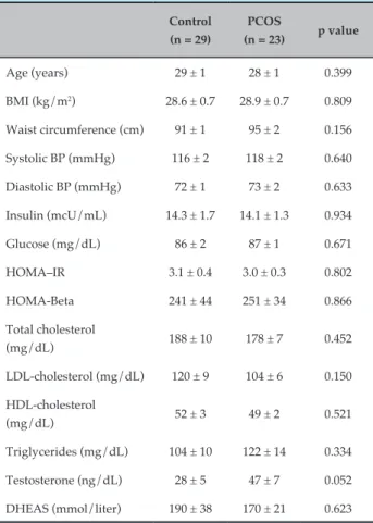
Table 1 - Clinical and demographic characteristics of patients with polycystic ovarian syndrome (PCOS) (n = 52)
Control (n = 29)
PCOS
(n = 23) p value
Age (years) 29 ± 1 28 ± 1 0.399
BMI (kg/m2) 28.6 ± 0.7 28.9 ± 0.7 0.809
Waist circumference (cm) 91 ± 1 95 ± 2 0.156 Systolic BP (mmHg) 116 ± 2 118 ± 2 0.640 Diastolic BP (mmHg) 72 ± 1 73 ± 2 0.633
Insulin (mcU/mL) 14.3 ± 1.7 14.1 ± 1.3 0.934 Glucose (mg/dL) 86 ± 2 87 ± 1 0.671 HOMA–IR 3.1 ± 0.4 3.0 ± 0.3 0.802 HOMA-Beta 241 ± 44 251 ± 34 0.866
Total cholesterol
(mg/dL) 188 ± 10 178 ± 7 0.452 LDL-cholesterol (mg/dL) 120 ± 9 104 ± 6 0.150 HDL-cholesterol
(mg/dL) 52 ± 3 49 ± 2 0.521 Triglycerides (mg/dL) 104 ± 10 122 ± 14 0.334
Testosterone (ng/dL) 28 ± 5 47 ± 7 0.052 DHEAS (mmol/liter) 190 ± 38 170 ± 21 0.623
BMI: body mass index; BP: blood pressure; HOMA: homeostatic model assessment; LDL: low-density lipoprotein; HDL: high density lipoprotein; DHEAS: dehydroepiandrosterone sulphate. Between-group differences were analyzed using unpaired Student’s t-test, considering a significance level of 5% and a two-tailed probability. Values expressed as mean ± standard error.
FMD was calculated as the percentage change of brachial artery diameter from baseline.
Pulse wave velocity
The same investigator measured the carotid-femoral PWV using a Complior device (Alam Medical, France) after the patients had rested for 10 minutes in supine position in a quiet room with a stable temperature.8
All measurements were performed between 8 a.m. and 11 a.m. During the measurements, speaking or sleeping was not allowed, and no meal, caffeine or smoking was allowed within 3h before measurement. Pulse waveforms were obtained transcutaneously from the right common carotid artery and femoral artery. Aortic PWV was calculated by dividing the distance traveled (DT) by the transit time (TT). TT was obtained by measuring the time difference between the arrival of the pulse wave at the femoral and at carotid arteries. DT was measured using a tape measure and estimated as 80% of the distance between carotid and femoral arteries. Carotid-femoral PWV was calculated as DT in meters divided by TT in seconds (PWV = DT/TT). The mean of two measurements was calculated and when the difference between them was more than 0.5 m/s, a third measurement was obtained. All PWV values were adjusted by mean arterial pressure (MAP) to obtain normalized PWV (PWV norm) as 100 x (PWV/MAP).
Central hemodynamic parameters
Applanation tonometry was performed with the SphygmoCor system (Atcor Medical, Sydney, Australia) with the patient in the sitting position, resting the arm on a rigid surface, and a sensor in the radial artery.20 Central
aortic pressure was calculated from the radial pulse wave analysis with the use of a validated transfer function. Wave reflection parameters, such as augmentation pressure (AP) and augmentation index (AIx), were also obtained by this method.
Statistical analysis
Continuous variables were expressed as mean ± standard error and categorical variables were described as absolute numbers and/or percentages. Kolmogorov-Smirnov test showed normal distribution of the variables. Differences between the two study groups were evaluated with unpaired Student’s t-tests for continuous variables. All tests were performed considering a significance
level of 5% and a two-tailed probability. Based on a recent study,12 assuming a 5% level of significance and
85% power, we estimated 18 patients in each group to detect 4% difference in FMD between the groups and 4% standard deviation.12 Statistical analysis was performed using the Statistical Package for Social Sciences (SPSS) version 18.0 for Windows (SPSS, Chicago, IL).
Results
Table 2. Vascular parameters of patients with polycystic ovarian syndrome (PCOS) (n = 52)
Control (n = 29)
PCOS
(n = 23) p value
Flow mediated
vasodilation (%) 12.8 ± 1.2 8.8 ± 1.0 0.021 CR PWV (m/s) 9.1 ± 0.3 8.8 ± 0.2 0.930 CF PWV (m/s) 7.5 ± 0.3 7.5 ± 0.2 0.671 Augmentation pressure
(mmHg) 6 ± 1 8 ± 2 0.337 Augmentation index (%) 20 ± 2 21 ± 1 0.716
Aortic systolic pressure
(mmHg) 106 ± 2 116 ± 5 0.320 Aortic pulse pressure
(mmHg) 31± 1 35 ± 4 0.335
Values expressed as mean ± standard error; CF- PWV: carotid-femoral pulse wave velocity; CR-PWV: carotid-radial pulse wave velocity. Between-group differences were analyzed using unpaired Student’s t-test, considering a significance level of 5% and a two-tailed probability.
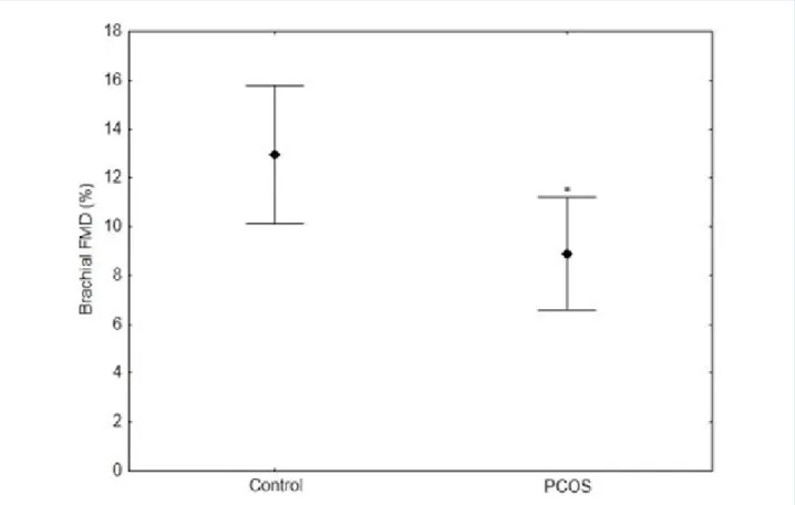
Figure 1 - Lower brachial flow-mediated dilation values in polycystic ovarian syndrome (PCOS) group (n = 29) compared with control group (n = 23). P value = 0.021 by Student’s t-test.
seborrheic dermatitis and acanthosis nigricans were observed in PCOS patients.
Brachial artery diameter was similar between the groups (3.13 ± 0.38 vs. 3.23 ± 0.37, p = 0.49). PCOS group had significant lower FMD than control group (Figure 1). PWV, AIx and aortic pressures were similar between the groups (Table 2).
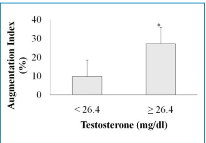
When PCOS individuals were divided into two groups according to the median of serum testosterone (46.4 ng/ dL), those with higher and lower testosterone levels had similar baseline clinical and laboratorial characteristics. PWV, FMD and aortic pressures were also similar between the groups. However, AIx was significantly higher in patients with higher testosterone levels (25 ± 2 vs. 17 ± 3%, p = 0.045; Figure 2).
Discussion
Figure 2 - Bar graph comparing augmentation index (mean ± standard error) by testosterone levels in polycystic ovarian syndrome (POS) women after division by the median; *P = 0.045 by Student’s t-test.
pulse wave reflection than those with PCOS and lower testosterone levels. Previous studies have shown that women with PCOS have high prevalence of CV risk factors, in addition to besides the clinical features of menstrual irregularity, hyperandrogenism and infertility. Thus, some studies have suggested an association between PCOS and accelerated CV disease.7,21,22 Recent
cohort studies had controversial results regarding CV events in PCOS patients.5,6 Both endothelial dysfunction
and arterial stiffness have been associated to worse CV outcomes in the general population and are proposed as complementary CV risk evaluation.10,11
Vascular endothelium plays a crucial role in maintaining vascular homeostasis and endothelial dysfunction is an important early step in the development of atherosclerosis and CV diseases.21 There are many pathophysiological
mechanisms that explain the relationship between PCOS and endothelial dysfunction.7,21 Insulin resistance
impairs intracellular signaling, which in endothelium may cause lower production of nitric oxide and increased secretion of endothelin-1, leading to vasoconstriction and decreased blood flow.23 Moreover, hyperinsulinemia
exerts a direct hypertrophic effect on the vascular wall, which deteriorates endothelial function and may lead to vascular stiffening.7,21,23 Other proposed mechanisms
involve atherogenic dyslipidemia, lipo-oxidative stress, products of glycation and glycoxidation, and inflammatory cytokines.7
One of the first studies relating PCOS and endothelial dysfunction was published in 2001 and enrolled 12 patients with PCOS and 13 age- and weight-matched controls.24
They observed that PCOS subjects had endothelial dysfunction that was related to hyperandrogenism and insulin resistance.24 These results have been confirmed
by subsequent studies and a recent meta-analysis, which showed that PCOS women had a pooled mean FMD 3.4% lower than controls.12,18,25 However, there are
controversies if endothelial dysfunction is a consequence of high androgen levels, hyperinsulinemia-obesity syndrome or both.18,24,26
A previous study compared PCOS women and controls, both groups with BMI < 30 kg/m2, and
observed that PCOS subjects had lower FMD and higher androgen levels despite no biochemical evidence of insulin resistance.18 A similar study involving overweight
young women with PCOS also observed endothelial dysfunction, but FMD was statistically correlated not only to high androgen levels but also to inflammatory markers and insulin resistance.26 In contrast, a previous
small study of PCOS women with high testosterone levels and normal insulin resistance did not observe endothelial dysfunction.27 A study of PCOS subjects with
high androgen levels also did not observe lower FMD when compared to age- and BMI- matched controls; in that study, the PCOS group had hyperinsulinemia.28 In
addition, a study comparing non-obese PCOS women and age- and BMI-matched controls showed similar FMD despite higher androgen levels in PCOS subjects; insulin levels and HOMA-IR indices were similar between the groups.29 In the present study, women with
PCOS had endothelial dysfunction, and our sample consisted essentially of young and overweight women. Furthermore, insulin levels and both HOMA-IR and HOMA-Beta were within normal range for their BMI, suggesting a non-significant insulin resistance. Thus, our results reinforce that PCOS women may have endothelial dysfunction due to mechanisms other than insulin resistance.
Changes in the arterial wall, with loss of elastin fibers and increase in collagen proteins, leads to vascular stiffening.30 Age and hypertension are two of the most
important factors that trigger these modifications.30
Endothelial dysfunction and arterial stiffening are related to each other. Carotid-femoral PWV is the gold-standard method to evaluate arterial stiffness and is a powerful and independent predictor of CV events.11,31
A previous study enrolled overweight women and demonstrated that subjects with PCOS had higher PWV and lower FMD than controls.32 However, other studies
1. Okoroh EM, Hooper WC, Atrash HK, Yusuf HR, Boulet SL. Prevalence of polycystic ovary syndrome among the privately insured, United States, 2003-2008. Am J Obstet Gynecol. 2012;207(4):299.e1–7.
2. Moran LJ, Misso ML, Wild RA, Norman RJ. Impaired glucose tolerance, type 2 diabetes and metabolic syndrome in polycystic ovary syndrome: a systematic review and meta-analysis. Hum Reprod Update. 2010;16(4):347–63.
3. Romano LGM, Bedoschi G, Melo AS, Albuquerque FO de, Rosa e Silva ACJ de S, Ferriani RA, et al. [Metabolic abnormalities in polycystic ovary
syndrome women: obese and non obese]. Rev Bras Ginecol Obstet. 2011;33(6):310–6.
4. Ramezani Tehrani F, Montazeri SA, Hosseinpanah F, Cheraghi L, Erfani H, Tohidi M, et al. Trend of Cardio-Metabolic Risk Factors in Polycystic Ovary Syndrome: A Population-Based Prospective Cohort Study. PLoS One. 2015;10(9):e0137609.
5. Mani H, Levy MJ, Davies MJ, Morris DH, Gray LJ, Bankart J, et al. Diabetes and cardiovascular events in women with polycystic ovary syndrome: a 20-year retrospective cohort study. Clin Endocrinol (Oxf). 2013;78(6):926–34.
References
women. In a young and non-obese sample of PCOS subjects, PWV and AIx were similar to healthy controls.18
A recent study of 84 women with PCOS and 95 healthy volunteers, aged 16-45 years, also reported similar PWV between groups.15 However, a small study showed that
PCOS non-obese women had higher AIx than healthy controls, although not measuring PWV.33 Our study
showed that PWV and AIx were similar between PCOS subjects and controls. On the other hand, those PCOS women with higher testosterone levels had higher AIx, indicating greater reflected pulse wave, which might represent an initial process of arterial stiffness. As PWV is largely influenced by age, young patients, including those with PCOS, tend to have normal values. AIx may be a better parameter in this population, as it reflects the influence, at the aortic level, of an increased stiffness of the arterial tree.30
This study has some limitations. First, sample size was small, so that we could have missed a small difference between PWV in PCOS and control groups. Second, as a cross-sectional study, we could not evaluate the occurrence of CV events, but merely suggest an association between FMD, AIx and PCOS. An important consideration of the study was to hypothesize a relationship between hyperandrogenism and an increased pulse wave reflection, maybe associated with arterial stiffness.
Conclusion
In summary, these PCOS women demonstrated endothelial dysfunction when compared to those young overweight women without the syndrome. Moreover, higher testosterone levels, even in the normal range, were associated with an increase in pulse wave reflection. Large prospective studies are needed to evaluate the prognostic value of FMD, PWV and AIx in this population. In addition, large trials can analyze if measurement of endothelial function and arterial stiffness
would improve CV risk stratification beyond traditional atherosclerotic risk factors.
Author contributions
Conception and design of the research: Burlá M, Oigman W, Neves MF, Medeiros F. Acquisition of data: Burlá M. Analysis and interpretation of the data: Burlá M, Cunha AR, Gismondi R, Oigman W, Neves MF, Medeiros F. Statistical analysis: Cunha AR, Gismondi R, Neves MF. Obtaining financing: Neves MF. Writing of the manuscript: Burlá M, Cunha AR, Gismondi R, Oigman W, Neves MF, Medeiros F. Critical revision of the manuscript for intellectual content: Burlá M, Cunha AR, Gismondi R, Oigman W, Neves MF, Medeiros F.
Potential conflict of interest
No potential conflict of interest relevant to this article was reported.
Sources of funding
There was no external funding for this study.
Study association
This article is part of the thesis submitted by Marcelo Burlá for fulfilment of the requirements for Master’s degree, University Hospital of Pedro Ernesto, Rio de Janeiro, Brazil.
Ethics approval and consent to participate
6. Iftikhar S, Collazo-Clavell ML, Roger VL, St Sauver J, Brown RD, Cha S, et al. Risk of cardiovascular events in patients with polycystic ovary syndrome. Neth J Med. 2012;70(2):74–80.
7. Conway G, Dewailly D, Diamanti-Kandarakis E, Escobar-Morreale HF, Franks S, Gambineri A, et al. The polycystic ovary syndrome: a position statement from the European Society of Endocrinology. Eur J Endocrinol. 2014;171(4):P1–29.
8. Laurent S, Cockcroft J, Van Bortel L, Boutouyrie P, Giannattasio C, Hayoz D, et al. Expert consensus document on arterial stiffness: methodological issues and clinical applications. Eur Heart J. 2006;27(21):2588–605.
9. Charakida M, Masi S, Luscher TF, Kastelein JJ, Deanfield JE. Assessment of atherosclerosis: the role of flow-mediated dilatation. Eur Heart J. 2010;31(23):2854–61.
10. Inaba Y, Chen JA, Bergmann SR. Prediction of future cardiovascular outcomes by flow-mediated vasodilatation of brachial artery: a meta-analysis. Int J Cardiovasc Imaging. 2010;26(6):631–40.
11. Vlachopoulos C, Aznaouridis K, O’Rourke MF, Safar ME, Baou K, Stefanadis C. Prediction of cardiovascular events and all-cause mortality with central haemodynamics: a systematic review and meta-analysis. Eur Heart J. 2010;31(15):1865–71.
12. Sprung VS, Atkinson G, Cuthbertson DJ, Pugh CJA, Aziz N, Green DJ, et al. Endothelial function measured using flow-mediated dilation in polycystic ovary syndrome: a meta-analysis of the observational studies. Clin Endocrinol (Oxf). 2013;78(3):438–46.
13. Armeni E, Stamatelopoulos K, Rizos D, Georgiopoulos G, Kazani M, Kazani A, et al. Arterial stiffness is increased in asymptomatic nondiabetic postmenopausal women with a polycystic ovary syndrome phenotype. J Hypertens. 2013;31(10):1998–2004.
14. Souza Dos Santos AC, Soares NP, Costa EC, de Sá JCF, Azevedo GD, Lemos TMAM. The impact of body mass on inflammatory markers and insulin resistance in polycystic ovary syndrome. Gynecol Endocrinol. 2015;31(3):225–8.
15. Rees E, Coulson R, Dunstan F, Evans WD, Blundell HL, Luzio SD, et al. Central arterial stiffness and diastolic dysfunction are associated with insulin resistance and abdominal obesity in young women but polycystic ovary syndrome does not confer additional risk. Hum Reprod. 2014;29(9):2041–9.
16. Hughan KS, Tfayli H, Warren-Ulanch JG, Barinas-Mitchell E, Arslanian SA. Early biomarkers of subclinical atherosclerosis in obese adolescent girls with polycystic ovary syndrome. J Pediatr. 2016;168:104–11.e1.
17. Ketel IJ, Stehouwer CD, Henry RM, Serné EH, Hompes P, Homburg R, et al. Greater arterial stiffness in polycystic ovary syndrome (PCOS) is an obesity--but not a PCOS-associated phenomenon. J Clin Endocrinol Metab. 2010;95(10):4566–75.
18. Cussons AJ, Watts GF, Stuckey BGA. Dissociation of endothelial function and arterial stiffness in nonobese women with polycystic ovary syndrome (PCOS). Clin Endocrinol (Oxf). 2009;71(6):808–14.
19. Corretti MC, Anderson TJ, Benjamin EJ, Celermajer D, Charbonneau F, Creager MA, et al. Guidelines for the ultrasound assessment of endothelial-dependent flow-mediated vasodilation of the brachial artery:
a report of the International Brachial Artery Reactivity Task Force. J Am Coll Cardiol. 2002;39(2):257–65.
20. Pauca AL, O’Rourke MF, Kon ND. Prospective evaluation of a method for estimating ascending aortic pressure from the radial artery pressure waveform. Hypertension. 2001;38(4):932–7.
21. Bajuk Studen K, Jensterle Sever M, Pfeifer M. Cardiovascular risk and subclinical cardiovascular disease in polycystic ovary syndrome. Front Horm Res. 2013;40:64–82.
22. Calderon-Margalit R, Siscovick D, Merkin SS, Wang E, Daviglus ML, Schreiner PJ, et al. Prospective association of polycystic ovary syndrome with coronary artery calcification and carotid-intima-media thickness: the Coronary Artery Risk Development in Young Adults Women’s study. Arterioscler Thromb Vasc Biol. 2014;34(12):2688–94.
23. Kim J, Montagnani M, Koh KK, Quon MJ. Reciprocal relationships between insulin resistance and endothelial dysfunction: molecular and pathophysiological mechanisms. Circulation. 2006;113(15):1888–904.
24. Paradisi G, Steinberg HO, Hempfling A, Cronin J, Hook G, Shepard MK, et al. Polycystic ovary syndrome is associated with endothelial dysfunction. Circulation. 2001;103(10):1410–5.
25. Cascella T, Palomba S, De Sio I, Manguso F, Giallauria F, De Simone B, et al. Visceral fat is associated with cardiovascular risk in women with polycystic ovary syndrome. Hum Reprod. 2008;23(1):153–9.
26. Diamanti-Kandarakis E, Alexandraki K, Piperi C, Protogerou A, Katsikis I, Paterakis T, et al. Inflammatory and endothelial markers in women with polycystic ovary syndrome. Eur J Clin Invest. 2006;36(10):691–7.
27. Brinkworth GD, Noakes M, Moran LJ, Norman R, Clifton PM. Flow-mediated dilatation in overweight and obese women with polycystic ovary syndrome. BJOG. 2006;113(11):1308–14.
28. Arikan S, Akay H, Bahceci M, Tuzcu A, Gokalp D. The evaluation of endothelial function with flow-mediated dilatation and carotid intima media thickness in young nonobese polycystic ovary syndrome patients; existence of insulin resistance alone may not represent an adequate condition for deterioration. Fertil Steril. 2009;91(2):450–5.
29. Soares GM, Vieira CS, Martins WP, Franceschini SA, dos Reis RM, Silva de Sá MF, et al. Increased arterial stiffness in nonobese women with polycystic ovary syndrome (PCOS) without comorbidities: one more characteristic inherent to the syndrome? Clin Endocrinol (Oxf). 2009;71(3):406–11.
30. Milan A, Tosello F, Fabbri A, Vairo A, Leone D, Chiarlo M, et al. Arterial stiffness: from physiology to clinical implications. High Blood Press Cardiovasc Prev. 2011;18(1):1–12.
31. Townsend RR, Wilkinson IB, Schiffrin EL, Avolio AP, Chirinos JA, Cockcroft JR, et al. Recommendations for Improving and Standardizing Vascular Research on Arterial Stiffness: A Scientific Statement From the American Heart Association. Hypertension. 2015;66(3):698–722.
32. Meyer C, McGrath BP, Teede HJ. Overweight women with polycystic ovary syndrome have evidence of subclinical cardiovascular disease. J Clin Endocrinol Metab. 2005;90(10):5711–6.
33. Dessapt-Baradez C, Reza M, Sivakumar G, Hernandez-Fuentes M, Markakis K, Gnudi L, et al. Circulating vascular progenitor cells and central arterial stiffness in polycystic ovary syndrome. PLoS One. 2011;6(5):e20317.