The Aquaculture of Corals
2017
The effects of temperature and modifications in photoperiod
1
João Pedro Gomes Meireles
Aquaculture of Corals
The effects of temperature and modifications in photoperiod in
performance and growth of Stylophora pistillata.
Mestrado em: Aquacultura e Pescas
(Especialidade em Aquacultura)
Trabalho efetuado sob a orientação de:
Ronald Osinga e Jorge Palma
2
The effects of temperature and modifications in photoperiod in
performance and growth of Stylophora pistillata.
Master thesis by João Pedro Gomes Meireles Supervisors: Ronald Osinga1 and Jorge Palma2
Keywords: Aquaculture, chlorophyll, light, bleaching, zooxanthellae, pigments, coral Institutions: 1Wageningen University and Research Center and 2Universidade do Algarve
3
Table of Contents
EN: Summary ... 4
PT: Resumo ... 5
1 - State of the Art ... 6
1.1 - Introduction ...6
1.2 – Coral’s Aquaculture ...7
1.3 - Coral’s Symbiosis: the marriage between an animal and a plant. ...8
1.4 – Coral Bleaching - When the marriage evolves into divorce. ...10
1.5 – Coral’s Growth ...11
1.6 – Factors that influence the Growth ...12
Light ...12
Water flow ...13
The Aragonite Saturation State ...13
Inorganic Nutrients ...13 Organic Nutrients ...14 Other factors ...14 1.7 – Temperature ...15 1.8 - Photoperiod ...17 2 – Objectives ... 17
3 - Materials and Methods ... 18
3.1 – The Corals...18
3.2 – Experimental design ...19
3.3 – Sampling: Parameters of coral’s growth and condition ...20
Growth Rate ...21
Metabolic Rates ...21
Chl a fluorescence measurements ...22
Zooxanthellae density ...22
Pigment content analysis ...23
3.4 – Statistical Analysis ...23
4 - Results ... 23
Disease outbreak ...23
Specific Growth rates ...24
Metabolic rates ...25
Photochemical efficiency of PSII. ...26
Zooxanthellae Density. ...27
5 - Discussion ... 35
6 - Conclusions and Final Remarks ... 40
4
EN: Summary:
Nowadays, corals have a large economic potential and the increasing demand places an enormous pressure on wild reefs. This issue brings new challenges for coral production in terms of increasing production and efficiency. Besides this, climate change and warmer oceans are threatening the future of corals, with several bleaching events occurring worldwide. Many factors that influence the growth and health of the corals have already been extensively studied, however, some factors, such as low temperatures and photoperiod require further research. Temperature and light also play a critical role in the phenomena of coral bleaching, which means that our knowledge about the interaction of these two factors is essential. The main objectives of this research were to find more information to improve the production protocols and better understand the physiology of stony corals under abnormal light and thermal conditions. In this study different combinations of temperatures (20º, 23º, 26º and 29ºC) and photoperiods (8L16D, 12L12D, 16L8D) were tested for a period of one month. Growth and metabolism measurements, zooxanthellae counts and pigments’ analysis were conducted to evaluate the condition, calcification and photosynthetic activity ofStylophora pistillata. No increase in growth was achieved with the extension of the photoperiod,
however, a shorter photoperiod revealed to be detrimental to growth after a significant reduction of 25% compared to control treatment. Colonies maintained at 20º and 29ºC suffered reductions on their growth rates independently of the photoperiod regime. Photosynthetic efficiency and concentration of pigments suffered a decrease under the 16h light regime while corals maintained at 8h regime kept their photosynthetic efficiency and increased their pigmentation. Zooxanthellar populations were strongly reduced by low temperatures. The interaction between photoperiod and temperature was observed in photosynthetic efficiency and pigments concentration. These results lead to conclude that the effects of photoperiod are similar to those of light intensity, cold stress presents analogous effects to heat stress, as, the combined effects of photoperiod and temperature are similar to light intensity and temperature.
5
PT: Resumo:
Na actualidade, os corais têm um potencial económico muito elevado e o aumento da sua procura coloca uma grande pressão sobre os recifes de coral. Este problema trás novos desafios para a produção de corais em termos de aumento da produção e da sua eficiência. Para além disto as alterações climáticas e oceanos mais quentes são uma ameaça ao futuro dos corais, com vários eventos de branqueamento a ocorrer em todo o mundo. Muitos dos factores queinfluenciam o crescimento e a saúde dos corais foram já extensamente estudados, no entanto, alguns factores como a temperatura ou o fotoperíodo requerem mais investigação. Boas performances a temperaturas de produção mais baixas ou em fotoperíodos mais curtos podem representar uma redução no consumo energético, aumentando a viabilidade económica da produção. Os principais objectivos deste estudo serão encontrar pistas para melhorar os protocolos de produção e melhorar o nosso entendimento sobre a fisiologia dos corais sob condições anormais de temperatura e luz. O propósito do procedimento experimental é testar diferentes combinações de temperaturas (20º, 23º, 26º e 29ºC) e fotoperíodos (8L16D, 12L12D, 16L8D) com a espécie Stylophora pistillata.
Medições de taxas de crescimento, de consumo e produção de oxigénio, contagem de zooxantelas e análises de pigmentos fotossintéticos e carotenoides serão levados a cabo para avaliar a condição, calcificação e actividade fotossintética de desta espécie de coral duro. A experiencia durou 1 mês e meio e foi interrompida devido ao surto de uma doença infeciosa e contagiosa que levou à morte dos corais. Os resultados não demonstraram incremento das taxas de crescimento em conjugação com a extensão do fotoperíodo. No entanto, uma redução do fotoperíodo demonstrou ser negativa para o crescimento com uma redução de 25% comparado com o grupo de controlo. Os corais mantidos a 20º e 29º sofreram uma redução na sua taxa de crescimento, independentemente do fotoperíodo a que estiveram expostos. A eficiência fotossintética e a concentração de pigmentos dos corais sofreram uma redução quando expostas a 16 horas de luz enquanto que corais expostos a 8 horas de luz mantiveram a sua eficiência fotossintética e aumentaram a sua pigmentação. A densidade de zooxantelas foi intensamente reduzida pelas temperaturas mais baixas. Ocorreu interação entre o fotoperíodo e as temperaturas na eficiência fotossintética e na concentração de pigmentos. Estas observação levam a concluir que os efeitos do fotoperíodo são similares aos da intensidade luminosa, que baixas temperaturas causam efeitos análogos aos das temperaturas elevadas, bem como os efeitos combinado do fotoperíodo e da temperatura são semelhantes ao da intensidade luminosa e da temperatura.
6
1 - State of the Art
1.1 - Introduction
Coral reefs are mainly found in tropical regions around the globe (Wilkinson, 2008). Corals typically live in large colonies of several identical polyps and many species live in symbiosis with photosynthetic unicellular dinoflagellates of the genus Symbiodinium, commonly known as zooxanthellae. These unicellular algae live within coral's tissue sharing a large proportion of organic carbon resultant from its photosynthetic activity with the host. In return, the coral provides shelter and nutrients (Osinga et al., 2011). The coral and the algae form an holobiont - assemblage of different species that form ecological units or acting like a unique organism.
Taxonomically, corals belong to the Phylum Cnidaria, class Anthozoa which is divided into three subclasses, Hexacorallia, Octocorallia, and Ceriantharia. These groups include all stony and soft corals, sea anemones, sea pens and gorgonians. A particular group, the order Scleractinia (subclass Hexacorallia), also known as stony corals, hard corals or even scleractinian corals, has an important role in coastal marine ecosystems since they build themselves a hard-calcified skeleton, and thus, they are responsible for the construction of corals reefs. Is this incredible ability, together with the symbiotic relationship with the zooxanthellae, involving recycling of nutrients and a close interaction between trophic levels that generates one of the richest, and most ecologically successful ecosystems on Earth (Smith et al., 2005).
Beyond their importance for the biodiversity, corals provide several benefits to humans, many of them with huge economic and social value (Wilkinson, 2008; Birkland, 2015). Coral reefs work as natural barriers that protect the shore from violent wave action and its biodiversity attracts tourism and promotes primary economic activities like fishing. These services guarantee a large support for many local populations. Also, our passion for the ocean together with its beautiful colours and shapes made corals a requested object for home aquariums and ornamental organism industry all around the globe (Delbeek, 2001). Furthermore, in the last decades an increased demand for natural products, for pharmaceutical or biotechnological purposes, also did mankind to look at marine organisms in search for new compounds and substances. Corals are one of the largest sources of these new products, giving them an increasing value for the future of our society (Leal et al., 2013). All these services and economic activities around the corals and the reefs created a large pressure and negative impact in natural populations of these organisms and in the ecosystem created by them, mainly due to, the direct collection and harvesting (Brukner 2001; Wilkinson, 2008; Birkland, 2015).
7
1.2 – Coral Aquaculture
Aquaculture of corals or coral farming seems to be a sustainable solution to avoid harvesting of wild populations and even to help in the process of reef restoration. However, the rearing of these organisms brings several challenges in provision and control of the components required for coral's growth. In the 80s the popularity of corals in home aquaria increased. Initially, this demand was supplied by a direct collection of coral fragments from nature causing a large pressure over the wild reefs due to unsustainable practices and large harvested volumes (Brukner 2001; Dee et al, 2014). The first cultivations through asexual propagation were made by some public aquaria in the early 80s and rapidly by hobbyists and retailers in their own aquaria and trading among themselves. However, this early production was not enough to supply the demand and new corals were still imported from nature. Nowadays, a large amount of corals is cultivated in in-situ, in Asia-Pacific, and ex-situ, in Europe and North America and they are one of the most lucrative organisms in the ornamental trade (US$7,000 per tonne (Wabnitz et al, 2003)). In the recent years, new purposes motivated the production of corals, such as conservation efforts or to the production of new products for the pharmaceutical and biotechnology industries. Nevertheless, coral aquaculture still requires the development of new techniques and protocols to optimize production.
Coral aquaculture can be divided in in-situ and ex-situ production; in-situ (production in the ocean; mariculture) requires low daily work and low maintenance costs, since all the conditions are provided by the natural environment. However, the unpredictability of local conditions, the production system exposure to predation, pollutants, sedimentation, diseases, natural disasters and fluctuations in food availability, along with the geographically limited areas to operate (tropical regions) are major issues that can limit production. Ex-situ production (land-based facilities) is considered an alternative to overcome these constraints, but many times, especially in northern latitudes, the high energy costs of lighting and heating of indoor systems, can compromise its profitability (Delbeek, 2001; Leal et al., 2013; Osinga et al., 2011; Olivotto et al., 2011). Like any other aquaculture production, coral’s aquaculture carries the risk of contracting diseases that can result in the lost of the entire production. The high density of individuals is one the main reasons for easy transmissibility of pathogens. Prevention or mitigation of potential pathologies plays a fundamental role in the survival of the production system (Sheridan et al. 2013).
8
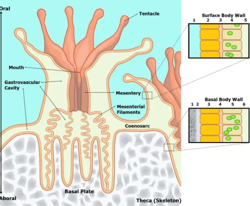
1.3 - Coral Symbiosis: the marriage between an animal and a plant. Scleractinian corals are
colonial animals, and their basic unit is the polyp. It has a simple shape of a cilindrum with an oral disk in the upper part and a basal plate in the opposite disk. In the oral disk, there is a mouth, in the middle, surrounded by tentacles used to capture preys. The mouth opens to the gastrovascular cavity, also known as coelenteron, divided by mesenteries forming compartments. In few words, the polyp resembles the sea anemones. Like all other Cnidaria, corals are diploblastic having the basic two layers’
organization; the endoderm and the ectoderm. In these organisms, the endoderm origins the gastrodermis and the ectoderm origins the epidermis. Usually, these layers are only one cell thick separated by a layer of connective tissue called mesoglea, consisting of collagen, mucus and “wandering cells”. The polyps are connected by horizontal sheets of tissue known as coenosarc extending over the superficial surface of the skeleton and completely covering it. These sheets are continuous with the body wall of the polyps and include extensions of the gastrovascular cavity of each polyp forming a common gastrovascular cavity that interconnects all the colony and it might work to transfer food, zooxanthellae, and waste compounds among polyps. In addition, the upper layer of the ectoderm (epidermis) is in contact with the sea water while the lower layer (aboral) is in contact with the skeleton, being these cells the responsible to secret the materials to build up the skeleton (Muller-Parker et al., 2015; Titlyanov and Titlyanova, 2002; Galloway et al., 2007) (Figure 1).
Figure 1 – Coral polyp anatomy.
Surface Body Wall: 1 – seawater; 2 – epidermis; 3 – mesoglea; 4 – gastrodermis with
zooxanthellae; 5 – gastrovascular cavity.
Basal Body Wall: 1 – Corallum (skeleton); 2 – calcifying medium; 3 - epidermis; 4 –
mesoglea; 5 – gastrodermis with zooxanthellae; 6 – gastrovascular cavity
Source: Adapted from http://www.dkfindout.com/uk/animals-and-nature/jellyfish-corals-and-anemones/inside-coral-polyp/
9
Although the name does not have taxonomic value, “zooxanthellae” is primarily used to describe the dinoflagellates that live-in symbiosis with the corals (Genus Symbiodinium), but a general use of the term is applied for every symbiont algae that live-in animals (Muller-Parker et al., 2015). Zooxanthellae inhabit in the endoderm cells of the coral’s tissue at heterogeneous densities of 0.5 to 5 million/ cm2 of coral (Smith et al., 2005). They are between 8-12 µm in diameter and have all the structural elements of a typical dinoflagellate. Their chloroplast contains all the characteristic photosynthetic pigments of a dinoflagellate: a and c2 chlorophylls, peridinin, diadinoxanthin, dinoxanthin, and β-caroten. Inside the host, the algae stay in a coccoid form, immobile, with or without a perisymbiotic membrane and reproducing by mitotic division. However, they can live freely outside of the host’s body acquiring two flagella and all the structural elements of a free-living dinoflagellate (Muller-Parker et al., 2015, Titlyanov and Titlyanova, 2002). Initially it was thought that the zooxanthellae that inhabit the corals belonged to a single species,
Symbiodinium microadriaticum, however, in the last years, with the advance of the new genetic
analysis, it was suggested that several zooxanthellae clades exist and it was proven that they have a large genetic variety with several taxa or genotypes living in the same host species or even in the same host organism (Muller-Parker et al., 2015; Little et al., 2004).
As previously said, when their metabolic needs are satisfied, microalgae zooxanthellae translocate the photosynthetic products to the host, thus the coral can use this resource for their own growth, both for soft tissues and for the calcified skeleton. Therefore, it can be said that the zooxanthellae take a large role in the coral's growth (Shutter et al., 2011). In contrast, the host provides shelter and important nutrients to the microalgae’s metabolism, such as nitrogenous compounds, phosphates, and CO2; that the coral itself acquires by its heterotrophic feeding (Smith et al., 2005). Furthermore, due to its optimal light reflection properties, the calcium carbonate skeleton also improves the light capture by the symbionts (Osinga et al., 2011a). About 90% of the food requirements of the coral can be derived from zooxanthellae photoassimilates – fatty acids, sugars, amino acids and even some vitamins (Titlyanov and Titlyanova, 2002). Corals also have heterotrophic feeding that comes from the predation of zooplankton, filtration of particles, absorption of dissolved organic substances (DOS) and the digestion of old or dead zooxanthellae (Titlyanov and Titlyanova, 2002). All these heterotrophic feeding strategies are the main source of organic compounds of nitrogen and phosphorus, nutrients that take a role in the metabolism of the coral, and of the zooxanthellae too. Additionally, it was found that nitrogen-fixing bacteria also inhabits corals and contributes as a source of nitrogen (Lesser et al., 2004). It was demonstrated that the coral cannot survive only by heterotrophic feeding since this source of food does not provide the amount of energy and certain substances that zooxanthellae can provide (Titlyanov and Titlyanova, 2002). Both
10
organisms obtain from seawater the inorganic compounds needed for their metabolism, such as CO2,
and bicarbonate for the plant symbiont’s photosynthesis and ions of calcium and carbonate for the coral’s skeleton. The CO2 is acquired not only from seawater but also from the respiratory metabolism
of the host. In brief, it was this close relationship, with large interchange and recycling of resources and energy that allowed corals and algae to successfully evolve and habit in an oligotrophic environment like tropical and subtropical water, and beyond that, they create a whole complex structure and ecosystem that is a true hotspot of biodiversity (Muscatine and Porter, 1977).
1.4 – Coral Bleaching - When the marriage evolves into divorce.
Under stress conditions, the coral-zooxanthellae symbiosis can be strongly affected resulting in the expulsion of the zooxanthellae and/or a loss of photosynthetic pigments, and subsequent reduction of pigmentation in corals, a phenomenon called coral bleaching. This bleaching is a reaction to abnormal environmental conditions and bleaching events have been highly correlated with the rising of seawater temperature. An increase of less than 2ºC above the average summer maxima is enough to cause coral bleaching (Jones et al.,1998; Goulet, 2006; Smith et al., 2005). The coral relies entirely on its heterotrophic feeding being able to survive for some weeks or even months. But, if the abnormal conditions persist, it leads to the coral’s death since the host is highly dependent on the food provided by the algae. Bleaching has been the biggest concern of the scientific community since global warming has caused changes in ocean’s temperatures. However, it has been hypothesized, that bleaching has an adaptive function that enables the coral to switch to a more suitable and adapted population of zooxanthellae, either by colonization of new genotypes or by rebalancing the dominant genotype in their tissue’s native populations (Goulet, 2006).
As referred before, bleaching is, in sensu lato, the interruption of the symbiotic relationship between the coral and the zooxanthellae, in the presence of stress. However, it is a complex mechanism that is triggered by a set of several factors at the metabolic, cellular and biochemical level. Zooxanthellae have the capacity to acclimatize to rapid and accentuated fluctuations on light regimes, however, when the rate of excitation (absorption of light) exceeds the photosynthetic rate the mechanisms to dissipate the excess of energy are activated to avoid damage of the photosynthetic apparatus. These mechanisms are energetically expensive, and so, the photosynthetic efficiency or yield is reduced, occurring what is called photoinhibition. But, since this energy dissipation ability is limited and other factors, such as temperature, UV radiation, pathogens, and others, can additionally increase the susceptibility to photoinhibition, then, over-excitation can lead to a total disruption of the photosynthetic system. Either when the Calvin cycle is interrupted or saturated, or the photosystem II (PSII) is damaged, or even when the fold/unfold mechanism of certain proteins of the
11
photosynthetic apparatus became slower, the whole system is no longer capable of dissipating or using all the energy/photons and these disruptions lead to the formation of reactive oxygen species (ROS) that are extremely harmful to the alga and the host. Both organisms are capable of reacting and neutralizing the damage, producing anti-oxidants or repairing proteins, but at a certain level, especially if there is a long-lasting stress, the host notices that is no longer advantageous to have this ROS producing symbiont and expels it. The release of zooxanthellae is also triggered if the photosynthesis is interrupted or reduced and the amount of photoassimilates shared with the host is lower than usual. This especially happens during extended darkness conditions, where none of the structures in the organisms are damaged but the photosynthesis stops (Douglas, 2003; Jones et al., 1998; Bhagooli and Hidaka, 2004; Smith et al., 2005; Baird et al., 2009; Obura, 2009; Roth, 2014). 1.5 – Coral’s Growth
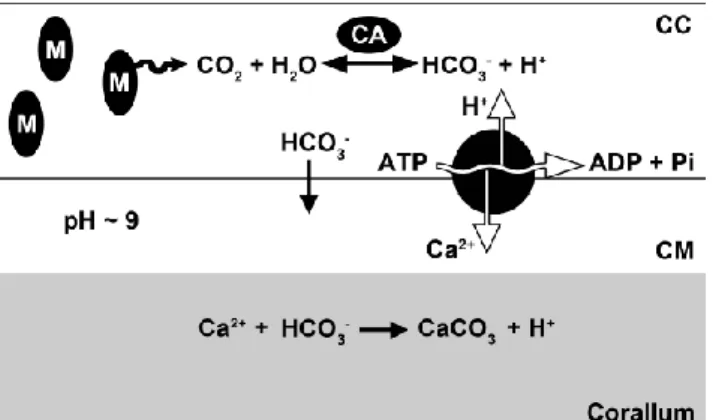
One important physiological process of scleractinian corals is the ability to build up a hard skeleton of calcium carbonite (aragonite), a process named calcification. This skeleton provides structural support for the whole colony, and each polyp lives in a small depression in the skeleton, the corallites, that allows to shelter against predators when polyps retract into them. It is the aboral ectoderm cells, also known as calicoblastic cells, that produce the corallum,
through the secretion of calcium ions. (Figure 2) This ions secretion is made by means of a Ca2+/H+ ATP-ases from the cytoplasm of the cell to a fluid layer between the cell and the corallum, known as the calcifying medium. The ATP-ases work in an antiport system extracting H+ from the calcifying medium in exchange for CA2+, and using ATP as energy to pump ions against the concentration gradient. The source of this ATP is the respiration of the compounds derived from the zooxanthellae’s photosynthesis or heterotrophic feeding. Consequently, the increasing concentration of calcium ions and high pH in the calcifying medium results in a supersaturation of calcium carbonate. Thus, the calcium carbonate precipitates and creates aragonite crystals producing the skeleton. Otherwise, the carbonate concentration mechanism in the calcifying medium is unknown, but, by removing protons and increasing the pH in the medium the equilibrium favors the CO32- instead of HCO3-
.
(Wijgerde,2013; Osinga et al, 2011). Zooxanthellae also take an indirect role in growth, beyond the energy provision to its host, as they increase the internal pH, due to their regular photosynthetic activity,
Figure 2 - Schematic overview of the calcification process in
scleractinian corals. Carbon dioxide produced by calicoblastic cells’ (CC) mitochondria (M) is converted to bicarbonate by the enzyme carbonic anhydrase (CA). Bicarbonate diffuses or is transported to the calcifying medium (CM). Source: Wijgerde, 2013
12
facilitating the formation of aragonite crystals, in what is called light-enhanced calcification (Osinga
et al, 2011; Pearse and Muscatine, 1971)
.
1.6 – Factors that influence the Growth
Four major factors are recognized to be fundamental for coral growth: Light, waterflow, aragonite saturation state and nutrients. There are other factors that can negative or positively influence this process, including, pH, temperature, competition or predation, trace elements, sedimentation, oil, pollutants, sunscreens, UV radiation, dissolved oxygen, genotype, etc. (Osinga et al, 2011). It is important to note that each coral species has a different response to different levels of each factor and that all these factors can interact among themselves. Following, some of this factors will be briefly described.
Light
Light is a key factor on coral’s growth since its symbiont is photosynthetic and that is the reason why in nature it is not usual to find photosynthetic scleractinian corals below 60m depth (Lesser et al., 2010). Higher photon flux densities are positively correlated with faster skeleton growth. Up to a certain limit, an increase in light quantity will enhance photosynthetic rate that leads to more energy translocated to the host. This effect is commonly referred to as light-enhanced calcification. Usually, calcification in light is found to be 3 to 4 times higher than in darkness (Shutter el al., 2011). Light also affects coral quality-related aspects such as physiological condition, shape, colour and metabolite content (Leal et al., 2014).
Light manipulation in ex-situ facilities comprises quantitative (irradiance), qualitative (light spectrum) and technological aspects (types of light sources). Changes in light regimes have demonstrated modifications in the population of zooxanthellae, the efficiency of photopigments, and all these affect the physiology and survival of the host (Rocha et al., 2013; Osinga et al., 2011). These photoacclimation mechanisms include modifications on zooxanthellae density, pigment concentration, pigment composition, production of photoprotectants etc (Osinga et al., 2011; Leal et al., 2014). Furthermore, a host growing under low light regims will try to expose more horizontal surface to the incoming light and will thus develop a more flattened shape than a specimen of the same species growing under high irradiance (Leal et al., 2014). Light variations also influence the colour. Corals under low light regimes may have an increase of pigments in their symbionts resulting in brighter and more intense colours. However, in moderate or high light regimes the same can happen, since occurs the production of photoprotective molecules, such as fluorescent proteins
(
Leal et al., 2014).13
Although photoperiod is an integral part of light conditions, it will be addressed in detail in the section 1.8.
Water flow
Scleractinian corals cannot generate actively water movements; thus, they are closely dependent on ambient water flow to facilitate their metabolism. All the exchanges from the external environment are reliant on the flow, that enhances the exchange of gasses, dissolved compounds, and food. Without a proper flow, the depletion of resources and the accumulation of toxic waste products compromise the survival of the coral. In addition, flow inhibits the settlement of sessile organisms on the coral’s body surface and removes any sediments and particles. Otherwise, high flows can have negative effects on corals, namely the deformation of the polyp’s shape reducing its predation efficiency (Osinga et al, 2011; Leal et al, 2014).
The Aragonite Saturation State
The Aragonite Saturation State is the product of the concentration of the dissolved calcium (Ca2+) and carbonate (CO
32-) ions divided by the temperature dependent solubility of the aragonite,
which is represented by Ω. Both these crucial components to calcification are actively concentrated into the calcifying fluid as it was explained above. Ω and pH are, in the calcifying medium, well above the sea water levels. Thus, it is stated that the concentration of this ions is positively correlated with calcification. Although in nature Ca2+ concentration is stable, in an aquarium, the amount of calcium is rapidly absorbed by the corals growing. In the case of carbonate, it is absorbed even faster and contrarily to calcium, the availability of carbonate in the seawater varies depending on biological and chemical processes that occur in the environment, particularly processes that change the pH. The pH strongly determines the concentration of carbonate, and that is why the monitoring of the pH is very important in an aquarium. For this reason, ocean acidification is threatening corals in nature because lower pH decreases CO32- availability in seawater (Osinga et al., 2011; Comeau et al., 2013;
Ohde and Hossain, 2004; Marubini et al., 2001).
Inorganic Nutrients
Organic and inorganic nutrients are very important for the metabolism and for the growth for the partners of the holobiont. The inorganic nutrients such as nitrogen and phosphorus are used as blocks for the synthesis of proteins and other organic components. Although the algae can obtain these nutrients directly from the sea water, the coral obtains them mainly by ingestion of food and by translocated compounds from zooxanthellae. But if the dissolved inorganic nitrogen (DIN) is limited the translocated food from the algae to the coral is poor in nitrogen and only provides metabolic energy to the host, missing the necessary building blocks for the biosynthesis. Many authors have
14
stated that the addition of DIN improves the overall performance of the holobiont by augmenting the zooxanthellae’s growth and increasing the concentration of pigments. However, if the concentration of DIN increases above the natural ambient concentrations it can have a negative effect on corals skeleton growth. The dissolved inorganic phosphorus (DIP) is important to be in balance with the DIN, otherwise, if added without a corresponding increase of DIN, it leads to the formation of polyphosphate crystals that have a negative impact on the coral’s growth. Beyond DIN and DIP, iron and zinc also influence the growth of the holobiont. Iron and zinc also benefit the coral’s growth when their amounts increase, but like DIN, when they reach concentrations above natural environmental levels they are harmful. Both play a role as components of many enzymes. Zinc, for example, makes part of carbonic anhydrase. This enzyme, in particular; that is used to capture dissolved inorganic carbon, is important both for photosynthesis and calcification. Nevertheless, high amounts of zinc can lead to adverse effects on growth due to the formation of toxic free radicals, that are harmful to zooxanthellae (Osinga et al., 2011).
Organic Nutrients
Heterotrophic feeding of scleractinian corals, as was previously summarised, takes the role of supplementing the holobiont with organic nitrogenous and phosphate compounds. Contrarily to the addition of DIN, that can inhibit the growth, feeding can stimulate positively both members of the holobiont, because organic food provides the nitrogen, carbon, and phosphorus in the right ratio and does not interfere with the nutritional balance (Osinga et al., 2011; Leal et al., 2014; Wijgerde, 2013). Several species were recorded to have improvements in their growth rates when fed, besides improving its resilience to stress and its tissue-skeleton ratio that is critical for drug production (Leal et al., 2014; Wijgerde, 2013).
Other factors
Others factors like competition, predation, pollutants, UV radiation etc, are especially important in nature or in in-situ production, and so, they will not be addressed in the present document. Furthermore, genetic factors also influence coral growth rate. In aquaculture facilities, corals are reproduced asexually by propagation, producing several identical clones from the original colony. All the clones, from the same original colony, are genetically identical, having the same set of genes – it means that all them belongs to the same genotype. Each genotype grants different responses to the environmental conditions, besides having different strategies, with some genotypes investing more in growth while others invest in disease and stress resistance. In production facilities it is common to use the same genotype to produce several clonal colonies what brings advantages for replicability purposes. In addition, the genetic variability of zooxanthellae also increases the variety
15
of responses of the holobiont to the environment (Osinga et al., 2011. Leal et al., 2014; Muller-Parker et al., 2015). Dissolved oxygen (DO) is a large lacuna in coral’s growth knowledge mainly, due to the complexity of working with low DO levels (Osinga et al., 2011). The other factors that can influence the coral’s growth, such as temperature and photoperiod, that are the main topic of this project are addressed in the following sections.
1.7 – Temperature
Comparatively to other factors, the effect of temperature on coral growth and metabolism was scarcely studied as most of the studies focused on natural environment events and bleaching of reef communities (Gates et al., 1992; Jones et al., 1998; Coles and Fadlallah, 1991; Hoegh-Guldberg and Fine, 2004; Smith et al., 2005). In general, all these studies refer to a typical bleaching event associated with thermal shocks and in some severe situations, mortality of the corals. Furthermore, most of this studies are focused on the impact of high-temperature stress, with low-temperature stress mostly ignored (Kemp et al., 2011). In production systems, lower temperatures can prevent possible disease outbreaks since pathogens increase their growth and virulence at higher temperatures (Sheridan et al., 2013). As stated previously (see section 1.4), the temperature shocks lead to zooxanthellae’s photoinhibition and photodamage and induce its release by the host, and this seems undoubtedly to be the major consequence of thermal stress (Hoegh-Guldberg and Smith, 1989, Jones et al., 1998). The same pattern was observed in other zooxanthellae symbiotic cnidarians (Steen and Muscatine, 1987; Muscatine et al., 1991; Hoegh-Guldberg et al., 2005) High temperatures reduce the tolerance to photoinhibition (Bhagooli and Hidaka, 2004) and the resistance to heat stress depends on zooxanthellae genotype and on the capability of the host to react to that stress and its associated damages, that varies from species to species (Fitt et al., 2009; Flores-Ramírez and Liñán-Cabello, 2007). The symbiotic corals are geographically limited to regions where the water temperature does not drop below 18ºC (Saxby et al., 2003). Most of the conducted studies with low-temperature stress reported similar responses as for high-temperature stress (Jokiel and Coles, 1977; Saxby et al., 2003; Hoegh-Guldberg et al., 2005; Roth et al., 2012). Published research on the effects of low temperatures in individual colonies under controlled conditions is to our knowledge scarce (Jokiel and Coles, 1977; Coles and Jokiel, 1977; Reynaud et al., 2004; Saxby et al., 2003; Al-horani, 2005; Roth et al., 2012) , however, the results are consistent with the field observations of cold condition events: bleaching effect (Hoegh-Guldberg and Fine, 2004; Coles and Fadlallah, 1991).
16
Overall, temperature-dependent growth in corals follows a bell-shaped curve (Figure 3) with a maximum that corresponds to the optimum and with the extreme temperatures having detrimental effects on growth. Jokiel and Coles (1977) observed lower growth rates at 21-22º C and rapid mortality at 18ºC in several Hawaiian coral species. In addition, the same authors observed strong regressions between temperature with photosynthesis and respiration (Coles and Jokiel, 1977). Saxby et al. (2003)
observed that cold temperatures have a negative effect on Montipora digitata, decreasing its photosynthetic efficiency, loss of zooxanthellae and pigments, and even bleaching and death. Nonetheless, moderate cold stress resulted in acclimatory responses of the holobiont. They also observed different responses according to the light regime, being the corals exposed to low light regime less affected by thermal stress (Saxby et al., 2003). Roth et al. (2012) observed similar patterns, with both, higher and lower temperatures (± 5ºC), resulting in slower growth, loss of zooxanthellae. However, they suggest that long-term high
temperatures exposure have stronger negative effects on Acropora yongei than long-term cold temperatures; the corals at higher temperatures stopped growing and bleached while the lower temperature ones kept a good number of zooxanthellae and grew. They also observed some level of acclimation by the coral at lower temperatures (Roth et al., 2012). In contrast, Jokiel and Coles (1977)
had opposite results when studying Hawaiian corals and observed stronger harmful effects to colder water stress than to higher temperatures (± 4º C) (Jokiel and Coles, 1977). Marshall and Clode (2004), compared the calcification rate of a zooxanthellae coral and an azooxanthellae coral along a temperature range (18-29ºC), observed a similar temperature dependence in both corals (Figure 3) and that the temperature influences calcification independently from light exposure. They suggest that the Calcium-ATPase has a temperature dependent activity and it shows consistency with the consensual thermic optimum to tropical coral’s growth (25-27ºC) (Marshall and Clode, 2004 and their references). Reynaud et al. (2004) observed higher growth rates and Strontium/Calcium incorporation with higher temperatures (20-29ºC) in Acropora verweyi (Reynaud et al., 2004). Many of these works also indicate that the time of exposure of the organism to the stress, the magnitude of the stress factor, and the thermal history of the specimens have and important role in the response of the organism to the thermic changes, with usually the sudden and long-term changes that cause
Figure 3 - Calcification rate (measured as calcium incorporation
per unit mass of skeleton) along a temperature range in a zooxanthellae coral (Galaxea fascicularis) and an azooxanthellae coral (Dendrophyllia sp.). Source: Marshall and Clode, 2004
17
serious effects (Jokiel and Coles, 1977; Coles and Jokiel, 1977; Coles and Fadlallah, 1991; Clausen and Roth, 1975; Saxby et al., 2003; Edmunds, 2009; Howe and Marshall, 2002; Roth et al., 2012).
1.8 - Photoperiod
The positive correlation between light and growth is already well studied: larger quantity of light results in better growth, until the photoinhibition trigger point. Higher photon flux densities are positively correlated with faster skeleton growth. Up to a certain limit, an increase in light quantity will enhance photosynthetic rate that leads to more energy translocated to the host.
To provide more light to a rearing system, usually, that can be achieved by increasing the light irradiance, however, the quantity of light available for zooxanthellae is not only established by the intensity of the source but also by the length of the stimulus – the photoperiod. At the moment, almost no research has been conducted on the effects of photoperiod on coral growth and physiology and there is no information about the optimal number of hours of light per day that corals need. Schutter
et al. (2011) did the first attempt to reveal such effects. The authors theorized that increasing the length
of the photoperiod corals would increase their daily growth rate. They tried different combinations of light irradiance and photoperiods, but, their results were not conclusive, since the growth was not significantly different between Galaxea fascicularis grown at 8 and 16 hours light. Nonetheless, they note that the corals had the ability to adapt to extended photoperiods (Schutter et al., 2011)
.
2 – Objectives
As was described previously, light and temperature play a strong role in the physiology of corals. According to the presented state of art, it can be observed that temperature and light regimes are closely related and interact in the coral’s physiology. Understanding the interaction between these factors is very important to improve the ex-situ production of these organisms. The aim of this thesis project was to assess the effects of different temperatures and modifications in the photoperiod, on the growth and physiology of corals. In addition, the interaction between these two factors were also analysed.
Saxby et al. (2003) observed that corals exposed to cold temperatures and low light regimes had less detrimental effects than their warmer temperatures or high light counterparties. Thus, is it possible to hypothesize that low light regimes increase the tolerance of corals to thermal stress and can shorter photoperiods be an approach to achieve this tolerance? The present work will address these questions and the obtained answers could help to provide better production protocols and increase our knowledge in corals physiology and capacity of acclimation. Several approaches, beyond the growth rate measures, will be taken into account to achieve these objectives. They will be, the
18
zooxanthellae’s density, to obtain the condition of the symbiotic relationship; chlorophyll fluorescence measurements, to assess the damage and efficiency of the photosynthetic apparatus, and pigments absorbance’s will be measured to determine the amount of different pigments: chlorophylls, carotenes, and xanthophylls. Many of these pigments not only take a role in photosynthesis but also work as photo protectors and antioxidants, which allows the evaluation of the response and aclimation to light and thermal stress.
The main questions of this project are:
- How do corals respond to different photoperiods? – This is largely unknown in contrast to variations in light intensity.
- How do corals respond to cold water induced stress? – Will they respond similarly to heat stress?
- Do photoperiods and temperatures have any interaction on coral’s physiology? – Are modified photoperiods able to neutralize the effects of thermal stress? Corals at higher latitudes are susceptible to suffer changes in these abiotic factors both in summer and in winter.
- Can different combinations of photoperiod and temperatures optimize coral aquaculture? – Improvement of growth rates and reduction of production costs.
- -
3 - Materials and Methods
3.1 – The Corals
One species of stony coral was chosen to be part of this experiment - Stylophora pistillata (Esper, 1797)) - an Indo-Pacific species. This S. pistillata clone was originally from Eilat Gulf (Israel) and the colonies were at coral’s lab of Wageningen University. They all belong to the same genotype. These coral colonies one year before, when nubbins (10 polyps clones), were fixed with aquarium epoxy to square shaped 5×5cm PVC plates and put in a 360L culture tank prior to start of the experiment. Light in this tank ranged from 100 to 200 μmol quanta m-2 s-1. with a 12L:12D
photoperiod (12 hours light:12 hours dark). Tanks were maintained at a constant temperature of 25-26ºC and a salinity of 34.0-35.5 ppt (artificial seawater - Tropic Marin: type = Zoomix). For the present experiment, 96 colonies were used and randomly distributed into the experimental tanks. Colonies are identified by a number written in their PVC plates.
19 Ethical Note: None of the specimens used in this experiment was collected from nature. All of them were captive breed in ex-situ facilities for research and/or educational purposes.
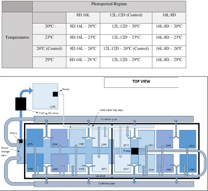
3.2 – Experimental design
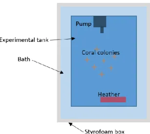
The experiment was conducted in the aquatic animal’s facilities at Wageningen University. In the present work 3 different photoperiod regimes and 4 different temperatures, were combined and tested resulting in 12 different treatments. Each treatment had two replicates, resulting into 24 tanks for the whole experiment. The photoperiod regimes were 8L:16D; 12L:12D and 16L:8D and the temperatures were 20º, 23º, 26º, and 29ºC (Table 1). The treatment 26ºC 12L:12D was set as control treatment since these are the same conditions kept in the nursery tanks. All experimental tanks were part of a single recirculation system. The recirculation system consisted of 24 experimental tanks (20L), a water storage tank (300L) and a bypass for chilling water (≈20ºC). Water was pumped from the water storage tank to a height above the experimental tanks where it is distributed along the tanks. The inflow in the tanks was set to accomplish a turnover rate of the tank’s volume per day (approximately 65 mL/min). The outflow was discharged by hoses to the storage tank by gravity. Water of the storage tank was maintained at ≈23ºC resorting to chilling machines. The tanks were placed in shelf and separated per photoperiod regime. Each tank was conditioned to the respective temperature. The tanks at 23ºC were at the same temperature of the storage tank water; tanks at 26º and 29ºC were heated using aquarium heaters (50W) while the 20ºC treatments were chilled by a chilling machine that makes part of a bypass in the system, specifically to chill the water for this treatment (Figure 4). To attain temperature stability, each tank was placed into a Styrofoam box, itself filled with water forming a bath (Figure 5). All the temperatures were monitored at every 2 days. The water storage tank had a foam fractionator (skimmer) and a UV filter. An irradiance level of 140-160 μmol quanta m-2 s-1 was provided by 187W Philips CoralCare LED lights and the photoperiod settled
using the software from the Philips’ lights. The water flow inside experimental tanks was created by an aquarium pump, compact 1000 (Eheim®, Germany). The corals were fed 3 times a week with 2 ml of Artemia nauplii hatched from cysts (Great Salt Lake Artemia cysts, Artemia International LLC, Fairview, USA; at a salinity of 25 gL−1 and a temperature of 28◦C and used immediately after hatching).
20 Table 1 – Experimental treatments
Photoperiod Regime Temperatures 8D:16L 12L:12D (Control) 16L:8D 20ºC 8D:16L – 20ºC 12L:12D – 20ºC 16L:8D – 20ºC 23ºC 8D:16L – 23ºC 12L:12D – 23ºC 16L:8D – 23ºC 26ºC (Control) 8D:16L – 26ºC 12L:12D – 26ºC (Control) 16L:8D – 26ºC 29ºC 8D:16L – 29 ºC 12L:12D – 29ºC 16L:8D – 29ºC
3.3 – Sampling: Parameters of coral’s growth and condition
Data collection was taken at the beginning of the experiment (time 0) and according to the analysis in intervals of 2 or 4 weeks. The measurements took a whole week since it was not possible to do all the measurements for all colonies in a single day. The experiment started after a 3-week period: first, one week of acclimation of the corals to the new system without treatment conditions. One week later, was used for the initial data collection (time 0) without treatment conditions, lastly, in the final week to slow transition to each experimental treatment. The experiment was planned to last for a minimum of 3 months (or 84 days – 7 days x 4 weeks x 3 months).
Figure 4 – Top view of the experimental setup. This scheme represents only 16 of the 24 experimental tanks. The shelf where the
21 Growth Rate
Buoyant weight (BW) is a practice to weight hanging the live sample by a threat (usually hooked to the PCV plate) connected to a balance. The coral is submerged in a water and the weight is recorded, avoiding stress due to air exposure. BW is good estimate of skeletal weight since coral tissue has a density which is close to that of seawater and does not contribute significantly to the buoyancy. Tissue represents only 1% of the total buoyant weight (Schutter et al., 2008). To measure coral skeletal growth, the increase in buoyant mass was used. In this
procedure, according to Schutter et al. (2008), coral colonies are taken from their experimental tank and suspended on a nylon thread with a hook, attached to an analytical balance in a predefined volume of seawater (35ppt) at a constant depth. The temperature of this seawater was at 25ºC to standardize the water density for all measurements. Averages of at least 3 measurements were used to create an estimate of buoyant mass. Prior to the attachment of coral nubbins to their PVC plate, the buoyant mass was measured and compared to the buoyant mass of coral nubbins attached to their PVC plate. This was done, in order to extract the mass of the PVC plate and glue from later measurements. Previous data of PVC plates was already available for the colonies of S. pistillata, to be used in this experiment. Buoyant mass was used to calculate the specific growth rate (μ) using the following formula;
Specific Growth Rate (μ) = (ln BMt – ln BMt0)/Δt
where the specific growth rate is expressed in BM day-1. BMt is the buoyant mass at the end
of a growth interval, BMt0 is the buoyant mass at the start of a growth interval and Δt is the time of
the growth interval in days.
Metabolic Rates
Net photosynthesis and dark respiration were measured by means of intermittent flow respirometry in a respirometric flow cell according to Schutter et al.
(2008)
. Three colonies of each tank, were randomly chosen and used for the measurements. Each colony was placed in the respirometric flow cell for 60 minutes. The water in the flow cell is originated from the experimental recirculatory system and it was kept at a temperature identical to each treatment and renewed after each measurement. A magnetic stirrer was used to ensure adequate mixing and to simulate the22
situation in the tank environment. Oxygen levels within the enclosure were recorded, by means of a luminescent oxygen probe (Hach). Oxygen consumption was measured in the dark and oxygen production was measured under similar light conditions to the experimental tanks (140-160 μmol quanta m-2 s-1), during 30 minutes each (total of 60 min of incubation). Oxygen consumption/production was then be calculated using the following formula:
Rd/Pnet = ((Vcell−Vcoral) x slope)/ BM (mg O2/gBW/min)
Where Rd/Pnet is the rate of dark respiration or the net photosynthesis (μmol O2/min/gBW);
Vcell is the volume of respirometric flowcell (l); Vcoral is the volume of coral; the slope is the regression
coefficient of dissolved oxygen against time (μmol O2/min), and BM is the buoyance weight of coral
(g). Surface-area was not used, as determination of the surface-area is believed to be very hard to measure for the branching S. pistillata. Later the daily photosynthesis/respiration ratio was calculated:
Daily
PR
ratio =
Pnet x hours of ligh day⁄ – Rd x hours on dark day⁄
Rd x hours on dark/day (Dimensionless)
The measurements were performed in the week before the beginning of the treatment conditions.
Chl a fluorescence measurements
Chl a fluorescence was measured using a pulse amplitude modulated (PAM) fluorometer according to Saxby et al. (2003). The fluorimeter was used to measure the minimal (F0) and maximal
(Fm) fluorescence yields. The measurements were done during the morning period, approximatey one
hour after the start of the photoperiod. Fluorimeter optical head was placed perpendicularly adjacent to the surface of the coral and fluorescence measured. 9 measurements were randomly obtained in the colonies of each tank. The measurements were performed at the start of the experiment and then, biweekly. The fluorimeter gives the quantum yield (Fv/Fm), the ration between variable fluorescence
(Fv) and maximum fluorescence (Fm). This value provides a good approximation of the maximum
photochemical efficiency of Photosystem II (PSII).
Zooxanthellae density
At the end of the experiment, 3 random branches of coral from each tank were cut and used as samples. Then the tissue from each branch was removed using high pressured air. The resulting slurry from each sample was homogenized by shaking it in 10ml of seawater. Samples were then centrifuged for 10 min at 3000 rpm and the supernatant was rejected after and the pellet was re-suspended in 2ml of seawater. Zooxanthellae were counted (3 replicate counts) using a counting chamber assembled in an inverted microscope and using an image software Fiji ImageJ.
23
Zooxanthellae density was expressed as the number of zooxanthellae per cm2 surface area. Surface area was determined by, covering the branches with a single layer of aluminium foil. The foil was removed and subsequently weighted. The area was then calculated comparing with the weight of a 1cm2 foil.
Pigment content analysis
Using the same homogenate obtained from zooxanthellae isolation, a 0.5ml aliquot homogenate was taken to determine the content of chlorophylls a, b and c, carotenes and xanthophylls. To obtain it, 4.5ml of acetone was addes to this aliquot and then was placed in a freezer (–20°C) for 24 h. The solution absorbance’s were determined at 664, 630 and 750 nm on a spectrophotometer to determine chl-a, chl-c, and turbidity, respectively. The same method was applied to measure peridinin, diadinoxanthin, dinoxanthin, and β-carotene concentrations with respective wavelengths; 442, 447, 466, and 454 nm (Jeffrey and Haxo, 1968). The concentrations of chlorophyll-a and chlorophyll-c were calculated according to the equations given by Jeffrey and Humphrey (1975) for dinoflagellates.
3.4 – Statistical Analysis
A 3-way repeated measures mixed ANOVA was conducted in SPSS software (IBM Corp., 2016) to determine differences between treatments for P/R ratio and measurements of photosynthetic efficiency. The three factors used in this test were photoperiod, temperature and, time. A 2-way ANOVA was used to test the significance of zooxanthellae density and pigments content, as well as to growth rate and hourly production/consumption of oxygen (one test for T0 and another for T30).
Before, a F-test and, subsequently, a T-test were performed to access the homogeneity of variances and differences in means, respectively, between replicates in order to evaluate the presence of tank effects. The 2-way ANOVAs used the tanks as experimental unit. Only zooxanthellae density data used each fragment sample as experimental unit, instead of the tanks, which due to, no significant tank effects were found. When significant differences were found, a Tukey’s post hoc test was used to attribute differences between specific treatments. A confidence level of 95% was used in all analyses.
4 - Results
Disease outbreak
On the 8th week of the experiment, the rapid tissue necrosis (RTN) was detected in some colonies. This unexpected event led to the abortion of the experiment, and to a fast collection of samples of remaining
24 healthy tissue. It also led to small changes in the zooxanthellae extraction protocol, since the samples were kept frozen, after collected, at -20ºC for later extraction and analysis. It is relevant to report that the disease was firstly detected in the tanks at 29ºC. In the following days, it was detected in the tanks at 26ºC. This disease is characterized by fast tissue degradation (peeling) and death of the colony. It is suggested to be caused by Vibrio harveyi, a bacteria that lives in the coral tissue (Luna et al., 2007).
Specific Growth rates
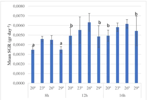
The overall mean specific growth rate (SGR) of the experiment was 0.005±0.00088 grams day-1 after
37 days. No later record of the BW was made because of the unexpected disease outbreak. The different experimental groups grew at different rates (Figure 6). A significant main effect of photoperiod was observed (Table 2): The 8 hours photoperiod treatments had a significant lower SGR compared with the other photoperiods; SGR at 8 hours light was 25,7% lower than at 12 hours light and 28,2% lower than at 16 hours light (p-value = 0.000 < 0.05 in Tukey’s test). S. pistillata grew similarly (p-value = 0.804 > 0.05) at the 12 and 16h light hour regimes, showing similar SGR, respectively.
The ANOVA test indicates a significant temperature effect (Table 2). Treatments at 20º and 29º were not significantly different as p-value = 0.957 > 0.05. These temperatures induced a lower SGR in all photoperiods. Statistically the for all photoperiods. SGR at 23º and 26º were not significantly different as p-value = 0.671 > 0.05 between them and both were different from 20ºC (p-p-value = 0.039 < 0.05 and p-p-value = 0.001 < 0.05). Additionally, 26º and 29º were also significantly different: p-value = 0.007 < 0.05). The highest mean of SGR was observed in the control treatment – 26ºC 12h – however, statically it did not differ from the other 23ºC or 16h treatments (Table 2). No interaction was found between photoperiods and temperatures (p-value = 0.891 > 0.05)
Figure 6 – Specific growth rates (SGR) for each experimental treatment after 37
days of experiment. n = 3. 0,0000 0,0010 0,0020 0,0030 0,0040 0,0050 0,0060 0,0070 0,0080 20º 23º 26º 29º 20º 23º 26º 29º 20º 23º 26º 29º 8h 12h 16h M ea n SG R (g r da y -1) a a b b b b
25 Metabolic rates
In the beginning of the experiment, photoperiods were significantly different for production and O2 consumption (Table 3).
The 8h photoperiod was significantly different from 12h in terms of oxygen production (p-value = 0.039 < 0.05), while 12h and 16h were different regarding consumption (p-value = 0.013 < 0.05) (Figure 7).
After 30 days, the longer photoperiod treatments (12 and 16h light) showed a great negative effect on metabolic rates. Hourly net photosynthetic rates in both cases were strongly reduced while respiratory rates, though they also decreased, were less strongly affected. (Figure 7 and Table 4). According to Tukey’s tests (Table 3) all comparisons between photoperiods were significant for O2 production. No
significant differences were detected between photoperiods regarding respiration rates. Initially, temperatures were significant for consumption and not significant for production (Table 3). Between 20º and 23ºC was detected significant differences (p-value = 0.035 < 0.05). 30 days later, temperatures were not significant for consumption and significant for production. Photosynthetic rates for treatments at 20ºC were different from 26ºC (p-value = 0.001 < 0.05)
The treatment 29ºC/16h was the only one with a negative photosynthetic rate after 30 days (Figure 7). It is also noteworthy that in the 12h photoperiod at the start of the experiment the several treatments showed strongly variable rates, and higher rates than the other photoperiods treatments. Moreover no treatments were, in fact, running at this point. Additionally, the control treatment (26ºC 12h light) was also affected, showing a
20º 23º 26º 29º 20º 23º 26º 29º 0 days 30 days 186,08 271,17 229,64 182,75 40,91 69,86 103,90 68,31 122,04 284,95 145,38 126,39 76,29 95,06 118,41 112,89 -50,00 50,00 150,00 250,00 350,00 450,00 O x y g en p ro d u ctio n /c o m sum p tio n m g O 2 /g B W /h o u r 12h Light treatment 20º 23º 26º 29º 20º 23º 26º 29º 0 days 30 days 145,91 175,08 144,90 176,07 73,17 153,37 151,98 150,53 120,97 147,86 112,77 134,78 112,82 111,09 85,58 100,16 -50,00 50,00 150,00 250,00 350,00 450,00 O x y g en p ro d u ctio n /co m su m p tio n m g O 2 /g B W /h o u r 8h Light treatment 20º 23º 26º 29º 20º 23º 26º 29º 0 days 30 days 161,17 160,04 175,72 180,55 16,04 55,13 39,48 -5,24 122,70 104,11 127,81 116,05 61,86 86,21 111,81 132,77 -50,00 50,00 150,00 250,00 350,00 450,00 O x y g en p ro d u ctio n /co m su m p tio n m g O 2 /g B W /h o u r 16hr Light treatment
Figure 7 – Hourly net photosynthesis for every treatment at T0 and after 30 days of experiment. In this analysis, the future treatment for each tank was predefined and they were analysed as separated treatments, though at T0 no treatment conditions were running. Blue = production; Orange = consumption; n = 3.
Production Comsumption Production Comsumption Production Comsumption
26 strong decrease after 30 days in
both photosynthetic and
respiratory rates. In the shorter photoperiod treatments(8h) the metabolic rates seem to be almost no affected. The only exception was the 20ºC treatments that had a relevant reduction in their photosynthetic rates.
Concerning the P/R ratio no significant effects both for temperature and photoperiod were observed, but there was a significant effect over time: p-value = 0.000 < 0.05. The test shows significant interaction
between temperature and
photoperiod with p-value = 0.012 < 0.05 (Table 3). Both values, in the beginning, and one month later, were quite variable (Figure 8). In the beginning, with no treatments running, one group displayed a negative ratio of -0.088 (23ºC 12L), and the remaining groups show a large range of values. The highest ratio was observed in the
treatment 23ºC 16h with a value of 0.87. The overall mean of the ratio between net photosynthesis and dark respiration was 0.406±0.251 After 30 days the major tendency observed is that almost all the treatments got a negative ratio. Only the treatments 23ºC 16h and 26ºC 8h kept a mean positive ratio. The treatment 29ºC 16h had the most negative value. The overall mean was -0.344±0.0344.
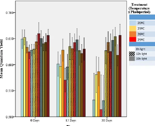
Photochemical efficiency of PSII.
The effects of both temperature and photoperiod were significant and a strong interaction occurred between both: p-value = 0.000 < 0.05 and significant differences between all the photoperiods and between the temperature 20ºC with the 23º and 26ºC were observed (Table 2).
Figure 8 – Mean P/R ratio at T0 and after 30 days of experiment for each treatment. In this analysis, the future treatment for each tank was predefined and they were analysed as separated treatments, though at T0 no treatment conditions were running. n = 3. 20º 23º 26º 29º 20º 23º 26º 29º 20º 23º 26º 29º 8h 12h 16h Série1 0,247 0,133 0,264 0,386 0,678 -0,08 0,678 0,433 0,343 0,870 0,372 0,559 -2,000 -1,500 -1,000 -0,500 0,000 0,500 1,000 1,500 2,000
T
0 20º 23º 26º 29º 20º 23º 26º 29º 20º 23º 26º 29º 8h 12h 16h Série1 -0,54 -0,25 0,188 -0,24 -0,47 -0,27 -0,13 -0,41 -0,51 0,256 -0,66 -1,06 -2,000 -1,500 -1,000 -0,500 0,000 0,500 1,000 1,500 2,00030 days
27 The average photochemical efficiency
(PE) of the PSII obtained in the beginning of the experiment was 0.64 ±0.03. Along the experiment was observed a great decay of the PE in the 16 hours treatments (Figure 9). At 30days it reached 0.55±0.098. This decrease was already observable after 15days of the experiment. Inside the 16hours photoperiod regime, the corals that seemed to be more affected were the ones exposed to 20º and 29ºC. The remaining photoperiods kept a high PE except for the treatment 20ºC 12h, that had a great decrease, similar to the 20ºC 16h treatment.
Zooxanthellae Density.
Strong effects of both temperature and photoperiod were found p-value = 0.000 < 0.05 (Table 5). The interaction between photoperiod and temperatures was significant (p-value = 0.036 < 0.05) The shorter photoperiod was different from 12 and 16h (p-value = 0.031 and 0.000 < 0.05). Treatments at 20ºC have differed from the other temperatures (p-value = 0.000 < 0.05) as well 23ºC was different from 26º and 29ºC, and the zooxanthellae density varied significantly between treatments (Figure 10). The 8h photoperiod was the one with higher mean density – 1003939.1 cells per cm2(±205493.81). The other
photoperiods had temperature groups that suffered a large reduction in the number of cells, which lead to lower mean values; 869962,9(±306666.74) and 751184.3(±307412.62) for the 12h and 16h respectively. It represents a reduction of 13.35% and 25.18% in zooxanthellae density respectively. Regarding the temperatures, is
Figure 9 – Quantum yield efficiency of each treatment at T0. 15 and 30 days of experiment. n = 9
Figure 10 – Zooxanthellae density for each treatment after 30 days of
experiment. n = 3
28 notorious that the lower temperature (20ºC) had the strongest reduction in the number of cells independently of the photoperiod. In addition, concerning this temperature it is visible the same effect of the photoperiods; the longer the photoperiod, the lower the zooxanthellae density. In the longer photoperiods (12 and 16h) a strong effect of the temperatures was observed, with a significant increase in the number of zooxanthellae following the increase in temperature.
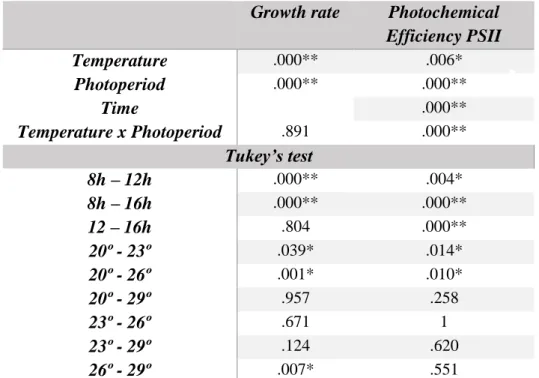
Table 2 – Statistical results of Growth rate and Photochemical Efficiency PSII
Growth rate Photochemical
Efficiency PSII Temperature .000** .006* Photoperiod .000** .000** Time .000** Temperature x Photoperiod .891 .000** Tukey’s test 8h – 12h .000** .004* 8h – 16h .000** .000** 12 – 16h .804 .000** 20º - 23º .039* .014* 20º - 26º .001* .010* 20º - 29º .957 .258 23º - 26º .671 1 23º - 29º .124 .620 26º - 29º .007* .551
Table 3 – Statistical results of O2 Production/Consumption and Photosynthesis/Respiration ratio. In this analysis, the future treatment for each tank was predefined and they were analysed as separated treatments, though at T0 no treatment conditions were running
O2 Production hr-1 O2 Consumption hr-1 P/R ratio
T0 30 days T0 30 days Temperature .557 .001* .022* .146 .157 Photoperiod .032* .000** .012* .939 .966 Time .000** Temperature x Photoperiod .656 .102 .007* .155 .012* Tukey’s test 8h – 12h .039* .000** .065 .988 .991 8h – 16h .919 .000** .796 .933 .962 12 – 16h .095 .001* .013* .976 .990 20º - 23º .481 .003* .035* .752 .530 20º - 26º .886 .001* .988 .408 .299 20º - 29º .936 .187 .998 .113 1 23º - 26º .892 .976 .077 .942 .977 23º - 29º .830 .399 .055 .575 .467 26º - 29º .999 .203 .999 .888 .251