José Avelino Martins Gonçalves
Development of protein-based formulations
for topical treatment of keratinaceous
appendices
Universidade do Minho
Escola de Ciências
José A velino Mar tins Gonçalv es De velopment of protein-based formulations for
topical treatment of k
José Avelino Martins Gonçalves
Development of protein-based formulations for
topical treatment of keratinaceous appendices
Tese de Mestrado em Biofísica e Bionanossistemas
Trabalho realizado sob a orientação de
Doutor Artur Jorge Araújo Magalhães Ribeiro
e coorientação de
Professora Doutora Andreia Ferreira Castro Gomes
Acknowledges/Agradecimentos
Durante este ano e meio que passou muitas pessoas foram fundamentais para que eu conseguisse atingir o objetivo de concluir esta etapa da minha vida.
Primeiro de tudo queria agradecer a ti, Artur, pela forma como me orientaste e pelo apoio que me deste tanto a nível científico como a nível pessoal. Conseguiste despertar a minha veia científica e todos os dias que ia para o laboratório ia com o pensamento de “O que vou descobrir hoje?”, isso fez com que eu tivesse sempre motivado para fazer o meu trabalho, MUITO obrigado mesmo não podia ter tido melhor orientador.
À professora Andreia também um muito obrigado pela sua disponibilidade, apoio e confiança que sempre demonstrou no trabalho que estava a realizar.
Ao professor Artur Cavaco Paulo queria agradecer por me ter aceite no seu grupo de trabalho e também pelas palavras que me dirigiu sempre a pensar no meu futuro como melhorar o decorrer das minhas experiências.
A todos os investigadores do BBRG que me inseriram de forma impecável no laboratório e sempre me trataram como parte deles, a vossa disponibilidade e companheirismo são notáveis. Em especial queria agradecer às minhas companheiras de bancada, Catarina e Filipa, que foram pessoas 5 estrelas no decorrer do meu trabalho e mostraram-se sempre disponíveis, até ao fim-de-semana, para me ajudarem em determinados assuntos, sem vocês teria sido muito mais difícil. Carla também te queria deixar uma palavra de agradecimento pela forma como me sempre ajudaste em determinadas técnicas que necessitei. À Filipa do Departamento de Física também um muito obrigado pela tua disponibilidade no procedimento e análise de algumas técnicas realizadas.
Telma minha companheira e amiga já nos conhecemos há alguns anos e continuas a ter um papel importante na minha vida. Foste imprescindível para que este ano e meio passasse de forma mais alegre e pelas ideias que discutimos de como será o nosso futuro no ramo científico.
Aos restantes meus amigos de Braga que tenho vindo a conhecer desde que entrei na Universidade vocês sabem o quanto foram importantes para mim e também pela ajuda que me deram sempre que necessitei, não vale a pena individualizar vocês sabem de quem falo.
Pessoal de Famalicão vocês também não foram esquecidos e, apesar de mais distantes, sempre que preciso vocês estão lá por mim, vocês são como irmãos para mim.
capacidades apesar das circunstâncias terríveis que nos abalaram. Estou cá por causa de vocês e quero que se orgulhem de mim para sempre tal como me orgulho de ser filho de quem sou.
Abstract
Keratinaceous appendices play an important role in the visual aspect of each individual. The chronic exposure of hair, nails and skin to external hazards have a negative impact on their structure. To prevent or revert the impact of such exposure, several cosmetic products have been developed and are now commercialized. Yet the use of cosmetic products is not restricted to this specific application as they are widely used in the adornment of the keratinaceous appendices, mainly for hair coloration.
In this thesis, two approaches for the development of new hair cosmetic formulations were tested. Keratin-based particles and two recombinant crystallins were designed and obtained. These were mainly focused on the restoration and improvement of hair mechanical properties.
Keratin-based particles composed by keratin (Kx), silk fibroin (SFy) and Poloxamer407
(Pz) (KxSFyPz) were obtained by high pressure homogenization. Particles size ranged between 150
and 250 nm with low polydispersity and a net negative surface, which was partially neutralized by the addition of poloxamer to particles’ formulation. Homogenization process resulted in an increase on the percentage of β-strands. All formulations do not show cytotoxic effect on human keratinocytes (NCTC 2544 cell line). The effects of keratin-based particles over virgin (VH) and overbleached (8xOB) Asian hair, were monitored regarding particles ability to bind to hair fibres and their effect over hair mechanical properties. Keratin-based particles increased hair fiber physically by 62.7% for VH and 83.4% for 8x OB, which was directly related with the quantity of β-strands present in the particle system as well their binding capability.
Two crystallin forms, a wild type (CrysWt) and a mutant (CrysMut), based on the sequence of human eye ɣD-Crystallin were expressed in E. coli using a commercial TB-AIM medium. The recombinant crystallins were obtained pure after purification with magnetic beads and the proteins had a molecular weight of 24.8 kDa and 24.6 kDa for wild type and mutant forms, confirmed by MALDI-TOF. Analysis of protein secondary structures revealed that the new crystallins retained the structure of human eye ɣD-Crystallin, which is rich in β-sheets. This type of structure
is typical of crystallin which belong to a family of proteins characterized by the presence of the Greek-key motif.
Outcome from protein formulation characterization supports the hypothesis that the keratin-based particles were able to recover and improve the mechanical properties of virgin and overbleached hair. Therefore, these particles can be considered as a very promising strengthening
Resumo
Os apêndices queratinosos desempenham um papel importante no aspeto visual de cada indivíduo. A constante exposição a perigos externos para cabelo, unhas e pele tem um impacto negativo na sua estrutura. Para evitar ou reverter o impacto dessa exposição, vários produtos cosméticos foram desenvolvidos e agora são comercializados. No entanto, o uso de produtos cosméticos não é restrito a esta aplicação específica, uma vez que são amplamente utilizados no adorno dos apêndices queratinosos, principalmente para a coloração do cabelo.
Nesta tese, foram testadas duas abordagens para o desenvolvimento de uma nova formulação cosmética para o cabelo. Partículas à base de queratina e duas cristalinas recombinantes foram projetadas e obtidas. Estas estratégias distintas são principalmente focadas no restauro e melhoramento das propriedades mecânicas do cabelo.
Partículas à base de queratina compostas por queratina (Kx), fibroína de seda (SFy) e
Poloxamer (Pz) (KxSFyPz) foram obtidas através de homogeneização de alta pressão. As partículas
possuíam tamanho entre 150 e 250 nm com baixa polidispersividade e carga superficial negativa, parcialmente neutralizada pela adição do poloxamer. Nenhuma formulação apresentou efeito citotóxico na linha celular NCTC 2544 de queratinócitos humanos. A capacidade das partículas se ligarem com as fibras e a sua deposição em cabelo virgem (VH) e cabelo descolorado (8xOB) foi analisada. A melhoria das propriedades mecânicas das fibras foi estudada através de testes de tensão. Em cabelo virgem houve um aumento de 62.7% e em cabelo descolorado de 83.4%. Estes aumentos estão diretamente ligados à quantidade de estruturas - no sistema particulado assim como a capacidade de ligação com a superfície da fibra do cabelo.
Os dois tipos de cristalinas, wild type (CrysWT) e mutante (CrysMut), baseadas na sequência da ɣD-Cristalina do olho humano foram expressas em E. coli em meio comercial
TB-AIM. As cristalinas recombinantes purificadas, obtidas após a purificação com “beads” magnéticas, apresentam peso molecular de 24.8 kDa e 24.6 kDa, para CrysWT e CrysMut respetivamente, confirmados pela técnica de MALDI-TOF. Análise à estrutura secundária destas proteínas puras revelara que estas mantêm uma estrutura rica em folhas , tal como é característico da estrutura em chave grega desta família de proteínas.
O resultado da caracterização da formulação proteica suporta a hipótese de que as partículas foram capazes de recuperar e melhorar as propriedades mecânicas dos cabelos virgens e excessivamente descolorados. Estas podem ser consideradas como um agente de fortalecimento muito promissor e seguro para o desenvolvimento de novos produtos reparadores para cabelo.
Table of Contents
Acknowledges/Agradecimentos ... iii Abstract ... v Resumo ... vi List of Figures ... x List of Tables ... xvList of Abbreviations ... xvii
Chapter 1: Introduction ... 1
1.1. Main Goal and Motivation ... 2
1.2. Keratinaceous Appendices ... 3
1.2.1. Hair ... 3
1.2.1.1. Types of Hair... 6
1.2.1.2. Effect of bleaching in hair ... 7
1.3. Nanotechnology for personal care ... 8
1.3.1. Nanoparticles in cosmetic formulations ... 8
1.3.2. Proteins in cosmetic formulations ... 11
1.3.2.1. Human eye D-cystallins ... 12
1.3.1.2. Human hair keratin ... 14
1.3.1.3. Keratin-based peptide (KP) ... 16
1.3.1.4. Silk fibroin ... 17
Chapter 2: Materials and Methods ... 19
2.1. Materials ... 20
2.1.1. Chemical Reagents ... 20
2.1.2. Buffers and Solutions ... 22
2.1.3. Culture media for bacteria ... 23
2.1.4. Biological Materials ... 23
2.1.4.1. Bacterial strains ... 23
2.1.4.2. Cell lines ... 24
2.2. Methods ... 24
2.2.1. Extraction of keratin and fibroin ... 24
2.2.1.1. Extraction and purification of keratin ... 24
2.2.1.2. Extraction and purification of silk fibroin (SF) ... 24
2.2.2.1. Lowry's method ... 26
2.2.2.2. DC method ... 26
2.2.2.3. Lyophilisation ... 27
2.2.3. Production of Protein-based Particles ... 27
2.2.4. Particles Characterization ... 27
2.2.4.1. Dynamic light scattering (DLS) ... 28
2.2.4.2. Scanning Transmission Electron Microscopy (STEM)... 28
2.2.4.3. Secondary structure analysis ... 28
2.2.5. Cellular viability assays ... 29
2.2.5.1. Cell culture maintenance ... 29
2.2.5.2. Cell viability assessed by MTS assay ... 29
2.2.6. Hair Fibres studies ... 30
2.2.6.1. Hair fibre washing ... 30
2.2.6.2. Hair fibre bleaching ... 30
2.2.6.3. Hair fibre treatment with keratin-based particles ... 30
2.2.6.4. Hair fibre Tensile Test ... 30
2.2.3. Recombinant Crystallins Production and Purification ... 32
2.2.3.1. Transformation of Competent Cells- TSS method ... 32
2.2.3.2. Glycerol Stock ... 33
2.2.4. Plasmid Purification ... 33
2.2.4.1. Plasmid Quantification ... 33
2.2.4.2. DNA digestion with restriction enzymes ... 33
2.2.5. Recombinant Crystallins Expression ... 34
2.2.5.1. Expression screening ... 34
2.2.5.2. Crystallins expression... 34
2.2.5.3. Analysis of soluble and insoluble protein fractions ... 35
2.2.6. SDS-Page ... 35
2.2.6.1. Protein Polyacrylamide Gel Electrophoresis Staining ... 36
2.2.7. Protein Purification ... 37
2.2.8. Determination of crystallins molecular weight by MALDI-TOF ... 38
Chapter 3: Results and Discussion... 39
3.1. Keratin-based particles... 40
3.1.1. Particle Formation Efficiency ... 41
3.1.2.1. Dynamic Light Scattering (DLS) ... 42
3.1.2.2. Particles Morphology ... 47
3.1.2.3. Particles’ proteins secondary structure ... 49
3.1.3. In vitro evaluation of keratin-based particles cytotoxic effect ... 56
3.1.4. Application of keratin-based particles to hair ... 62
3.1.4.1. Keratin-based particles binding to hair ... 62
3.1.4.2. Effect of keratin-based particles treatment on hair’s mechanical properties ... 66
3.2. Recombinant Crystallins ... 69
3.2.1. Plasmid analyses ... 70
3.2.1.1. Plasmids Concentration ... 70
3.2.1.2. Plasmids Restriction ... 71
3.2.2. Expression of recombinant crystallins ... 73
3.2.2. Purification of recombinant crystallins ... 75
3.2.4. Crystallins characterization ... 78
3.2.4.1. Analyses of recombinant crystallins by MALDI-TOF Mass Spectrometry... 78
3.2.4.2. Analyses of recombinant crystallins secondary structure ... 79
Chapter 4: Conclusion and Future perspectives ... 82
List of Figures
Figure 1 – Scheme of hair structures.
Figure 2 – Illustrative image of cuticle layer structure as well its 5 different sublayers.
Figure 3 – Scheme of 18-MEA structure present on the epicuticle.
Figure 4 – Different hair fibres types with respective cross-sections. A - Asian; B - Caucasian; C - African.
Figure 5 – Surface electron microscopy (SEM) micrographs of normal Asian virgin hair (A) and overbleached hair (B).
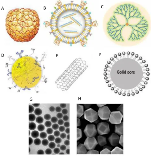
Figure 6 – Examples of nanoparticles used in the cosmetic industry. Polymeric nanoparticles (A); liposomes (B); dendrimers (C); mineral nanoparticles (D); nanotubes (E); SLNs (F); nanoemulsions (G) and nanocrystals (H).
Figure 7 – Micrographs of damage hair fibres before (A) and after (B) treatment with seracin nanoemulsion.
Figure 8 – Hair fibres before (left) and after (right) treatment with gold nanoparticles.
Figure 9 – Greek key motif structure of crystallins. Pattern from Greek pottery (A) and bi-dimensional representation of crystallin motif (B).
Figure 10 – Schematic ribbon diagram depicting the overall structure of human D-crystallin (-Crystallin. A) Tri-dimensional representation of Greek Key motif, each domain has a respective colour; B) Amino acid sequence highlighting each domain sequence.
Figure 11 – Molecular structure of α-keratin (A) and β-keratin (B). Hydrogen bonds are highlighted (red circle) for each conformation.
Figure 12 – Standard stress-strain curve profile for hair fibre tensile-test.
Figure 13 – Keratin-based peptide [ KP(X3CX5CX3)] surface charge analysis, from N- to
C- terminal, as well as some KP characteristics at table. Red zones are identified for negative residual amino acids and at blue are the positive ones
Figure 14 – Silk Fibroin β-sheet with hydrogen bonds (green) and van der Walls forces between C and N terminal (red) highlighted.
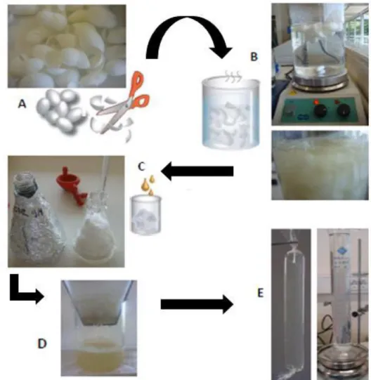
Figure 15 – Schematic representation of silk fibroin processing. Bombyx mori silkworm cocoons (A); Degumming process (B); Dissolution of degummed silk in LiBr 9 M (C); Filtration process (D); Dialysis process (E).
Figure 16 – Schematic representation of protein purification by Magnetic Beads (Biotool).
Figure 17 – Polymeric structure of Poloxamer 407.
Figure 18 – Characterization of keratin-based particles, during storage at 4 ºC: A) Z-average and PDI; B) Zeta-potential. Data were analysed by one way-ANOVA: * p-value≤0.05, ● p-value≤0.01; ♦ p-value≤ 0.001; ■ p-value≤0.0001, compared to day 1.
Figure 19 – STEM microphotographs (50000x magnification) of keratin-based particles obtained by high pressure homogenization.
Figure 20 – FTIR spectra of keratin (solid line) and silk fibroin (dash line). The three amides regions are highlight: Amide I (1600-1700 cm-1) C=O stretch; Amide II
(1510-1580 cm-1) N-H bending; Amide III (1230-1270cm-1) C-O stretch; as well the
peak at 1400 cm-1 peak (A).
Figure 21 – FTIR spectra of extracted keratin (solid line) and K100 particle (dash line).
The three amides regions are highlight: Amide I (1600-1700 cm-1) C=O stretch;
Amide II (1510-1580 cm-1) N-H bending; Amide III (1230-1270cm-1) C-O stretch;
as well the 1095cm-1 and 1400 cm-1 peaks, B and A respectively.
Figure 22 – FTIR spectra of K100 particles (solid line), K95SF5 particles (dash line), K90SF10
particles (dash-dot line) and K80SF20 particles (dot line). The three amides regions
are highlight: Amide I (1600-1700 cm-1) C=O stretch; Amide II (1510-1580 cm-1)
N-H bending; Amide III (1230-1270cm-1) C-O stretch; as well the 1095 cm-1, 1168
Figure 23 – FTIR spectra of K100 (solid line) and K100P (dash line) particles. The three
amides regions are highlight: Amide I (1600-1700 cm-1) C=O stretch; Amide II
(1510-1580 cm-1) N-H bending; Amide III (1230-1270cm-1) C-O stretch; as well the
peak at 1400 cm-1 peak (A).
Figure 24 – FTIR spectra of K100P particles (solid line), K95SF5P particles (dash line),
K90SF10Pparticles (dash-dot line) and K80SF20Pparticles (dot line). The three amides
regions are highlight: Amide I (1600-1700 cm-1) C=O stretch; Amide II (1510-1580
cm-1) N-H bending; Amide III (1230-1270cm-1) C-O stretch; as well the 1095 cm-1,
1400 cm-1 , 1465 cm-1 and 1738 cm-1 peaks, B, A, C and D respectively.
Figure 25 – Relative viability of NCTC-2544 human keratinocytes (evaluated by the MTS assay) after 24 h and 48 h incubation in medium containing control solutions (1mg/mL) of keratin, fibroin and poloxamer. Cells only incubated with medium were used as life control (+) and cells incubated with 30 % DMSO as control of death (-). Data were determined in relation to the life control. Results are the mean ± SD of triplicate of three independent experiments. Statistical significant differences from the life control are indicated: ■ p-value≤0.001.
Figure 26 – Relative viability of NCTC-2544 human keratinocytes (evaluated by the MTS assay) after 24 h incubation in medium containing K100, K100P, K95SF5, K95SF5P,
K90SF10, K90SF10P, K80SF20 and K80SF20P particles. Cells only incubated with medium
were used as life control (+) and cells incubated with 30 % DMSO as control of death (-). Data were determined in relation to the life control. Results are the mean ± SD of triplicate of three independent experiments. Statistical significant differences from the life control are indicated as: ■ p-value≤0.001.
Figure 27 – Relative viability of NCTC-2544 human keratinocytes (evaluated by the MTS assay) after 48 h incubation in medium containing K100, K100P, K95SF5, K95SF5P,
K90SF10, K90SF10P, K80SF20 and K80SF20P particles. Cells only incubated with medium
were used as life control (+) and cells incubated with 30 % DMSO as control of death (-). Data were determined in relation to the life control. Results are the mean ± SD of triplicate of three independent experiments. Statistical significant differences from the life control are indicated as: ■ p-value≤0.001.
Figure 28 – SEM micrographs of normal Asian virgin hair after treatment with the keratin-based particles.
Figure 29 – SEM micrographs of 8x overbleached Asian hair after treatment with the keratin-based particles.
Figure 30 – SEM micrographs of overbleached Asian virgin hair a treated with K80SF20
particles.
Figure 31 – Mechanical resistance parameters shown by Young’s Modulus. Values are mean of ± SD of thirty independent measurements. Statistical significant differences from the control (untreated virgin air) are indicated as: * p-value ≤0.05, ● p-value ≤0.01; ♦ p-value ≤0.001; ■ p-value ≤0.0001.
Figure 32 – Mechanical resistance parameters shown by Young’s Modulus. Values are mean of ± SD of thirty independent measurements. Statistical significant differences from the control (untreated overbleached hair) are indicated as: * p-value ≤0.05, ● p-p-value ≤0.01; ♦ p-value ≤0.001; ■ p-value ≤0.0001.
Figure 33 – Plasmid digestion of pet-28a (+)::KP-(GA)5-CrysWt and pet-28a
(+)::KP-(GA)5-CrysMut using NdeI and NotI restriction enzymes. Agarose Gel 1% in the
left: M – 100bp DNA ladder Plus; 1 – KP-(GA)5-CrysWt; 2 – KP-(GA)5-CrysMut.
SnapGene simulated gel in the right: 3 – KP-(GA)5-CrysWt; 4 – KP-(GA)5-CrysMut.
Figure 34 – A - Total protein fractions analysis of CrysWt producing colonies. B - Total protein fractions analysis of CrysMut producing colonies. M – Protein Marker; Lanes 1 to 8 – total protein fraction of eight B21 (DE3) producing transformants after overnight induction in AIM medium; Lane ∅ - negative control of protein expression.
Figure 35 – A - Total protein fractions analysis of CrysWt expression. B - Total protein fractions analysis of CrysMut expression. M – Protein Marker; Lanes LB, TB, TB-AIM, LB IPTG and TB IPTG – total protein fraction of best producing transformant B21 (DE3) after induction in the corresponding culture media; Lane ∅ - negative control of protein expression performed in TB-AIM.
Figure 36 – Fraction analysis for expressed crystallins. M – Protein Marker; Lanes SF: Soluble Fraction; Lanes IF: Insoluble Fraction; Lane ∅ - negative control of protein expression performed in TB-AIM.
Figure 37 – A – Purification fractions of CrysWt; B – Purification fractions of CrysMut M: Protein Marker ; Lane F1 – Binding fraction (10 mM imidazole); Lane F2 – Washing fraction 1 (50 mM imidazole); Lane F3 – Washing fraction 2 (80 mM imidazole); Lane F4 – Elution fraction 1 (100 mM imidazole); Lane F5 – Elution fraction 2 (250 mM imidazole); Lane F6 – Elution fraction 3 (500 mM imidazole); Lane F7 – Elution fraction 4 (500 mM imidazole).
Figure 38 – SDS-PAGE of pure crystallins. M – Protein marker; Lane 1 – CrysWt and Lane 2 – CrysMut.
Figure 39 – Determination of CrysWt (A) and CrysMut (B) molecular weight by MALDI-TOF mass spectrometry.
Figure 40 - FTIR spectra of recombinant Crystallins: CrysWt (dash line) and CrysMut (solid line). The three amides are highlight: Amide I (1600-1700 cm-1) C=O stretch; Amide II (1510-1580 cm-1) N-H bending; Amide III (1230-1270cm-1) C-O stretch
List of Tables
Table I - Hair shaft composition
Table II – Reagents used in the work.
Table III - Buffers and solutions used in experimental work presented in this thesis.
Table IV – Growth media for E. coli cultures.
Table V – Steps of Lowry’s protocol.
Table VI - Composition of resolving and stacking gel in SDS-Page. Volumes described are for one gel of 1.5mm thickness
Table VII – Particle formation efficiency determined by Lowry’s method.
Table VIII - Values of Z-Average, PDI and Zeta-Potential for keratin-based particles after 3 months of storage at 4 ºC. Data were analysed by one way-ANOVA: * p-value≤0.05, ● p-value≤0.01; ♦ p-value≤0.001; ■ p-value≤0.0001 compared to K100 particles.
Table IX – Secondary structural assignments on keratin solution and on K100, K95SF5,
K90SF10 and K80SF20 particles obtained by HPH. The results of Amide I
deconvolution, expressed in percentages, were analyzed using OriginPro 8.5 software.
Table X - Secondary structural assignments on keratin solution and on K100, K100P
K95SF5P, K90SF10P and K80SF20P particles obtained by HPH. The results of Amide
I deconvolution, expressed in percentages, were analyzed using OriginPro 8.5 software.
Table XI - Improvement (I) and recovery (R) of hair’s mechanical properties measured in terms of Youngs’ modulus and expressed in percentage (%), treated with keratin-based particles, compared with untreated virgin and overbleached hairs.
Table XII - Recombinant wild-type crystallin (CrysWT) and mutant crystallin (CrysMut) amino acidic sequences. Characters in bold correspond to the
in dark blue and light correspond to the His-Tag and trombine cleavage site sequences, respectively. Characters in Green correspond to the KP peptide sequence and characters in red correspond to the linker sequence.
Table XIII – Concentration and purity in terms of ⋌260/280 and ⋌260/230 ratio of pet-28a (+)::KP-(GA)5-CrysWt and pet-28a (+)::KP-(GA)5-CrysMut plasmids.
Table XIV – Secondary structural assignments on CrysWt and CrysMut proteins. The results of Amide I deconvolution, expressed in percentages, were analyzed using OriginPro 8.5 software.
List of Abbreviations
BSA Bovine Serum Albumin
CrysWt Wild-type Crystallin
CrysMut Mutant Crystallin
DLS Dynamic Light Scattering
DMEM Dulbecco’s modified Eagle’s medium
DMSO Dimethyl Sulphoxide
DOC Deoxycholate
FWHM Full Width Half Maximum
HPH High Pressure Homogenization
K Keratin
KAPs Keratin Associated Proteins
KP Keratin Peptide
LB Lysogeny Broth medium
MALDI-TOF Matrix Assisted Laser Desorption Ionization Time-of-Flight mass spectroscopy.
MTS 3- (4,5-dimethylthiazol-2-yl) -5- (3-carboxymethoxyphenyl) -2- (4-sulfophenyl) -2H-tetrazolium
PBS Phosphate-buffered saline
P Poloxamer 407
SDS Sodium Dodecyl Sulfate
SDS-PAGE Sodium Dodecyl Sulfate Polyacrylamide Gel Electrophoresis
SEM Scanning Eletron Microscopy
STEM Scanning Transmission Eletron Microscopy
SF Silk Fibroin
TB Terrific Broth medium
TB-AIM Terrific Broth Auto Inducible medium
TEMED Tetramethylethylenediamine
1.1. Main Goal and Motivation
Many cosmetic products for keratinaceous appendices like skin, hair and nails do not show viable results regarding their final application. This is mostly related to how products are applied and how they interact with the keratinaceous appendices. Barriers like hair cuticles and nails plate play an important role in preventing the entry of compounds in aqueous solutions and thus the use of solvents becomes necessary.
In this work two strategies for the treatment of damaged appendices were pursued. The new approaches are solvent free and constituted by biological materials. Keratin was extracted from hair waste of barbershops while fibroin was extracted from Bombyx mori cocoons and recombinant crystallins, based on the sequence of human D-crystallin, were expressed in Escherichia coli.
The work presented here was carried out at the BBRG laboratory at Department of Engineering Biological and was divided in two parts:
1- Production, characterization and application of keratin-based particles 2- Production and characterization of recombinant crystallins
The production of keratin-based nanoparticles for hair restoration and strengthening was pursued due to the affinity of keratin and keratin-based peptides to hair fibres. [1]
The effect of γD-crystallin on the properties of virgin and damage asian hair [2], was the motivation for producing two new recombinant crystallins. To increase the anchorage and the film forming properties of the proteins on hair fibres a keratin-like peptide (KP) was included in the crystallins sequences (wild type and mutant forms). The KP peptide will improve the crystallins polymerization on hair surface creating a protein film. This film could be used for the anchorage of other functionalities like chromogenic proteins for hair colouring.
1.2. Keratinaceous Appendices
Keratinous appendices are structures characterized by the presence of high amounts of keratin on their constitution or have keratinocytes which are responsible for the production and accumulation of keratin.
These appendices, apart from conferring protection, regulate body temperature and act like sense organs, have a central role in the esthetic of the human being. Thus, the development of new formulations for the treatment and improvement of the properties of these appendices is of great importance with a significant economic and social impact. [3]
Hair, nails and skin are differentiated by the structural type and the type of keratins. [4] Skin on stratum corneum have a variety of “soft” epithelial keratins opposite to the keratins found in hair and nails. These are built up from a separate subfamily of hard keratins which are rich in sulfur content, in their non- -helical head and tail domains, that are responsible for the high degree of filamentous cross-linking by keratin associated proteins (KAPs). [5–7]
1.2.1. Hair
Human hair has an important and undeniable relevance in society due to its important role in visual appearance and social communication. Hair is mainly composed of structural proteins mainly keratin, keratin associated proteins, water, lipids, trace elements and pigments (Table I).[3]
Table I - Hair shaft composition
Structural proteins Keratin 90% Water Trace elements Pigments Melanine Lipids 18-MEA 10%
Hair is generally divided into two main parts: the hair follicle and the hair shaft. The study of the hair follicle is mainly addressed by biological sciences while the hair fibre is mainly studied from a physicochemical perspective by cosmetic sciences. The hair shaft is divided in cuticle and cortex with hard keratins, and the medulla made by soft keratins. These three regions act like a unit and are represented at Figure 1. [3,8]
Figure 1- Scheme of hair structures.[8]
The cortex is surrounded by the cuticle, has a thickness of 45–90 µm and corresponds to 90% of the total hair mass. It is composed of elongated cortical cells, which enclose melanossomes, and the cell membrane complex (CMC). The cortical cells are tightly packed contain macrofibrils which are parallel and longitudinal oriented to the fiber axis. These macrofibrils are constituted by microfibrils (intermediate filaments (IF)) and a matrix with keratin associated proteins (KAPs). IFs are surrounded by KAPs and are constituted by high sulphur proteins, ultra-high sulphur proteins and high glycinetyrosine proteins. The KAPs, rich in cysteines, interact with IF through intermolecular disulphide bonds, providing high mechanical strength, inertness and rigidity to the keratin fibres.[3,8,9]
The medulla (5–10 µm) is located on the core of the hair fiber composed by spherical hollow vacuoles, surrounded by CMC, and do not play an important role in hair mechanical properties. They can be continuous or discontinuous and absent. In coarse hair, like beard, it is usually continuous, but in fine hairs it appears discontinuous while is absent in African hair. [8,10] The hair cuticle is a protecting barrier of cortex from external environmental damage, containing 2-6 layers of overlapping scales. The cuticle’s proximal end is firmly attached to the cortex (CMC cells) and the distal open end of the overlapping tiles points towards the tip of the fibre.
Figure 2- Illustrative image of the cuticle’s layered structure with its 5 different sublayers.[11]
The cuticle is divided into 5 different strata (Figure 2), among which stand out A-Layer, Exocutile and Endocuticle. Compositional differences between them render the cuticle a very reactional structure to external elements like water, heat, chemicals and others.
The A-layer is the region of the hair fiber most highly enriched in disulfide (cysteine) and isopeptide (amide) cross-links and has an average thickness of 110 nm. This layer is attached tothe epicuticle which supports 18-MEA (18-methyl-eicosanoic acid). The presence of 18-MEA is associated with cuticles’ hydrophobicity (figure 3) [11–13].
The exocuticle usually has a thickness of 200 nm and is composed essentially by cysteines and non-polar amino acids and virtually does not show any isopeptide cross-links. However in interaction with A-Layer, the exocuticle will be fitting in the role of protecting the other surfaces of the hair from mechanical damage [14].
Endocuticle consists of the remaining cellular debris. This layer is very poor in cysteine residues, does not present cross-linked sections, but the presence of high levels of acid and basic amino acids makes this section the most prone to swelling.[12,14]
1.2.1.1. Types of Hair
Hair is a unique characteristic of each individual and differs in terms of growing rate, size and shape. It is possible to distinguish hair from different ethnic backgrounds such as Asian, Caucasian and African. Despite this phenotype variation, the amino acid composition appears to be uniform. In other cases, the percentage and location of lipids differs from each ethnical group [3,7,12]. In Figure 4 are presented the three types of ethnic hair and corresponding cross-sections.
Hair fibre shape is dependent on hair follicle structure. Large and straight follicles with a circular cross section produce thick straight hair, and flat follicles make curly elliptical/ ribbon shaped cross section hair.[15,16]
Figure 4 - Different hair fibres types with respective cross-sections. A - Asian; B - Caucasian; C - African.
Asian hair presents higher density and a rougher surface when compared with the other types of hair. It is usually straight with a round cross-section and is very similar to the caucasian hair regarding the lipidic constitution.
Caucasian hair can be quite variable in its shape going from straight to wavy or curly. The fibre is elliptical and is, on average, thinner than the Asian fibre. African hair is frequently tightly looped and twisted. In cross section, it is almost flat and ribbon-like in some cases. Presents higher concentration of lipids which is related with the higher surface hydrophobicity of this type of hair.[3,12,17]
The mechanical properties of hair are also influenced by the hair pattern. Curly hair, due to the smaller curve diameter, is more likely to break under external forces, than straight hair.[18]
1.2.1.2. Effect of bleaching in hair
Chemical bleaching is commonly used in cosmetics, which is apply to lighter the hair fibres and, more usually, to prepare the hair for dyeing. However, repeated hair bleaching results in hair damage.[10]
Many studies report the effect of chemical oxidation on hair structure and composition. After chemical treatment, there is a change in hairs’ amino acid composition, hydrophobicity, melanin content, matrix and fibre diameter. Moreover the process due to the oxidation of cysteines results in an increase of the negative groups on hair surface. [12]
The overbleaching using H2O2 results in very fragile hair fibres with lower fibre resistance
mainly related with the disruption of cross-linked cysteines in the cortex region along with the appearance of cracks and holes in the cuticle (Figure 5) [19].
Figure 5- Surface electron microscopy (SEM) micrographs of normal Asian virgin hair (A) and overbleached hair (B). [19]
It is important to develop new strategies for hair colouring and products that reverse the undesired collateral effects of hair bleaching. Protein-based products are now seen as excellent candidates to restore hair fibre aspect and strength.
1.3. Nanotechnology for personal care
Engineered nanomaterials such as fullerenes, nanotubes, liposomes, quantum dots, solid lipid nanoparticles (SLN), starch lipid composites (SLC), nanoemulsions, among others are employed nowadays in cosmetic industry due to their nano-scale unique properties. Cosmetic industry is replacing the cosmeceuticals for nanocosmeceuticals by incorporating nanotechnology in the manufacturing processes resulting in nanotech based cosmetics and some of these new products are already available to the consumer.[20–22]
Nanotechnology can be defined as the study of matter on an atomic or molecular scale handling with exact size and shape structures which must be measured on a nanometric scale. Nanotechnology aims to create products with both special and improved chemical–physic features compared to conventional products. Consequently, the goal of nanobiotechnology is the development of nanoscale biomolecular components and analytical instruments for the investigation of cell. [20]
Nanomaterials may exhibit unique biochemical properties when compared with microparticles of the same chemical compound. Due to larger surface area to mass ratio they could have unique bioactivity profiles. Nanocosmetics are cosmetic products with nano-based structures used as delivery vehicles. There is an increasing interest in this type of cosmetic formulations, and major cosmetic companies such as L’Oréal, Christian Dior and Estee Lauder are focused on the development and commercialization of new functional nanocosmetics. [23,24]
1.3.1. Nanoparticles in cosmetic formulations
The use of nanoparticles brings many advantages for the cosmetic industry. Several advantages can be pointed out: large scale production, long-term stability, possibility to lyophilize and controlled drug release. Nowadays cosmetic industry applies engineered bio-nano-systems in cosmetic products, which are mostly created by nanoemulsion because of its transparency and pleasant to touch. However, there is a variety of nanostructures that are used for personal care purposes like liposomes, solid lipid nanoparticles (SLN), nanocapsules, dendrimers, cubossomes,
hydrogels, mineral nanoparticles and polymeric nanoparticles. Some examples of nanostructures are represented in Figure 6.[20,22]
Figure 6 - Examples of nanoparticles used in the cosmetic industry. Polymeric nanoparticles (A); liposomes (B); dendrimers (C); mineral nanoparticles (D); nanotubes (E); SLNs (F); nanoemulsions (G) and nanocrystals (H).
Nanoemulsions are normally oi-in-water dispersions of nanoscale droplets and are used in odorants, shampoos and skin/hair care products. These are suitable for drug delivery since their small particle size provide higher stability, which also increases the shelf life of the cosmetic product. Some products with nanoemulsions are used for hair care improving cuticle structure and maintaining hair lubrification. Cationic nanoemulsions with silicone oil and seracin were tested and results showed a significant improvement of the cuticle’s aspect even after several washes (Figure 7). A B C F E D G H
Figure 7- Micrographs of damage hair fibres before (A) and after (B) treatment with seracin nanoemulsion.[25]
Nanoemulsions structure can be manipulated based on the method of preparation (high pressure homogenization and ultrasound homogenization) and the nature of components.[25–27] Gold nanoparticles (Figure 8) and hyaluronic nanoparticles with p-phenylenediamine dye had already been used as new hair colouring reducing the toxicity and enhancing cuticle penetration. Also, luminescent silica quantum dots were applied to hair resulting in the lightening of the hair shafts. [25]
Figure 8 - Hair fibres before (left) and after (right) treatment with gold nanoparticles. [25] B
1.3.2. Proteins in cosmetic formulations
The use of proteins (from the Greek word proteios = of primary importance) in cosmetic products is easy to understand as these play dominant roles in almost all biological processes. They act as enzyme catalysis; are involved in the transfer of small molecules and ions; coordinated movement, are involved in mechanical support and are associated with the control of growth and differentiation. The proteins can also be applied in the development of protein-based cosmetic products for the treatment of skin and/or hair. [28]
Since great ancient civilizations protein materials are used for cosmetics purposes. There are several examples in history like Cleopatra’s legendary use of donkey milk and the use of soy flour to prepare facial masks by Hokkaido island fishers. Nevertheless, the first rational use of proteins on cosmetics dates back to the 1950’s. Since then, many authors have recognized the effects of protein-rich substances for the topical treatment. Proteins were quickly considered useful ingredients to produce a suitable environment for healthy skin, hair and nail (i.e. conditioning agents).[28,29]
Protein derivatives are used for a variety of cosmetic formulations like emulsions, lotions, gels, creams, make-ups among others. Native proteins and hydrolysates with high molecular weight usually tend to form biofilms that result from protein polymerization providing continuous softness and smoothness. The possibility to anchor other types of proteins or peptides to the films, improving film properties like stability and visual appearance makes this type of approach very attractive in the development of new protein-based cosmetics. Some examples of proteins used in cosmetic products are: collagen, elastin, silk fibroin, keratin and casein.[30]
1.3.2.1. Human eye D-cystallins
Crystallins are a group of proteins located in the cytoplasm of fibrous cells of the ocular lens and are divided in two super families: gamma (γ) and beta ( ). Both have an antiparallel -sheet protein structure, also known as Greek key motif due to the similar pattern with pottery from ancient Greece. The crystalline structure and the Greek key motif are represented in Figure 9. The Greek key structure is represented in pairs and in two domains that interact between themselves. This interaction leads to a high thermodynamic stability which is not usual for proteins [2,31,32]. Gamma-Crystallin superfamily can be clearly differentiated in a chromatographic gel of the ocular lens compounds, appearing in the last peak, due to its monomeric structure and low molecular mass. This superfamily has an isoelectric point between pH 7.1 e 8.6 and are characterized by the existence of cysteine residues that are highly sensitive to the formation of disulfides in oxidative stress. Complementary studies distinguished this crystallins in two groups, γA/B/C-Crys and γD/E/F-Crys, which are differentiated by state of hydration. These states correspond to the critical temperature of separation which is superior in the γD-Crys group and it is explained by the presence of three amino acid residues (Leu51, Ile103 and His115) [33,34].
Figure 9 - Greek key motif structure of crystallins. Pattern from Greek pottery (A) and bi-dimensional representation of crystallin motif (B).[35]
The γD-Crystallins contain 173 amino acids and two terminal domains (NH2- e COOH-)
unified by an amino acidic sequence of 6 amino acids and interact by contact between lateral chain and domain interface. Studies realized in physiological conditions that tested the capacity of this protein to fold and unfold showed that the termination COOH reacquire the natural form faster and consequently these domains do not present the same stability [36,37]. In Figure 10 is represented
the ribbon scheme of γD-crystallin (A) and the amino acidic sequence with color differentiation for the four structures of Greek key motif (B).
The γD-crystalins are found in high concentration in the nuclear lens region [>400 mg/mL] and some studies indicate that its intermolecular interaction are directly related with lens transparency. In the occurrence of mutations, diseases like ocular cataract can be developed due to the opacity of the lens. Owing to the Greek key structure, these proteins are involved in the formation of coatings, like in the protein S present in the coating of spores of Myxococcus xanthus, which can be useful for the development of cosmetic formulations. This was already demonstrated in the production of crystallin-based films on hair[2,35,38,39].
Figure 10 - Schematic ribbon diagram depicting the overall structure of human D-crystallin (-Crystallin. A) Tri-dimensional representation of Greek Key motif, each domain has a respective colour; B) Amino acid sequence highlighting each domain sequence.[35]
1.3.1.2. Human hair keratin
Keratin is an abundant biological material, represents a group of insoluble, usually with high-sulphur content and filament-forming proteins. They constitute the bulk of animal epidermal appendices such as hair, nails, claws, horns, beaks, and feathers. These “dead tissues” will end up in the trash or simply degrade so recovering keratin from “waste” could make it a bio-recoverable material with unique characteristics. It has been reported that approximately 300,000 tons of keratin-rich resources are discarded from salons, hospitals and slaughterhouses.[40,41]
Hair keratins, also known as hard keratins, are a family of structural proteins present in hair and fingernails. These are self-assembled into fibres in hair follicle and are controlled by many growth factors (≈30) in a highly proliferative process. They are sub-divided into two large groups: type I and type II, distinguished by their isoelectric point (Type I: acid, Type II: basic/neutral).[42,43]
Figure 11 - Molecular structure of α-keratin (A) and β-keratin (B). Hydrogen bonds are highlighted (red circle) for each conformation.
Keratin in human hair is composed by 400 to 600 amino acids and contains a variety of cell-binding sites like LDV (Leu-Asp-Val) and RGD (Arg-Gly-Asp). It can be classified into three groups depending on the localization and structure. α-keratins are mainly structural and are the most abundant component of human hair. They present a α-helical structure, are low in sulfur content and have molecular weights between 60 and 80 kDa. The α-helical structure consists in two helical
polypeptide chains, usually found in right-handed direction, stabilized by the hydrogen bonds causing chain twisting (Figure 11 A). β-keratins form most of the cuticle and when extracted do not form any type of useful reconstituted structures. These involves laterally packed β-strands which can be parallel or antiparallel, held by hydrogen bonds stabilizing pleated sheet structures (Figure 11 B). [40,41,44]
The contribution of α- and β- strands to the mechanical properties of hair fibre was studied (Figure 12). Between 2 – 5 % strain, a near linear Hookean region, α-helices are stretched but no substantial structural changes happen. A transition from α-helix to β-sheet occurs in the yield region (5 – 50 %), at this point α-coiled coil conformation starts to disentanglement and β-sheets formed. In the post-yield region, there is a total refolding of α to β conformation. An increase in the area of stress-strain curve shows that these conformations enhance the energy-absorption capability of hair fibre. [40]
Figure 12 - Standard stress-strain curve profile for hair fibre tensile-test.[40]
Keratins extracted from human hair, wool or chicken feathers are reported as biocompatible, biostable and biodegradable materials. Keratins possess the physical and chemical characteristics for spontaneous self-assembly and polymerization, which allows the development of various types of biomaterials like scaffolds, films and hydrogels.[45]
Materials for cell culture coated with keratin improved cell adhesion and cell proliferation through cell-binding sequences such as LDV and RGD. [41] Keratin-based hydrogels were developed as hemostatic agents, wound healing dressings and as nerve conduit supports for
peripheral nerve regeneration. [46] Also, keratin thin films were developed to be used as in vitro nail plate models in permeability tests. [47]
1.3.1.3. Keratin-based peptide (KP)
Keratin-based peptide (KP) is based in an amino acidic sequence of KRT85 (Keratin, type II cuticular HB5) with peptide sequence X3CX5CX3.It is composed by 13 amino acids with two
residues of cysteine separated by 4 amino acids. In 2012, Fernandes & Cavaco-Paulo studied the ability of KP peptide to penetrate and restore some of the properties of damaged hair. [19]
Figure 13 – Keratin-based peptide [ KP(X3CX5CX3)] surface charge analysis, from N- to C- terminal,
as well as some KP characteristics at table. Red zones are identified for negative residual amino acids and at blue are the positive ones.[9]
In a posterior study, the effect of KP was tested in the mechanic and thermic properties of hair as well as the toxicity for skin in order to develop a new hair product. The results showed that KP penetrated in the hair fiber and improved its properties, even when the hair was not damaged. The easy penetration was related with the similar composition between KP and hair. Being like type II α-keratin, it can transfer its elasticity and malleability to the cortex of hair fibers. The toxicity results reveal that, when tested alone, irritation towards the skin was not present which could lead to the development of a cosmetic hair product. The structure of KP is depicted in Figure 13.[9]
1.3.1.4. Silk fibroin
Silks are fibrous proteins that are spun into fibres by some arthropods such as silkworms, spiders, scorpions, mites and fleas. Silk Fibroin (MW≈ 391kDa) as a natural protein polymer presents advantages over synthetic polymers. It has unique qualities such as biocompatibility, biodegradability and bioresorbality which makes it environmentally friendly. Silk is involved in cocoon (i.e. B. moori) and web spider formations and in raw state it is composed by two main proteins: Sericin (glue-like) and Fibroin (filaments).[48–51]
Silk fibroin protein is mostly constituted by layers of antiparallel β-sheets with a regular amino acid sequence (Gly-Ser-Gly-Ala-Gly-Ala)n. It has a high content of glycine and alanine and a
tight packaging which improves silk’s rigidity and tensile strength. Silk fibroin rigidity is also related with hydrophobic domain predominance as result of homogeneity in secondary structure. Combining stiffness and hardness make this a material that is used in several areas like biotechnology, biomedicine and cosmetics.[52,53]
Figure 14 - Silk Fibroin β-sheet with hydrogen bonds (green) and van der Walls forces between C and N terminal (red) highlighted.
Three different crystalline structures have been described for silk fibroin: glandular state (silk I); crystalline silk structure and air/water interface (silk II and III respectively). Silk II presents a β-sheet conformation with the chains in an antiparallel orientation linked by hydrogen bonds between hydrogen side chains of glycine and methyl side chain of alanine, establishing strong intermolecular bonds and Van der Walls forces that will lead to a thermodynamically stable structure (Figure 14). [54]
Unique properties of fibroin make it a very versatile material for producing biofilms, modified textiles, scaffolds, contact lenses, hydrogels, micro and nanoparticles. [52]
2.1. Materials
2.1.1. Chemical Reagents
All reagents used in this work are listed in Table II. Table II – Reagents used in the work presented in this thesis.
Product Brand
Acetic Acid glacial Chem-Lab
Acetone PanReac AppliChem
Acrylamide/Bisacrylamide (30 %/0.8 % (w/v)) Bio-Rad
Antibiotic Antimycotic Solution (100×) Sigma
-mercaptoetanol Bio-Rad
Bovine Serum Albumin (BSA) Sigma
Coomassie Blue Merck
DC Protein Assay Bio-Rad
Deoxycholate (DOC) Sigma
Dimethyl Dulphoxide (DMSO) Fisher Scientific
Dulbecco’s modified Eagle’s medium (DMEM) Sigma
Ethylenediamine Tetraacetic Acid (EDTA)
Fluka
Ethanol Absolute PanReac AppliChem
Fetal Bovine Serum (FBS) Biochrom
Glycerol 99 % Sigma
Hydrochloric Acid (HCL) Fisher Cientific
Hydrogen peroxide (H2O2) Sigma
Imidazole Sigma
IPTG Fluka
L- Glutamine Sigma
Lithium bromide (LiBr) Sigma
Lowry Reagent Sigma
Magnetic Beads Biotools
Methanol Fisher Cientific
MTS Promega
Protease cocktail inhibitor Biotool
Plus Protein All Blue Standard Bio-Rad
Poloxamer 407 Sigma
Potassium Hydrogen Phosphate (K2HPO4) Merck
Shampoo Pantene
Sodium carbonate (Na2CO3) Sigma
Sodium Chloride (NaCl) Panreac
Sodium metabissulfite Sigma
Tetramethylethylenediamine (TEMED) Bio-Rad
Trichloroacetic Acid (TCA) PanReacQuimica
Tris(hydroxymethyl)aminomethane (Tris) Fisher Cientific
Trypsin Biochrom
Tryptone PanReac
Urea Sigma
Vegetable oil Pingo Doce
Yeast extract Sigma
2.1.2. Buffers and Solutions
Table III – Buffers and solutions used in experimental work presented in this thesis.
Running Buffer (1x) 25 mM Tris-HCl (pH 7.4), 192 mM glycine, 0.1% SDS
Laemmli Sample Buffer (5x) 250 mM Tris-HCl (pH 6.8), 50% Glycerol, 10% SDS, 0.1% Bromophenol Blue.
Binding buffer 20 mM Sodium Phosphate, 500 mM NaCl,
10 mM Imidazole, pH 7.4.
Washing buffer 0 mM Sodium Phosphate, 500 mM NaCl,
20-100 mM Imidazole, pH 7.4.
Elution buffer 20 mM Sodium Phosphate, 500 mM NaCl,
Hair keratin extraction solution 8 M Urea, 0.2 M SDS, 0.5 M Na2S2O5
Phosphate Buffer Saline (PBS 1x, pH 7.4) 137 mM NaCl, 2.7 mM KCl, 8.5 mM Na2HPO4, 1.5mM KH2PO
2.1.3. Culture media for bacteria
Several culture media were used throughout this work and are listed in Table IV. Table IV – Growth media for E. coli cultures.
2.1.4. Biological Materials 2.1.4.1. Bacterial strains
The E. coli strains used on this work have the following genotypes: Composition for 1liter of dH2O,
pH=7.0 Used for LB -10 g of Tryptone; -10 g of NaCl; -5 g of Yeast Extract Overnight pre-inocules
LB-IPTG 0,1M -LB composition; -100µm IPTG Protein expression
TB -12 g of Tryptone; -24 g of Yeast Extract; -2.5 mL of Glycerol; Protein expression TB AIM -12 g of Tryptone; -24 g of Yeast Extract; -150 mg of MgSO4; -3.3 g of (NH4)2SO4; -6.5 g of KH2PO4; -7.1 g of Na2HPO4; -0.5 g of Glucose; -2 g of alpha-lactose Protein expression TB-IPTG 0.1M -12 g of Tryptone; -24 g of Yeast Extract; -2.5 mL of Glycerol; -100µm IPTG Protein expression
XL1-Blue (Stratagene): endA1 supE44 hsdR17 thi1 recA1 gyrA96 relA1 lac [F’ proAB lacIq
ZΔ M15 Tn (Tetr)]
BL21 (DE3) (Novagen): F- ompT hsdSB (rB- mB-) gal dcm (DE3) 2.1.4.2. Cell lines
In this project the human skin keratinocytes NCTC2544 cell line was used. The NCTC2544 cell line was supplied by Instituto Zooprofilattico Sperimentale Della Lombardia e Dell’Emilia-Romagna, Pavia (Italy).
2.2. Methods
2.2.1. Extraction of keratin and fibroin
2.2.1.1. Extraction and purification of keratin
Keratin was used as the core constituent of keratin-based particles. It was extracted from donated human hair obtained from a local barbershop. The hair samples were first washed according the IAEA/RL/50 1978 recommendations to remove contaminants and lipids. Hair washing consisted of cycles of distilled water and acetone with continuous agitation. After washing hair samples were kept at 40 ºC until completely dried.
For the keratin extraction, a solution of 8 M urea; 0.2 M SDS; 0.5 M sodium metabissulfite in a ratio of 10:1 (solution to dry hair) was used. The mixture was heated at 100 ºC for 45 minutes and then incubated overnight at 37 ºC with constant agitation.
After extraction, the hair/keratin solution was centrifuged at 5000 rpm for 10 min and the supernatant was then filtered to remove hair fragments. The keratin solution was then dialysed for 5 days in distilled water using a dialysis membrane with a 14 kDa cut-off. The water was renovated twice per day. After dialysis, the keratin solution was quantified by Lowry's method and DC method .
2.2.1.2
.
Extraction and purification of silk fibroin (SF)The silk fibroin was extracted from Bombyx mori cocoons donated by Dr. Silvia Cappellozza from “Sezione Specializzata per la Bachicoltura” (Padova).
Obtaining a pure solution of fibroin is a time-consuming process in which the withdrawal of sericin by a degumming process, increases the biocompatible properties of the protein.
About 5 g of cocoons were immersed in 1 L of boiling Na2CO3 (0.05 M) solution for 10
minutes. The Na2CO3 solution was changed several times until the cocoons get fragmented. The
degummed SF was then kept at 40 ºC until completely dried.
The degummed silk fibroin fibres were then incubated in a solution of 9.3 M lithium bromide at 60ºC until total solubilisation. The soluble SF was then filtered to remove undissolved fibres and dialysed for 5 days in distilled water using a dialysis membrane with a 14 kDa cut-off. The water was changed two times per day. After dialysis, the keratin solution was quantified by Lowry's method and DC method. Processing of silk fibroin is summarized in Figure 15.
Figure 15 – Schematic representation of silk fibroin processing. Bombyx mori silkworm cocoons (A); Degumming process (B); Dissolution of degummed silk in LiBr 9 M (C); Filtration process (D); Dialysis process (E).
2.2.2. Protein quantification
Keratin and silk fibroin solutions were quantified by three methods: Lowry's method, DC method and Lyophilisation.
2.2.2.1. Lowry's method
The Lowry’s method quantifies protein by its absorbance at 750nm against a standard curve. To quantify the extracted keratin and the SF, diluted solutions of both proteins were used as well as other solutions: DOC (1,5 mg/ml), TCA 72% (v/v), Vial of Lowry's reagent and Follin 20% (v/v).[55]
The steps of Lowry’s protocol are resumed on Table V. The protein concentrations were expressed in mg/ml.
Table V – Steps of Lowry’s protocol.
Step 1 Add 50 µl of DOC solution to 400 µl of diluted protein solutions Step 2 Vortex and rest for 10 min
Step 3 Add 50 µL of TCA
Step 4 Centrifuge for 10 min at 5000 rpm and discard supernatant
Step 5 Add 400 µL of distilled water and 400 µL of Lowry's reagent
Step 6 Vortex and rest for 20 min
Step 7 Add 200 µL of Follin reagent
Step 8 Vortex and rest for 30 min
Step 9 Place 200 µL of Lowry treated solution in a 96 well microplate
Step 10 Read the absorbance at 750 nm
2.2.2.2. DC method
The DC method (Biorad) uses two different solutions: 200 µl of solution A and 25 µl of solution B and 20 µl sample of protein solution to be quantified. This process has a range from 0.2 to 1.5 mg/mL and sample dilution was required. Protein samples were prepared in a 96 well microplate and after 15 minutes of incubation in dark at room temperature, the absorbance was measured at 750nm. The protein concentrations were determined against a BSA standard curve.[56]
2.2.2.3. Lyophilisation
The lyophilisation or freeze-drying was invented in 1906 by Arséne s'Arsonval and consists in a dehydration process that works by freezing the material and reducing surrounding pressure to allow frozen water in the material to sublimate directly from solid phase to gas phase.[57] 1 ml of protein solutions were stored in previous weighted centrifuge tubes and sent to lyophilisation process. After three days, the tubes containing the lyophilized proteins were weighted and the difference was expressed as the final protein concentration in mg/ml.
2.2.3. Production of Protein-based Particles
Eight different formulations of keratin-based particles were obtained by High Pressure Homogenization (HPH). The particles had the general formula:
Keratinx SilkFibroiny Poloxamer407z (KxSFyPz)
where x = 80, 90, 95 and 100%; y = 0, 5, 10 and 20%; z = 0 and 5 mg/mL.
The protein solutions were prepared in phosphate buffer saline (PBS 1x) pH 7.4 to a final concentration of 10 mg/mL with 0.5% (v/v) of vegetable oil and 0.05% of Poloxamer 407, when present. The homogenization process occurred at 240 – 580 bar pressure during 10 – 13 mins (3 – 5 mins of open cycles and 7 – 8 mins of close cycles) for each particle. The nanoemulsions obtained after homogenization cycles were centrifuged at 4000 rpm for 10 mins at 4 ºC in Amicon tubes (Amicon Ultra-15 Millipore) to separate the free protein from particles. The free protein was quantified by Lowry's method and the nanoparticle formation efficiency was estimated for each formulation.
2.2.4. Particles Characterization
To study the physical properties and stability along time the keratin-based particles were characterized regarding their size, net charge and structure.
2.2.4.1. Dynamic light scattering (DLS)
Dynamic light scattering is a technique used to get size distribution, polidispersivity index (PDI) and Zeta-Potential of particles.[58] Particles samples were analysed, by correlation spectroscopy using a Nanosizer Malvern ZS, at 25 ºC with values of viscosity and refractive index of 0.8872cP and 1.330, respectively, for protein standards (pH = 7.4).
Each sample was measured in triplicate and presented as mean value with standard deviation. Keratin-based particles were kept at 4 ºC and measured weekly until month one and then were measured monthly until month three.
2.2.4.2. Scanning Transmission Electron Microscopy (STEM)
To study size and surface distribution, keratin-based particles and treated hair fibres were coated with 80% Au and 20% Pd and observed at 5.0 kV with SEM (NOVA NanSEM 200 FEI).
2.2.4.3. Secondary structure analysis
Infrared (FTIR) spectra were acquired at room temperature on a NICOLET-AVATAR 360 FTIR spectrometer using KBr disks made with 10 bar pressure. FTIR spectra were collected after 16 scans with a resolution of 32 cm−1 from 4000 to 600 cm−1.
For protein, secondary structure Gaussian deconvolution of Amide I band region (wavenumber between 1600 and1700 cm–1) was analysed using OriginPro 8.5 software (OriginLab
Corporation, MA, USA).
In deconvolution analyses, a linear baseline was fitted; the number of components and their peak position were determined according to the second derivative spectrum of this same region; and the secondary structure content was calculated from the areas of the assigned peak as percentage fraction of the total area of the Amide I range. Using Gaussian function, all data were treated manually in three fitting modes: first, the base line was held fixed and fitted while intensity and bandwidth was allowed to vary, then, the base line and bandwidth were fixed and fitted again, and finally, the base line and centre peaks were fixed and fitted once more. The deconvoluted frequencies were then assigned to the respective secondary structure -sheet, -turns, random-coil and -helix.
FTIR characterization was performed for keratin particles formulations and for recombinant proteins with the same specifications.
2.2.5. Cellular viability assays
2.2.5.1. Cell culture maintenance
The NCTC 2544 cell line (human skin keratinocytes) was cultured in DMEM medium, supplemented with 7.5 % FBS, 1 % Glutamine (2 mM) and 1 % (v/v) penicillin/streptomycin solution. Cells were maintained in 25 and 75 cm2 tissue culture flasks at 37 ºC in a humidified
atmosphere with 5 % CO2.
The cell culture medium was renewed twice per week. For subcultures and plating, the adherent cells were detached with a trypsin solution 0.05%, and fresh medium was added in order to neutralize the trypsin. The cell suspension was centrifuged 5 min at 160 g. The supernatant was discard and fresh medium was added to obtain a new cell suspension. The cell suspension was loaded in a Neubaeur chamber and the concentration of cells present in the suspension was estimated.
2.2.5.2. Cell viability assessed by MTS assay
The MTS assay is a colorimetric method used to measure the toxicity of different substances in cells. The principle of this assay is that, in viable cells, mitochondrial activity is constant and therefore an increase or decrease in the number of viable cells is linearly related to mitochondrial activity. MTS is a tetrazolium reagent that can be reduced by viable cells resulting in formazan products soluble in cell culture medium. This conversion is thought to be carried out by NAD(P)H-dependent dehydrogenase enzymes in metabolically active cells. [59][60]
Cells were seeded at a density of 22500 cells/well on a 48-well tissue culture plate, the day before the experiments. The cells were then exposed to 6 concentrations (0.01, 0.05, 0.1, 0.25, 0.5 and 1 mg/mL) of keratin-based particles. Cells incubated with DMSO (30 % of the total volume) and cells without the addition of the compounds were used as controls; control of death and control of life respectively.
NCTC 2544 cells were incubated at 37 ºC in a humidified atmosphere with 5 % CO2. At
incubation, the medium with the particles was removed and 100 µl of fresh medium with 100 µl of MTS were added to each well, and cells were incubated at 37 ºC for 2 h. The medium with reduced MTS was transferred to a 96 microwell plate and colour intensity was measured with 96-well plate reader at 490 nm in a microplate reader SpectraMax Plus (Molecular Devices).
2.2.6. Hair Fibres studies
2.2.6.1. Hair fibre washing
Black Asian hair fibre samples (0.1 g) were washed before treatment with a classic commercial shampoo (Pantene). After washing, hair fibres were placed at 30 ºC until completely dry.
2.2.6.2. Hair fibre bleaching
After washing, hair samples were subjected to eight cycles of bleaching (8xB). Bleaching consisted on the application of 12% H2O2 (v/v) in the presence of 0.1 M Na2CO3/NaHCO3, pH 9.0
buffer at 50 ºC for 1 h, in a bath ratio of 1:10 at 20 rpm, using a Rota-wash laboratory machine (MKII Series 7227, Shirley Developments). Treatments were applied to the same tresses of hair.
2.2.6.3. Hair fibre treatment with keratin-based particles
Hair samples (virgin and overbleached Asian hair) were subjected to the treatment with keratin-based particles. Treatments were performed for 1 h at 37 ºC with 0.5 mg/mL of particles in 0.05 M phosphate buffer pH 7.5. Subsequently, all samples were thoroughly washed in tap water with a commercial shampoo. Treated hair samples were analysed by SEM as described previously.
2.2.6.4. Hair fibre Tensile Test
The effect of keratin-based particles on virgin and overbleached Asian hair was assessed by the differences in mechanical properties following guidelines outlined in ASTM D1145-95 for fibre tensile testing. For this test, a set of hair fibres with low variability in diameter was selected using a light microscope.
The measurements were performed using a 250 N dynamometer Hounsfield machine. For each measurement, 30 single hair fibres were randomly taken from the tress. Each hair was individually mounted in the tensile jig by means of a paper template with a fixed gauge length of 20 mm. Before the tensile test, the paper template was cut across. Measurements were performed under controlled conditions (25 ± 0.8 ºC; 70 ± 5% humidity) at a rate of 2.5 mm/min with 0.01 N preload force, and stretched until fibres broke. All measurements were made in the middle part of the hair fibre.
Results were presented in Young's module (mPa), and define the relationship between stress and strain, which is related to material strength and elasticity.[61]
2.2.3. Recombinant Crystallins Production and Purification
For the wild-type crystallin (Cwt) and mutant crystallin (Cmut) biosynthesis, the genes based on the sequence of human eye D-crystallin were synthesized by GenScript USA Inc. (Piscataway, NJ, U.S.A.) and cloned on the expression vector pET-28a (+). E.coli XL1-Blue and BL21 (DE3) strains were transformed with the recombinant vectors, pet28a (+)::KP-(GA)5-CrysWt
and pet28a (+)::KP-(GA)5-CrysMut.
2.2.3.1. Transformation of Competent Cells- TSS method
The TSS “Transformation and Storage Solution” method is a combination of two steps from the transformation procedure, firstly competent cells are obtained and secondly the cells, using the same solution, are transformed resulting in transformation efficiency that goes up to 107
-108 cfus (colony forming units) per microgram of DNA. This method was applied for the preparation
of competent cells of the E. coli BL21(DE3) and for XL1-Blue MRF’ cells.
A single colony, isolated and grown in a LB-agar plate, was used to inoculate 10 mL of LB medium and was grown at 37 ºC with shaking (250 rpm), until a OD600 =0.3-0.4 was reached. At
this point the metabolism and cell growth was stopped by incubation on ice for 5 minutes. The cell suspension was centrifuged at 3000 rpm for 10 min at 4 ºC. The supernatant was discarded and the pellet was ressuspended in 1 mL of cold x1TSS solution. About 1-10 ng of plasmid in final volume of 1-10 L were added to the mix. The cellular suspension plus the plasmidic DNA were kept on ice 1 h and subsequently the cells were subject to a heat shock by exposition of the mix at 42 ºC for 2 minutes. The heat shock was stopped by immersion on ice for 2 min. After it 1 mL of warm LB medium was added and the suspension was incubated 1 h at 37 ºC with shaking (250rpm) in order to activate and express the gene that confers resistance to the selective marker (kanamycin).
For last, 50-200 L of the transformation mix were plated in LB-agar-kanamycin plates and were incubated for 16-20 h at 37 ºC.
![Figure 2- Illustrative image of the cuticle’s layered structure with its 5 different sublayers.[11]](https://thumb-eu.123doks.com/thumbv2/123dok_br/17574417.818269/23.892.132.752.132.458/figure-illustrative-image-cuticle-layered-structure-different-sublayers.webp)

![Figure 7- Micrographs of damage hair fibres before (A) and after (B) treatment with seracin nanoemulsion.[25]](https://thumb-eu.123doks.com/thumbv2/123dok_br/17574417.818269/28.892.141.729.112.335/figure-micrographs-damage-hair-fibres-treatment-seracin-nanoemulsion.webp)
![Figure 9 - Greek key motif structure of crystallins. Pattern from Greek pottery (A) and bi-dimensional representation of crystallin motif (B).[35]](https://thumb-eu.123doks.com/thumbv2/123dok_br/17574417.818269/30.892.246.673.616.852/figure-structure-crystallins-pattern-pottery-dimensional-representation-crystallin.webp)

![Figure 12 - Standard stress-strain curve profile for hair fibre tensile-test.[40]](https://thumb-eu.123doks.com/thumbv2/123dok_br/17574417.818269/33.892.282.637.558.811/figure-standard-stress-strain-curve-profile-fibre-tensile.webp)