junho de 2015
Mélanie Fernandes Gonçalves
Structural Connectivity of the Default
Mode Network in Obsessive-Compulsive
Personality Disorder
Universidade do Minho
Escola de Psicologia
Dissertação de Mestrado
Mestrado Integrado em Psicologia
Trabalho realizado sob orientação da
Doutora Joana Fernandes Pereira Coutinho
junho de 2015
Mélanie Fernandes Gonçalves
Structural Connectivity of the Default
Mode Network in Obsessive-Compulsive
Personality Disorder
Universidade do Minho
Escola de Psicologia
DECLARAÇÃO
Nome: Mélanie Fernandes Gonçalves
Endereço eletrónico: [email protected]
Número do Cartão de Cidadão: 14107620
Título da dissertação: Structural Connectivity of the Default Mode Network in Obsessive-Compulsive Personality Disorder
Orientadora: Doutora Joana Fernandes Pereira Coutinho
Ano de conclusão: 2015
Designação do Mestrado: Mestrado Integrado em Psicologia
É AUTORIZADA A REPRODUÇÃO INTEGRAL DESTA DISSERTAÇÃO APENAS PARA EFEITOS DE INVESTIGAÇÃO, MEDIANTE DECLARAÇÃO ESCRITA DO INTERESSADO, QUE A TAL SE COMPROMETE.
Universidade do Minho, 12/06/2015
ii INDEX RESUMO ... iv ABSTRACT ... v Introduction ... 6 Methods ... 11 Participants ... 11 Psychological Assessment ... 12 Image acquisition ... 13 Data processing... 13 Statistical analysis... 14 Results ... 15 Discussion ... 19
Limitations and future directions ... 21
References ... 22
Appendix A ... 31
INDEX FOR TABLES Table 1. Means and Standard Deviations of personality measures of the NEO-FFI...15
Table 2. Results from Independent Samples T-Test of the fiber tracts present at least on 50% of the sample...16
Table 3. Means and Standard Deviations of DMN white matter fiber tracts with significant differences ...17
Table 4. Results from Independent Samples T-Test of the fiber tracts present under 50% of the sample...19
INDEX FOR FIGURES Figure 1. Three-dimensional tractography images of the cingulum bundle, respectively in sagittal, coronal and axial anatomical planes...17
Figure 2. Three-dimensional tractography images of the tract that connects the precuneus to the lateral occipital cortex in the right hemisphere, respectively in sagittal, coronal and axial anatomical planes...18
Figure 3. Three-dimensional tractography images of the tract that connects the precuneus to the middle temporal gyrus in the left hemisphere, respectively in sagittal, coronal and axial anatomical planes...18
iii Agradecimentos
À minha orientadora, Doutora Joana Coutinho, agradeço a disponibilidade permanente, incentivo, apoio e partilha de conhecimentos ao longo deste ano.
Aos Engenheiros Liliana Maia, Miguel Soares e Paulo Marques por toda a disponibilidade, ajuda e partilha de conhecimentos fundamentais para a concretização da minha tese.
Ao meu Pai, por me ter tornado na pessoa que sou hoje e por me ter mostrado o que é a verdadeira coragem e persistência. És a minha alma gêmea e a minha estrela que me guia todos os dias.
À minha Mãe, agradeço por todo o esforço que fez nos últimos dois anos, pelo apoio e carinho incondicional e por tudo o que teve de abdicar para me ajudar.
Ao Rafael, por estar sempre presente e acreditar em mim, mais do que eu própria. Por toda a paciência, apoio e incentivo que me deu ao longo deste ano.
Aos meus afilhados, Rafael e Pires, por me conseguirem fazer sorrir a qualquer momento e por me fazerem esquecer o pior lado do dia.
À minha segunda família, Luís, Nelinha, Sara, Maguie, Rita, Simão e Marco por toda a amizade, carinho e apoio.
À Joana Alegria, por ser a minha companheira de tese ao longo deste ano, pelas risadas e brincadeiras, por toda a paciência e amizade.
À Raquel e Roxita, por me acompanharem há tantos anos e serem imprescindíveis na minha vida.
À Lilas, por ser a minha companheira em muitas peripécias da vida e pela “diversion” constante das nossas aventuras.
“Cada pessoa que passa na nossa vida, passa sozinha pois cada pessoa é única e nenhuma subtitui a outra. Cada pessoa que passa sozinha na nossa vida, passa sozinha e não nos deixa só, deixa um pouco
de si e leva um pouquinho de nós” Charlie Chaplin
iv Conectividade Estrutural da Default Mode Network na Perturbação de Personalidade
Obsessiva-Compulsiva
RESUMO
O principal objetivo do presente estudo é analisar a arquitetura da substância branca em pacientes diagnosticados com perturbação de personalidade obsessiva-compulsiva. Recorreu-se a uma técnica de neuroimagen, diffusion tensor imaging, aliada a uma estratégia de análise multimodal, usando-se para esse efeito o BrainCAT, com o intuito de estabelecer uma relação entre os padrões de conectividade estrutural da default mode network e a sintomatologia clínica da perturbação de personalidade obsessiva-compulsiva. Imagens de diffusion tensor imaging foram adquiridas em nove pacientes com perturbação de personalidade obsessiva-compulsiva e nove controlos, emparelhados por idade e sexo. Foram extraídos índices de diffusion tensor imaging como a anisotropia fracionada, difusividade média, difusividade radial e difusividade axial. Os pacientes com perturbação de personalidade obsessiva-compulsiva exibiram um aumento significativo de difusividade média no lado direito do feixe do cíngulo e aumento de difusividade axial no feixe que conecta o precuneus ao córtex lateral occipital. Os nossos resultados apontam assim para possíveis alterações na conectividade estrutural em dois feixes de subtância branca que conectam áreas chave da default mode network na perturbação de personalidade obsessiva-compulsiva. O possível papel destas alterações na patofisiologia da perturbação de personalidade obsessiva-compulsiva é discutido.
Palavras-chave: Perturbação de personalidade obsessiva-compulsiva, default mode network,
v Structural Connectivity of the Default Mode Network in Obsessive-Compulsive Personality
Disorder
ABSTRACT
The main objective of the present study is to analyze white matter architecture in patients with obsessive-compulsive personality disorder. Using diffusion tensor imaging techniques, together with a multimodal analysis approach, using for this purpose the BrainCAT, in order to establish a relation between neuronal correlates of the default mode network and obsessive-compulsive personality disorder clinical symptomatology. Diffusion tensor imaging images were acquired from nine patients with obsessive-compulsive personality disorder and nine healthy control subjects, matched on age, sex and handedness. Diffusion tensor imaging indexes such as fractional anisotropy, mean diffusivity, radial diffusivity and axial diffusivity were reported. Obsessive-compulsive personality disorder patients exhibited significantly higher mean diffusivity in the right side of the cingulum bundle and higher axial diffusivity in the fiber tract that connects the precuneus to the lateral occipital cortex. Our results points to the possibility of structural connectivity alterations in two white matter fiber tracts that connect key regions of default mode network in the obsessive-compulsive personality disorder. The possible role of these alterations in the pathophysiology of the obsessive-compulsive personality disorder is discussed.
Keywords: Obsessive-compulsive personality disorder, default mode network, cingulum bundle,
6 Introduction
The Diagnostic and Statistical Manual of Mental Disorders (DSM-V; American Psychiatric Association, 2013) defines personality disorder as an enduring maladaptive pattern of inner experience, cognition and behavior, exhibited across many contexts, that deviates markedly from the expectations of the individual's culture. Beginning in adolescence or early adulthood, a personality disorder is characterized by stability over time and can cause a significantly impaired functioning or subjective distress, (American Psychiatric Association, 2013). This pattern is manifested in two or more of the following areas: cognition, affectivity, interpersonal functioning and impulse control (American Psychiatric Association, 2000). Together with avoidant personality disorder and dependent personality disorder, obsessive-compulsive personality disorder form the cluster C – anxious or fearful (American Psychiatric Association, 2013).
The obsessive-compulsive personality disorder (OCPD) is a maladaptive, chronic and invasive pattern characterized by excessive perfectionism, preoccupation with the organization, mental and interpersonal control, inflexibility, excessive devotion to work and productivity present in a variety of contexts, affecting all areas of functioning (Light & Mulder, 2009). This disorder is characterized by eight main clinical criteria: concern with details, perfectionism, excessive devotion to work, hypermorality, inability of discard worthless objects and to delegate tasks, rigidity and stubbornness (American Psychiatric Association, 2000). The OCPD patients have excessive fixations with lists, rules and minor details, unwillingness to give responsibilities to others, lack of generosity and hoarding behaviors. All combined, these clinical symptoms can result in a significant impairment in social, work and/or family functioning, where these patients presents difficulties in establish and sustain close relationships (American Psychiatric Association, 2013). These personality traits are stable over time, early and ego-syntonic, that means that they are congruent with the wishes and goals of the individual. Typically, these individuals experience some degree of satisfaction or pleasure when they dive in perfectionists behaviors, even if those behaviors have negative consequences in social and occupational functioning (Livesley, 2003). The internal speech is marked by self-blame associated with self-referential perfectionist and ruminative thinking interfering with task conclusion, efficiency and openness to new experiences, in which the individual strictly follows moral and ethical codes (Skodol et al., 2011). In patients
7 with psychiatric disorders, OCPD is the second most prevalent disorder within the personality disorders (28.3%); De Reus and Emmelkamp, 2010). The prevalence of OCPD in the general population is estimated to be high, up to almost 8% and in outpatient settings between 8% - 9% (Cain, Ansell, Simpson & Pinto, 2014).
In this study we aim to look at the neuronal correlates of this disorder, specifically at the level of a resting brain network - the Default Mode Network (DMN). The DMN is an unique organized functional network of several brain regions, characterized by decreased activity while performing external cognitively demanding tasks, compared to the level of activation in the resting state (Raichle & Snyder, 2007). In addition, this resting brain network increased activity during social cognitive higher-order tasks such as attributing mental states to others (Harrison et al., 2008; Mars, Neubert, Noonan, Sallet, Toni, & Rushworth, 2012), self-reference tasks (Gusnard & Raichle, 2001), mind wandering (Mason, Norton, Van Horn, Wegner, Grafton & Macrae, 2007), and social cognition (Spreng & Grady, 2010).
The DMN is mostly composed by cortical structures of the posterior cingulate cortex (PCC)/precuneus, the medial prefrontal cortex (mPFC), the medial temporal lobes (MTL) and angular gyrus (AG). The PCC is involved in the response to salient stimuli from the world around us (Corbetta, Kincade, Ollinger, McAvoy & Shulman, 2000; Van den Heuvel, Mandl, Luigjes & Hulshoff Pol, 2008) and it is closely linked to autobiographical episodic memories, as well as to self-reference tasks and plans for the future. It is a highly heterogeneous region and plays an important role in regulating the focus of attention (Leech & Sharp, 2014). In contrast, the mPFC, involved in the emotional processing and monitoring of the state of mind, is more related to the expression of personality, decision making and planning behaviors. The MTL is engaged in episodic memory (Milner, 2005) and the AG implicated in semantic processing and attention (Binder, Desai, Graves & Conant, 2009; Chambers, Payne, Stokes & Mattingley, 2004). In short, the DMN is characterized by mental holdings based on personal introspection, autobiographical memories and thoughts about the future, not only involved in the processing of self-reference, but also in our memory of the past and future planning (Spreng, Mar & Kim, 2009). These psychological dimensions have a key role in clinical symptoms of several psychopathologies including the OCPD.
The contribution of the DMN for the development of models of mental illness has been empirically supported by evidence of changes in this network associated with different neurological
8 and neuropsychiatric disorders (e.g.Whitfield-Gabrieli & Ford, 2012). These changes may be evident both in the activation patterns in the resting state and in the transition to an externally oriented task (Zhao et al, 2007; Zhou et al., 2007). This neural network can be modulated by different factors, such as the individual’s emotional state (Saxe, Moran, Scholz & Gabrieli, 2006; Harrison, Pujol, Ortiz, Fornito, Pantelis & Yücel, 2008) or the cognitive load of the active task (Esposito et al., 2006). Recently, there has been an exponential increase of research about the DMN alterations, in which different functional studies have evidences that psychopathological conditions such as schizophrenia (Garrity, Pearlson, McKiernan, Lloyd, Kiehl & Calhoun, 2007), depression (Grimm et al., 2009) or anxiety (Zhao et al., 2007) may alter the pattern of neuronal activity in brain networks, particularly in the DMN.
A limitation of the aforementioned studies is that they are mostly fMRI studies, existing however, less empirical studies analyzing the presence of changes in terms of structural connectivity. In addition to this gap in structural connectivity studies, there is also a gap in the literature about the neuronal changes in the DMN in OCPD, forwarding these two conditions for our purposes.
Specifically, the aim of this study is to examine the integrity of white matter in the main bundle that connects the areas of DMN: the cingulum bundle and explore other fiber tracts that connects other regions of interest in the DMN such as the parahippocampal gyrus, the lateral occipital cortex, the middle frontal gyrus or even the middle frontal gyrus.
For that we did a whole brain diffusion tensor imaging study and used a multimodal approach which combines functional (activation patterns) and structural (white matter pathways) neuroimaging techniques. We assessed the presence of structural connections between the DMN regions, which have been previously identified through the functional activation maps of each subject. Diffusion tensor imaging (DTI) enables comprehensive whole-brain mapping of the white matter tracts that link regions throughout the entire brain. DTI is a technique that was presented in 1986and has been used to study the architecture of white matter and its integrity in normal brains and with diseases (Assaf & Pasternak, 2008). The white matter is the region of the brain that underlies the gray matter cortex and it is composed of neuronal fibers coated with an electrical isolation called myelin, consisting mostly of myelinated axons and glial cells (Fields, 2008). It plays a key role in supporting, electrical isolation and nutrition of neurons and glial cells. There is evidence that the white matter is also involved in learning, information processing and neurological
9 and neuropsychiatric disorders (Fields, 2008). This neuroimaging technique measures the direction of water diffusion, in the brain tissue, in order to estimate the organization of the axons in the brain.
One of the DTI indexes used in brain research is the fractional anisotropy (FA). This index was created from the notion that the diffusion of water molecules in the brain tissue or in different tissues has no equal amplitude in all directions, that means, it is anisotropic (Pierpaoli, Jezzard, Basser, Barnett & Di Chiro, 1996). This scalar measures ranges from 0 to 1, where 0 represents no preferential direction (isotropic diffusion), and 1 means unidirectional movement (anisotropic diffusion) (Thomason & Thompson, 2011). Kim and Whalen (2010) assume that the degree of anisotropy is modulated by the degree of myelination, thickness and diameter of the membrane of the axon and/or the amount of organization parallel to the axon, interpreted as a white matter integrity indicator. High values of FA indicate constrained diffusion in one dominant direction along the white matter fiber tract and are associated with fiber integrity or connectivity. In addition to this index, there is also the mean diffusivity (MD, also called the apparent diffusion coefficient or ADC) that is obtained by the arithmetic average of the three eigenvalues (𝜆1, 𝜆2 and 𝜆3). This index reports the presence of barriers to free diffusion (Alexander, Lee, Lazar & Field, 2007). Other DTI index is radial diffusivity (RD), also called perpendicular diffusivity, it results from the average of the second and third eigenvalues of the diffusion tensor. This index is related with white matter myelination and can be influenced by axonal diameter or density. At last, axial diffusivity (AD) also called longitudinal diffusivity or even parallel diffusivity is the principal eigenvalue, it detects diffusion along the axons and it is related with white matter degeneration (Thomason &Thompson, 2011).
The cingulum bundle is a set of white matter bundles that connects regions of the frontal lobe to precuneus, posterior cingulate cortex, hippocampus and parahippocampus, being one of the most important white matter bundles that mediates the functional connectivity between the two main poles of the DMN (Lawes et al., 2008). It extends longitudinally above the corpus callosum. At its rostral limit the cingulum curves around the front of the genu of the corpus callosum while caudally it curves behind the splenium (Jones, Christiansen, Chapman & Aggleton, 2012). The anterior segment of the cingulum bundle extends to the orbitofrontal cortex, dorsolateral prefrontal cortex, amygdala, hippocampus, striatum and hypothalamus (Beckmann, Johansen-Berg & Rushworth, 2009). This segment has a key role in emotion, pain perception, attention and conflict-control functions. The middle segment of the cingulum bundle connects to the dorsolateral prefrontal
10 cortex, primary motor cortex, premotor cortex, parietal cortex and dorsolateral striatum. This region has an important role in the execution of motor and attention related tasks. Finally, the posterior and inferior segments of the cingulum bundle are connected to the hippocampus, parahippocampus and parietal cortex and are associated with episodic memory, spatial navigation and attention-shifting tasks (Lin et al., 2014). However, there are other white matter fiber tracts that have a relevant role in the DMN. A study conducted by Teipel and colleagues (2009), where they combined resting state fMRI and DTI data in healthy elderly subjects, found a pattern of white matter tract showing an anatomical link between the posterior cingulate and the hippocampus and between the posterior cingulate gyrus with lateral temporal lobes, medial temporal lobes, including hippocampus and parahippocampal gyrus.
Due to the absence of previous studies conducted in OCPD we analyzed previous findings in related psychopathology disorders such as obsessive-compulsive disorder (OCD), schizotypal personality disorder (SPD), and Major Depressive Disorder (MDD).
A strong association between OCPD and OCD is empirically based on the evidence that OCPD occurs more frequently in individuals with OCD compared to individuals with other anxiety disorder, such as social phobia or panic disorder (Lochner et al ., 2011). Recent studies have found differences in FA values on the left side of the cingulum bundle, with higher values in subjects with OCD, compared to a sample of healthy subjects. As previously mentioned, this psychological disorder has a high co-morbidity with the disorder under study, the OCPD (Cannistraro et al., 2007). Other study with OCD patients found decreased FA and increased RD in the body of the corpus callosum compared with healthy control group (Bora et al., 2011). The meaning of higher FA values remains unclear, but many studies suggests that high levels of FA indicate an increase of connectivity in white matter bundles (Dong et al., 2004). In addition to these findings, evidence was also found differences in MD values in patients with OCD I, showing that there is a decrease of this value in the anterior cingulate region (Lochner et al., 2012).
On the other hand, recent studies investigated the white matter abnormalities in the cingulum bundle in other personality disorders than OCPD. As an example Hazlett et al. (2011) compared healthy controls with schizotypal personality disorder (SPD) group and found that FA values in the cingulum were lower in the SPD group in the posterior regions (Broadman Areas (BA) 31 and 23), higher in the anterior (BA 25) regions and lower overall in the right but not the left cingulum. Among the SPD group, lower FA in the cingulum was associated with more severe negative
11 symptoms (e.g., odd speech). Other study investigated the cingulum bundle, in unmedicated subjects with SPD, and found that there was no significant group differences for FA or MD measures (Nakamura et al., 2005).
Finally, previous studies conducted in axis I disorders, such as depressive or anxiety disorders, showed that these disorders are linked to OCPD due to the similarity of the symptoms. A study showed that OCPD symptomology such as perfectionism, excessive devotion to work and preoccupation with minor details was associated with depression (Starcevic & Brakoulias, 2014). Depressive states include cognitive symptoms such as rumination, feeling of guilt and difficulty in decision making (Mor & Winquist, 2002). A study with adolescents diagnosed with Major Depressive Disorder (MDD) revealed significantly lower FA compared with the healthy group in the right and left uncinate and supragenual cingulum and also found lower FA in the white matter tract connecting subgenual anterior cingulate cortex to amygdala in the right hemisphere (Cullen et al., 2010).
We explored the hypothesis that OCPD patients would exhibit structural alterations when compared with healthy controls in important fiber tracts connecting DMN regions, specifically in the cingulum bundle.
Methods
Participants
The initial study included ten patients with OCPD (5 males and 5 females) and eleven control participants (5 males and 6 females). However, due to the presence of artifacts, the final sample included nine patients with OCPD (5 males and 4 females, mean age 40.11 (SD = 9.99), ranging from 23 to 52 years) and nine control participants (5 males and 4 females, mean age 36.0 (SD = 9.2) ranging from 23 to 51 years), matched in age and gender. Patients were recruited if they fulfilled the clinical criteria for the diagnosis of an OCPD. Exclusion criteria for this sample were as follow: presence of an OCDP diagnosis, left handedness, under 18 years old, history of neurological illness, alcohol or drug dependence, claustrophobia, pregnancy or breastfeeding and consumption of psychotropic medication. The study was approved by the Institutional Review
12 Board of University of Minho and by the Ethics Committee of Centro Hospitalar do Porto (Portugal). The experimental protocol applied in this study complied with the ethical principles expressed in the Declaration of Helsinki (World Medical Association, 1964). The study goals and tests were explained to all participants and all gave informed written consent.
Psychological Assessment
Each participant underwent a clinical assessment by psychologists that included the administration of the Structural Clinical Interview for Axis I and II Disorders (SCID-II; First, Gibbon, Spitzer, Williams & Benjamin, 1997) and the NEO - Five Factor Inventory (NEO - FFI, Costa & McCrae, 1987; portuguese version, Lima & Simões, 2000),
- The SCID-II (Spitzer, Williams & Gibbon, 1997) are a semi-structured diagnostic interview and can be used both for research and clinical purposes. These instruments begins with an overview that characterizes the usual behavior and relationships of the subject followed by a global assessment of past and present OCPD symptoms and absence of symptoms of other psychological disorder.
- The NEO – FFI, portuguese version, was administered to evaluate the five personality dimensions (neuroticism, extraversion, agreeableness, conscientiousness and openness to experience). It is based on the five-factor model (FFM; McCrae & Costa, 2004) of personality that is a hierarchical organization of personality traits and is composed by 60 items (12 per dimension) answered on a five-point Likert scale. The internal consistency, which is acceptable and similar compared with the original version, rates between .69 and .81 (Magalhães et al., 2014). The validity and reliability of the NEO-FFI have been demonstrated, with a Cronbach’s alpha between .75 and .82 (McCrae & Costa, 2004).
- Handedness was assessed with the Edinburgh Handedness Inventory (Oldfield, 1971). It is a questionnaire used to measure self-reported handedness preference and only participants with a laterality quotient greater than 0.4, right handed, were included in this study.
13 Image acquisition
Subjects were scanned on a clinically approved Philips Achieva 3-T at Centro Hospitalar do Porto, Portugal, equipped with an eight-channel head coil.
All the subjects underwent a task free functional scan (participants were instructed to keep their eyes closed and to think about nothing in particular) acquired with the following parameters: 90 volumes; repetition time (TR) = 3000 ms; echo time (TE) = 40ms; flip angle = 90⁰; 45 interleaved ascendant slices with no gap; voxel size: 2.9375 x 2.9375; slice thickness = 3.2 mm; field of view (FOV) = 235 mm; imaging matrix 80x80.
The DWI (diffusion weighted images) were acquired with TR = 10934 ms; TE = 55 ms; 64 axial slices with voxel size 2 x 2 x 2 mm³ and no gap; FOV = 224 (2 x imaging matrix) mm; imaging matrix 112x112; Following acquisition with b = 0 s/mm² and 700 s/mm² as maximum, along 32 non-collinear directions.
Data processing
All imaging preprocessing and processing was performed using the Brain Connectivity Analysis Tool (Marques, Soares, Alves & Sousa, 2013).
The fMRI data preprocessing started with the removal of the first three volumes of the acquisition, using a tool called fslroi, part of the FSL software package, in order to ensure that the instability of the magnetic field at the beginning of the acquisition does not interfere with the results. Then we proceeded with the motion correction to correct involuntary head movement, ensuring that each anatomical landmark stays at the same position across all the volumes of the acquisition, using for that purpose the FSL’s mcflirt tool. The next step performed was slice timing correction, using another FSL tool named slicetimer, which allowed correct the timing differences between slices since each slice is acquired in a different time-point. After that steps, skull stripping was performed using FSL’s bet program and spatial normalization which spatially transformed all subject’s data in a common standard space, allowing that each anatomical landmark is in the same position across all the subjects (Evans, Janke, Collins & Baillet, 2012) using for that the FSL’s flirt tool with interpolation to 2 mm isotropic voxel size. Smoothing with a 8 mm FWHM kernel was
14 performed in order to reduce residual inter-subject misregistrations and increase signal-to-noise ratio (SNR), and band-pass temporal filtering (0.01 – 0.08 Hz) using the FSL’s fslmaths tool.
After preprocessing, the fMRI images underwent group ICA with automatic estimation of the number of independent components using the command line version of MELODIC, ICA tool from FSL that allowed the extraction of ROI’s from the ICA results of the fMRI data.
After finishing the preprocessing of the fMRI data, BrainCAT did the DTI preprocessing. The DWI images underwent motion and eddy current distortions correction since these currents produced distortions to the images. For that purpose eddy_correct script distributed with FSL was implemented. Skull stripping was also performed in a similar manner to the skull-stripping process of the functional images.
Then BrainCAT performed the tractography using the preprocessed DWI images with the
dti_recon command line program to fit the tensors and estimate the scalar metrics (Marques,
Soares, Alves & Sousa, 2013). The “Interpolated Streamline” algorithm was use to reconstruct the tracks and ten seeds were randomly place in each voxel. Fiber tracking was stopped when voxels with FA values lower than 0.2 were reached or when the angle change (from one voxel to the adjacent one) was greater than 35⁰. In order to combine the ROI’s from de ICA analysis with the tractography of each subject, BrainCAT transformed them to the native space of the DWI acquisition using TrackVis command line version to filter the tracts that link each possible pairs of ROI’s.
The last step of data processing involved the extraction of the DTI indexes. For that purpose we used the Diffusion Toolkit where the track data file can be transformed to a new target space using given registration matrix. After that step we used TrackVis that allowed the extraction of the mean and standard deviation of the different DTI indexes for each subject.
Statistical analysis
To perform the statistical analysis of the data we used IBM® SPSS® (Statistical Package for
Social Sciences version 22.0 for Windows).
Initially, we performed descriptive statistics that describe the basic features of the data in the study. Then, we needed to verify if the assumptions underlying the use of parametric tests were fulfilled. For that we assessed the normality of data and homogeneity of variances.
15 Independent t-tests were used in order to assess differences between OCPD patients and controls in the microstructural white matter measures (FA, MD, AD, RD) for all the white matter fibers tracts of the DMN.
Results
Independent samples t-tests analyses revealed significant between group differences between OCPD and HC in the MD of the right side of the cingulum bundle, t (16) = -2.515, p = .023.
We also found significant between group differences in the tract that connects the posterior cingulate cortex and the lateral occipital cortex (right hemisphere) in the DTI index AD t (16) = -.2.439, p = .027.
Table 1.
Means and Standard Deviations of personality measures of the NEO-FFI
NEO-FI_N NEO-FI_C NEO-FI_O NEO-FI_A NEO-FI_E
Mean SD Mean SD Mean SD Mean SD Mean SD
OCPD 22.67 7.18 36.00 6.80 28.78 6.59 30.22 6.32 27.89 3.79
HC 20.44 4.90 31.22 3.53 25.89 3.72 28.78 7.53 29.22 4.60 HC – healthy controls
16 Table 2.
Results from Independent Samples T-Test of the fiber tracts present at least on 50% of the sample
Fractional anisotropy Mean diffusivity Radial diffusivity Axial diffusivity
t value p value t value p value t value p value t value p value
CB WS -.830 .419 -1.465 .162 .332 .744 -1.578 .134 RH -.018 .986 -2.515 .023* -.543 .594 -1.119 .280 LH -1.116 .281 -.776 .449 .559 .584 -1.694 .110 MFG-FP .946 .358 U= 31.00 .402 U= 32.00 .453 U=26.00 .200 LOC-MTG -1.112 .283 .627 .540 1.712 .106 -.685 .503 FP-MFG -1.231 .236 .110 .913 .636 .534 -1.198 .248 PCC-TFC .878 .393 -.705 .491 -.796 .438 .076 .941 PCC- MFG .517 .612 -1.144 .269 -.973 .345 -.183 .857 PCC-LOC (LH) U=31.00 .402 -.002 .998 .075 .941 -.142 .889 PCC-LOC (RH) -.415 .684 -1.672 .114 -1.135 .273 -2.439 .027*
*p<.05; CB – cingulum bundle; WS – whole structure; RH – right hemisphere ; LH – left hemisphere; PCC – posterior cingulate cortex/precuneus; LOC - Lateral Occipital Cortex, superior division; FP - Frontal Pole; MFG - Middle Frontal Gyrus; TFC - Temporal Fusiform Cortex / Parahipocampal Gyrus, posterior division; MTG - Middle Temporal Gyrus, posterior division ;
Note. We performed Mann-Whitney Tests when the assumptions underlying the use of parametric tests were not fulfilled.
17 Table 3.
Means and Standard Deviations of DMN white matter fiber tracts with significant differences
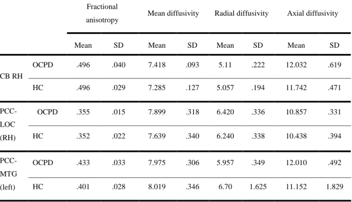
Fractional
anisotropy Mean diffusivity Radial diffusivity Axial diffusivity
Mean SD Mean SD Mean SD Mean SD
CB RH OCPD .496 .040 7.418 .093 5.11 .222 12.032 .619 HC .496 .029 7.285 .127 5.057 .194 11.742 .471 PCC-LOC (RH) OCPD .355 .015 7.899 .318 6.420 .336 10.857 .331 HC .352 .022 7.639 .340 6.240 .338 10.438 .394 PCC-MTG (left) OCPD .433 .033 7.975 .306 5.957 .349 12.010 .492 HC .401 .028 8.019 .346 6.70 1.625 11.152 1.829
*values of MD, RD and AD are multiply for 10−4.
Figure 1. Three-dimensional tractography images of the cingulum bundle, respectively in sagittal,
coronal and axial anatomical planes.
18
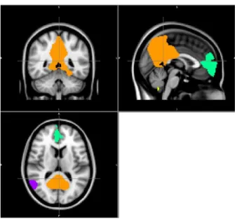
Figure 2. Three-dimensional tractography images of the tract that connects the precuneus to the
lateral occipital cortex in the right hemisphere, respectively in sagittal, coronal and axial anatomical planes.
Figure 3. Three-dimensional tractography images of the precuneus to the middle temporal gyrus
in the left hemisphere, respectively in sagittal, coronal and axial anatomical planes.
No significant differences were found between groups for FA, MD, RD and AD in other white matter fiber track analysed.
Posterior Cingulate Cortex Lateral Occipital Cortex
19 Table 4.
Results from Independent Samples T-Test of the fiber tracts present under 50% of the sample
Fractional anisotropy Mean diffusivity Radial diffusivity Axial diffusivity
t value p value t value p value t value p value t value p value LOC-MTG (right) -1.151 .267 .34 .736 .88 .393 U=37.00 .757 PCC-MTG (left) -2.204 .043* .28 .785 U= 23.00 .122 U=26.00 .200 PCC-MFG (left) -.681 .505 -.571 .576 .365 .720 -1.340 .199 *p<.05
Note. We performed Mann-Whitney Tests when the assumptions underlying the use of parametric tests were not
fulfilled.
Discussion
To the best of our knowledge, this is the first study which analyzed structural alterations in the white matter fiber tracts that connect different nodes of the DMN in individuals diagnosed with obsessive-compulsive personality disorder.
The results of the study revealed that patients diagnosed with obsessive-compulsive personality disorder exhibited significantly higher MD in the right side of the cingulum bundle and significantly higher AD in the tract that connects the posterior cingulate cortex to the lateral occipital cortex (right hemisphere). In addition, we also found a significant increase in FA in the fiber tract that connects the precuneus to the middle temporal gyrus in the analyses we performed of the fiber tracts present in less than fifty per cent of the sample.
As mentioned before, there are not studies that analyze white matter abnormalities in the DMN in OCPD patients, however several previous studies have used DTI in order to identify structural abnormalities in the white matter in patients with obsessive-compulsive disorder, schizotypal personality disorder or major depressive disorder. The results on MD found in the literature are quite controversial, with some studies reporting a decrease in MD in clinical populations (Lochner
20 et al., 2012) when compared to controls, while others report the opposite finding. In our study we found an increase of MD which is consistent with preceding finding where values of MD were significantly higher in OCD patients in the left cingulum bundle compared with healthy control group (Fontenelle, Bramati, Moll, Mendlowicz, Oliveira-Souza & Tovar-Moll, 2011). These results suggest the presence of white matter structural abnormalities in the cingulum bundle associated with damaged or atrophied white matter due of the increased free diffusion in this diagnostic group compare with healthy controls. In addition, the difference in the MD of the cingulum bundle found in our study was localized in the right side of the brain. The right hemisphere is specialized in the perception and expression of emotion, and is thought to be responsible for the regulation of experience emotions (Demaree, Everhart, Youngstrom & Harrison, 2005). Previous studies also suggest that the right hemisphere is specialized in the processing of negative emotions (Silberman & Weingartner, 1986) which have a key role in psychopathology in general, and in this case in the obsessive-compulsive personality disorder. As mentioned before, the cingulum bundle is one of the most important white matter bundles that mediates the functional connectivity between the two main nodes of the DMN, respectively the PCC/precuneus and the mPFC. The precuneus is involved in the response to salient stimuli from the world around us and it is closely linked to self-reference tasks and plans for the future. On the other hand, the mPFC is involved in the emotional processing and monitoring of the state of mind, is related to the expression of personality, decision making and planning behaviors. Thus we may speculate that the alteration found in this white matter fiber tract can be related with an altered communication between these two main nodes of the DMN that have a relevant role in the psychological dimensions underlying the symptomatology of the OCPD.
In addition to these findings, we also found increased AD values in the fiber tract that connects the posterior cingulate cortex and the lateral occipital cortex. To the best of our knowledge there are no studies reporting differences in this particular white matter tract, however we hypothesize that since this tract is connecting one of the most important regions (PCC) for the psychological disorder under study it might play an important role in the pathophysiology of OCPD. Li and colleagues (2011) found higher FA and AD among individuals with OCD in the truncus and genu of the corpus callosum and in the right superior frontal gyrus. Thomason and Thompson (2011) suggests that these high values of AD are associated with axonal degeneration and a restricted diffusion since the diffusion occurs in the fiber direction.
21 Lastly, in the analyze we did of the fiber tracts that were present in less than fifty per cent of the sample, we found higher values of FA in the OCPD patients in the tract that connects the posterior cingulate cortex to the middle temporal gyrus. The temporal lobe has a significant role in anxiety and depression disorders. A study conducted by Qiu et al. (2014) found significantly decreased FA values in the white matter of the left insula, left inferior frontal gyrus, left middle temporal gyrus, and left inferior parietal gyrus in patients with social anxiety disorder. Other study is consistent with the study by Qui and colleagues (2014), which found significant reductions in the FA values in regions of the frontal and temporal lobes in patients with depression compared with healthy controls (Nobuhara, et al., 2006). This result is a more exploratory one since it was obtained from the analysis of a fiber tract presented in less than fifty per cent of the sample.
In summary, the present study identified structural white matter abnormalities, quantified with de DTI metrics of Mean Diffusivity and Axial Diffusivity, respectively in the right side of the cingulum bundle and the tracts that connects the posterior cingulate cortex to the lateral occipital cortex.
Limitations and future directions
Although the promising data found in our study, our results should be interpreted with caution due to some limitations. The small sample size, which was due to the exclusion of some participants due to the presence of artifacts such as head movement and the inherent difficulties in recruiting OCPD patients is one of the limitations. It would be advantageous if the results were replicated in a larger sample of participants.
Since this is the first study that analyzed the structural connectivity of the default mode network in obsessive-compulsive personality disorder, it should be replicated using other analyze tools. In this study, we used the BrainCAT, an automated and optimized tool to help in the processing and analysis of combined fMRI/DTI data. BrainCAT allows the user to go from fMRI and DWI data to results without the need to know how manually perform image-processing and requires minimal user intervention which diminish the probability of human mistake in the pre-processing steps. Using this tool, we assume that functional connectivity may be related with the pattern of white matter microstructure within the resting state DMN. However, our research question could be analyzed using other tools such as the FreeSurfer that is a software package for automated surface
22 reconstruction and analysis, which extracts white matter, computes measures such as cortical thickness and sulcal depth, and performs cross-subjects analysis using spherical registration (Fischl et al., 2004) and also includes an automated segmentation algorithm. Another tool which could be used for this purpose is the 3D Slicer that is a software package for images visualization and analysis using an manual or automated approach. In future studies it would be important to analyze these same data using alternative methods such as 3D slicer or Freesurfer in order to evaluate the consistency of the results.
References
Alexander, A. L., Lee, J. E., Lazar, M., & Field, A. S. (2007). Diffusion tensor imaging of the
brain. Neurotherapeutics, 4, 316–329. doi:10.1016/j.nurt.2007.05.011
American Psychiatric Association (Ed.). (2000). Diagnostic and statistical manual of mental
disorders DSM-IV-TR (4th ed., text rev.). Washington, DC: Author.
American Psychiatric Association (Ed.). (2013). Diagnostic and statistical manual of mental
disorders DSM-V (1st ed.). Washington, DC: Author.
Assaf, Y., & Pasternak, O. (2008). Diffusion tensor imaging (DTI) - based white matter mapping
in brain research: a review. Journal of molecular neuroscience, 34, 51–61.
doi:10.1007/s12031-007-0029-0
Beckmann, M., Johansen-Berg, H., & Rushworth, M. F. S. (2009). Connectivity-based
parcellation of human cingulate cortex and its relation to functional specialization. The
Journal of Neuroscience, 29, 1175–1190. doi:10.1523/JNEUROSCI.3328-08.2009
Binder, J. R., Desai, R., H., Graves, W. W., & Conant, L. L. (2009). Where is semantic system?
A critical review and meta-analysis of 120 functional neuroimaging studies. Cerebral Cortex,
23 Bora, E., Harrison, B. J., Fornito, A., Cocchi, L., Pujol, J., Fontenelle, L. F., Velakoulis, D., …
Yücel, M. (2011). White matter microstructure in patients with obsessive-compulsive
disorder. Journal of Psychiatry and Neuroscience, 36, 42–46. doi:10.1503/jpn.100082
Cannistraro, P. A., Makris, N., D., Howard, J. D., Wedig, M. M., Hodge, S. M., Wilhelm, S., …
Rauch S. L. (2007). A diffusion tensor imaging study of white matter in obsessive-compulsive
disorder. Depression and Anxiety, 24, 440–446. doi:10.1002/da.20246
Cain, N. M., Ansell, E. B., Simpson, H. B., & Pinto, A. (2014). Interpersonal functioning in
obsessive–compulsive personality disorder. Journal of Personality Assessment, 97, 90–99.
doi:10.1080/00223891.2014.934376
Chambers, C. D., Payne, J. M., Stokes, M. G., & Mattingley, J. B. (2004). Fast and parietal
pathways mediate spatial attention. Nature Neuroscience, 7, 217–218. doi:10.1038/nn1203
Corbetta, M., Kincade, J. M., Ollinger, J. M., McAvoy, M. P., & Shulman, G. L. (2000). Voluntary
orienting is dissociated from target detection in human posterior parietal cortex. Nature
Neuroscience, 3, 292–297. doi:10.1038/73009
Cullen, K. R., Klimes-Dougan, B., Muetzel, R., Mueller, B. A, Camchong, J., Houri, A., … Lim,
K. O. (2010). Altered white matter microstructure in adolescents with major depression: A
preliminary study. Journal of the American Academy of Child and Adolescent Psychiatry, 49,
173–183. doi:10.1016/j.jaac.2009.11.005
De Reus, R. J. M., & Emmelkamp, P. M. G. (2010). Obsessive – compulsive personality disorder :
A review of current empirical findings. Personality and Mental Health, 6, 1–21.
doi:10.1002/pmh.144
Demaree, H. A., Everhart, D. E., Youngstrom, E. A., & Harrison, D. W. (2005). Brain
24
“dominance”. Behavioral and Cognitive Neuroscience Reviews, 4, 3–20.
doi:10.1177/1534582305276837
Dong, Q., Welsh, R. C., Chenevert, T. L., Carlos, R. C., Maly-Sundgren, P., Gomez-Hassan, D.
M., & Mukherji, S. K. (2004). Clinical applications of diffusion tensor imaging. Journal of
Magnetic Resonance Imaging, 19, 6–18. doi:10.1002/jmri.10424
Esposito, F., Bertolino, A., Scarabino, T., Latorre, V., Blasi, G., Popolizio, T., Tedeschi, G., ... Di
Salle, F. (2006). Independent component model of the default-mode brain function: Assessing
the impact of active thinking. Brain Research Bulletin, 70, 263–9.
doi:10.1016/j.brainresbull.2006.06.012
Evans, A. C., Janke, A. L., Collins, D. L., & Baillet, S. (2012). Brain templates and atlases.
Neuroimage, 62, 911–922. doi:10.1016/j.neuroimge.2012.01.024
Fields, R. D. (2008). White matter in learning, cognition and psychiatric disorders. Trends in
Neurosciences, 31, 361–370. doi:10.1016/j.tins.2008.04.001
First, M. B., Gibbon, M., Spitzer, R. L., Williams, J. B. M., & Benjamin, L. S. (1997). Structured
Clinical Interview for DSM-IV Axis II Personality Disorders, (SCID-II). Washington, D.C.:
American Psychiatric Press, Inc.
Fischl, B., Van Der Kouwe, A., Destrieux, C., Halgren, E., Ségonne, F., Salat, D. H., … Dale, A.
M. (2004). Automatically parcellating the human cerebral cortex. Cerebral Cortex, 14, 11–
22. doi:10.1093/cercor/bhg087
Fontenelle, L. F., Bramati, I. E., Moll, J., Mendlowicz, M. V., de Oliveira-Souza, R., &
Tovar-Moll, F. (2011). White Matter Changes in OCD Revealed by Diffusion Tensor Imaging. CNS
25 Garrity, A. G., Pearlson, G. D., McKiernan, K., Lloyd, D., Kiehl, K. A., & Calhoun, V. D. (2007). Aberrant “default mode” functional connectivity in schizophrenia. The American journal of
psychiatry, 164, 450–7. doi:10.1176/appi.ajp.164.3.450
Gusnard, D. A., & Raichle, M. E. (2001). Searching for a baseline: Functional imaging and the
resting human brain. Nature Reviews Neuroscience, 2, 685–694. doi:10.1038/35094500
Grimm, S., Boesiger, P., Beck, J., Schuepbach, D., Bermpohl, F., Walter, … Northoff, G. (2009).
Altered negative BOLD responses in the default-mode network during emotion processing in
depressed subjects. Neuropsychopharmacology, 34, 932–43. doi:10.1038/npp.2008.81
Harrison, B. J., Pujol, J., Ortiz, H., Fornito, A., Pantelis, C., & Yücel, M. (2008). Modulation of
brain resting-state networks by sad mood induction. PloS One, 3, e1794.
doi:10.1371/journal.pone.0001794
Harrison, B. J., Pujol, J., López-Solà, M., Hernández-Ribas, R., Deus, J., Ortiz, H., ... Cardoner,
N. (2008). Consistency and functional specialization in the default mode brain network.
Proceedings of the National Academy of Sciences of the United States of America, 105, 9781–
9786. doi:10.1073/pnas.0711791105
Hazlett, E. A., Goldstein, K. E., Tajima-Pozo, K., Speidel, E. R., Zelmanova, Y., Entis, J. J., …
Siever, L. J. (2011). Cingulate and temporal lobe fractional anisotropy –in schizotypal
personality disorder. Neuroimage, 55, 900–908. doi:10.1016/j.neuroimage.2010.12.082
Jones, D. K., Cristiansen, K. F., Chapman, R. J. & Aggleton, J. P. (2012). Distinct subdivisions of
the cingulum bundle revealed by diffusion MRI fibre tracking: Implications for
neuropsychological. Neuropsychologia, 51, 67–78.
26 Kim, M. J., & Whalen, P. J. (2010). The structural integrity of an amygdala-prefrontal pathway
predicts trait anxiety. The Journal of neuroscience, 29, 11614–11618.
doi:10.1523/JNEUROSCI.2335-09.2009
Lawes, I. N., Barrick, T. R., Murugan, V., Spierings, N., Evans, D. R., Song, M., & Clark, C. A.
(2008). Atlas-based segmentation of white matter tracts of the human brain using diffusion
tensor tractography and comparison with classical dissection. NeuroImage, 39, 62–79.
doi:10.1016/j.neuroimage.2007.06.041
Leech R., & Sharp DJ. (2014). The role of the posterior cingulate cortex in cognition and disease,
Brain, 137, 12–32. doi:10.1093/brain/awt162
Li, F., Huang, X., Yang, Y., Li, B., Wu, Q., Zhang, T., … Chiu, M. J. (2011). Microstructural
brain abnormalities in patients with obsessive-compulsive disorder: Diffusion-tensor MR
imaging study at 3.0 T. Radiology, 260, 216–223. doi:10.1148/radiol.11101971
Light, K., & Mulder, R. (2009). Obsessive-compulsive personality disorder. Encyclopedia of
Molecular Mechanisms of Disease, 27, 1507–1508. doi:10.1007/978-3-540-29676-8_3188
Lin, Y. C., Shih, Y. C., Tseng, W. Y., Chu, Y. H., Wu, M. T., Chen, … Chiu, M. J. (2014).
Cingulum correlates of cognitive functions in patients with mild cognitive impairment and early alzheimer’s disease: A diffusion spectrum imaging study. Brain topography, 27, 393– 402. doi:10.1007/s10548-013-0346-2
Livesley, W. J. (2003). Practical management of personality disorder. Guilford Press.
Lochner, C., Serebro, P., Van der Merwe, L., Hemmings, S., Kinnear, C., Seedat, S., & Stein, D.
J. (2011). Comorbid obsessive-compulsive personality disorder in obsessive-compulsive
disorder (OCD): a marker of severity. Progress in Neuro-Psychopharmacology & Biological
27 Lochner, C., Fouché, J. P., Du Plessis, S., Spottiswoode, B., Seedat, S., Fineberg, N., … Stein, D.
J. (2012). Evidence for fractional anisotropy and mean diffusivity white matter abnormalities
in the internal capsule and cingulum in patients with obsessive-compulsive disorder. Journal
of psychiatry & neuroscience, 37, 193–9. doi:10.1503/jpn.110059
Magalhães, E., Salgueira, A., Gonzalez, A. J., Costa, J. J., Costa, M. J., Costa, P., & Pedroso de
Lima, M. (2014). NEO-FFI: Psychometric properties of a short personality inventory in
Portuguese context. Psicologia: Reflexão e Crítica, 27, 642–657.
doi:10.1590/1678-7153.201427405
Marques, P., Soares, J. M., Alves, V., & Sousa, N. (2013). BrainCAT - a tool for automated and
combined functional magnetic resonance imaging and diffusion tensor imaging brain
connectivity analysis. Frontiers in Human Neuroscience, 7, 1–11.
doi:10.3389/fnhum.2013.00794
Mars, R. B., Neubert, F.-X., Noonan, M. P., Sallet, J., Toni, I., & Rushworth, M. F. S. (2012). On the relationship between the “default mode network” and the “social brain.” Frontiers in
Human Neuroscience, 6, 1–9. doi:10.3389/fnhum.2012.00189
Mason, M. F., Norton, M. I., Van Horn, J. D., Wegner, D. M., Grafton, S. T., & Macrae, C. N.
(2007). Wandering minds: The default mode network and stimulus-independent thought.
Science, 315, 393–395. doi:10.1126/science.1131295
McCrae, R. R., & Costa, P. T. (1987). Validation of the five-factor model of personality across
instruments and observers. Journal of Personality and Social Psychology, 52, 81–90.
doi:10.1037/0022-3514.52.1.81
McCrae, R. R., & Costa, P. T. (2004). A contemplated revision of the NEO five-factor inventory.
28 Milner, B. (2005). The medial temporal-lobe amnesic syndrome. Psychiatric Clinics of North
America, 28, 599–611. doi:10.1016/j.psc.2005.06.002
Mor, N., & Winquist, J. (2002). Self-focused attention and negative affect: A meta-analysis.
Psychological Bulletin, 128, 638–662. doi:10.1037//0033-2909.128.4.638
Nakamura, M., Mccarley, R. W., Kubicki, M., Dickey, C. C., Niznikiewicz, M. A., Voglmaier,
M. M., … Shenton, M. E. (2005). Fronto-temporal disconnectivity in schizotypal personality
disorder: A diffusion tensor imaging study. Biol Psychiatry, 77, 468–478.
doi:10.1016/j.biopsych.2005.04.016
Nobuhara, K., Okugawa, G., Sugimoto, T., Minami, T., Tamagaki, C., Takase, K., ... Kinoshita,
T. (2006). Frontal white matter anisotropy and symptom severity of late-life depression: A
magnetic resonance diffusion tensor imaging study. Journal of Neurology, Neurosurgery &
Psychiatry, 58, 120–122. doi:10.1136/jnnp.2004.055129
Oldfield, R. C. (1971). The assessment and analysis of handedness: The Edinburgh inventory.
Neuropsychologia, 9(1), 97-113. Retrieved from
http://www.sciencedirect.com/science/article/pii/0028393271900674
Pedroso de Lima, M., Magalhães, E., Salgueira, A., Gonzalez, A., Costa, J. J., Costa, M. J., &
Costa, P. (2014). A versão portuguesa do NEO-FFI : Caracterização em função da idade ,
género e escolaridade. Psicologia: Revista da Associação Portuguesa de Psicologia. 28(2),
1–10. Retrieved from http://web.b.ebscohost.com/abstract?site=ehost&scope=site&jrnl=08742049&AN=9995671 7&h=QpMkhGUswLvyqXyBmER4Zf0R9CoqjLxwx8lCvoCqVtc78omsrpbhOoS3KLjsYY kRBrpz5VKqjyqFRVwYQHdfTw%3d%3d&crl=c&resultLocal=ErrCrlNoResults&resultNs =Ehost&crlhashurl=login.aspx%3fdirect%3dtrue%26profile%3dehost%26scope%3dsite%2 6authtype%3dcrawler%26jrnl%3d08742049%26AN%3d99956717
29 Pierpaoli, C., Jezzard, P., Basser, P. J., Barnett, A., & Di Chiro, G. (1996). Diffusion tensor MR
imaging of the human brain. Radiology, 201, 637-648. doi:10.1148/radiology.201.3.8939209
Qiu, C., Zhu, C., Zhang, J., Nie, X., Feng, Y., Meng, Y., … Gong, Q. (2014). Diffusion tensor
imaging studies on Chinese patients with social anxiety disorder. BioMed Research
International, 2014. doi:10.1155/2014/860658
Raichle, M. E., & Snyder, A. Z. (2007). A default mode of brain function: A brief history of an
evolving idea. NeuroImage, 37, 1083–1090. doi:10.1016/j.neuroimage.2007.02.041.
Saxe, R., Moran, J. M., Scholz, J., & Gabrieli, J. (2006). Overlapping and non-overlapping brain
regions for theory of mind and self reflection in individual subjects. Social Cognitive and
Affective Neuroscience, 1, 229–34. doi:10.1093/scan/nsl034
Silberman, E. K., & Weingartner, H. (1986). Hemispheric lateralization of functions related to
emotion. Brain and Cognition, 5, 322–353. doi:10.1016/0278-2626(86)90035-7
Skodol, A. E., Clark, L. A., Bender, D. S., Krueger, R. F., Morey, L. C., Verheul, R., … Oldham,
J. M. (2011). Proposed changes in personality and personality disorder assessment and
diagnosis for DSM-5 Part I: Description and rationale. Personality disorders, 2, 4–22.
doi:10.1037/a0021891
Spreng, R. N., & Grady, C. L. (2010). Patterns of brain activity supporting autobiographical
memory, prospection, and theory of mind, and their relationship to the default mode network.
Journal of Cognitive Neuroscience, 22, 1112–1123. doi:10.1162/jocn.2009.21282.
Spreng, R. N., Mar, R. A., & Kim, A. S. (2009). The common neural basis of autobiographical
memory, prospection, navigation, theory of mind, and the default mode: A quantitative
30 Starcevic, V., & Brakoulias, V. (2014). New diagnostic perspectives on obsessive-compulsive
personality disorder and its links with other conditions. Current Opinion in Psychiatry, 27,
62–67. doi:10.1097/YCO.0000000000000030
Teipel, S. J., Bokde, A. L. W., Meindl, T., Amaro Jr, E., Soldner, J., Reiser, M. F., ... Hampel, H.
(2009). White matter microstructure underlying default mode network connectivity in the
human brain. NeuroImage, 49, 2021–2032. doi:10.1016/j.neuroimage.2009.10.067
Thomason, M. E., & Thompson, P. M. (2011). Diffusion imaging, white matter, and
psychopathology. Annual Review of Clinical Psychology, 7, 63–85.
doi:10.1146/annurev-clinpsy-032210-104507
Van den Heuvel, M., Mandl, R., Luigjes, J., & Hulshoff Pol, H. (2008). Microstructural
organization of the cingulum tract and the level of default mode functional connectivity. The
Journal of neuroscience, 28, 10844–51. doi:10.1523/JNEUROSCI.2964-08.2008
Whitfield-Gabrieli, S., & Ford, J. M. (2012). Default mode network activity and connectivity in
psychopathology. Annual Review of Clinical Psychology, 8, 49–76.
doi:10.1146/annurev-clinpsy-032511-143049
Zhao, X. H., Wang, P. J., Li, C. B., Hu, Z. H., Xi, Q., Wu, W. Y., & Tang, X. W. (2007). Altered
default mode network activity in patient with anxiety disorders: an fMRI study. European
Journal of Radiology, 63, 373-378. doi:10.1016/j.ejrad.2007.02.00
Zhou, Y., Liang, M., Tian, L., Wang, K., Hao, Y., Liu, H., … Liang, T. (2007). Functional
disintegration in paranoid schizophrenia using resting-state fMRI. Schizophrenia research,
31
Appendix A