Ana Sofia da Costa e Brito
Saccharomyces cerevisiae strains
expressing human
KRAS as tools for
targeting therapeutic anti-EGFR/RAS
pathway antibody/drugs
Ana Sofia da Costa e Brito
Saccharomyces cerevisiae strains
expressing human
KRAS as tools for
targeting therapeutic anti-EGFR/RAS
pathway antibody/drugs
Dissertação de Mestrado
Mestrado em Genética Molecular
Trabalho realizado sob a orientação da
Professora Doutora Cândida Lucas Professora Doutora Célia Ferreira
Nome: Ana Sofia da Costa e Brito
Endereço Electrónico: anasofiabrito_23@hotmail.com Número do Bilhete de Identidade: 13583510
Título da Tese: Saccharomyces cerevisiae strains expressing human KRAS as tools for
targeting therapeutic anti-EGFR/RAS pathway antibody/drugs
Orientador (es):
Professora Doutora Cândida Lucas Professora Doutora Célia Ferreira
Ano de Conclusão: 2015
Designação do Mestrado: Mestrado em Genética Molecular
É AUTORIZADA A REPRODUÇÃO PARCIAL DESTA TESE/TRABALHO APENAS PARA EFEITOS DE INVESTIGAÇÃO, MEDIANTE DECLARAÇÃO ESCRITA DO INTERESSADO, QUE A TAL SE COMPROMETE
Universidade do Minho, ___/___/___
iii
AGRADECIMENTOS
Gostaria de manifestar o meu agradecimento a todos que, directa e indirectamente, contribuíram para a realização desta tese:
Às minhas orientadoras, a Doutora Cândida Lucas e Doutora Célia Ferreira, por me terem dado a possibilidade de realização deste trabalho. E mais ainda, por toda a paciência, apoio, disponibilidade e ensinamentos transmitidos que tanto contribuíram para a minha evolução.
À Joana Tulha por toda a paciência e compreensão que teve comigo, pelo apoio incansável, incentivo, pelas discussões científicas e suporte dado ao longo desta etapa.
À Giulia Cazzanelli pelos conhecimentos trocados e apoio prestado ao longo deste ano.
À Eliana Carneiro, minha amiguinha nesta etapa, por todas as conversas, pelos bons e “maus” momentos passados juntas e sobretudo pela amizade.
Ao Aureliano, à Simone e ao Diogo pela alegria, companhia e por tornarem o ambiente laboratorial ainda mais agradável.
À Diana, Cristiana, Nanda e Carla e aos restantes colegas do mestrado pela amizade, apoio moral demonstrado e pelas inúmeras vezes que me elevarem o espírito.
Aos restantes colegas, professores, técnicos e funcionários do departamento de Biologia pela disponibilidade para ajudar sempre que foi preciso.
Um especial reconhecimento aos meus amigos, principalmente Emília, Adriana, Patrícia, Marisol e Liliana e aos familiares pela amizade incondicional, apoio constante e acima de tudo por acreditarem em mim.
Aos meus irmãos por estarem lá sempre que preciso.
Aos meus pais pelo apoio, por todos os sacrifícios, por tudo aquilo que me ensinaram e me ajudaram a alcançar.
v
ABSTRACT
Fundamental cellular processes appear to be highly conserved between Saccharomyces
cerevisiae and other more complex Eukaryotic species, including humans. “Humanized yeast
systems” emerged as a tool to study molecular aspects of human pathologies. The present work aimed at contributing to build and validate a large high throughput platform of yeast strains displaying phenotypes that can enable further testing galectin-related drugs and peptides. This platform was designed to consist of two types of strains, the ones expressing human galectins and the ones expressing these together with the human KRAS cDNA. The rationale behind this relates with the putative dialogue between Galectins and RAS signaling pathway in mammals. Considering that EGFR mediates KRAS signaling and that yeast also harbors a RAS signaling pathway, the “humanized yeasts” expressing KRAS were used to identify the yeast target of anti-EGFR. Furthermore, it was also used for phenotyping the most well-known biological processes known to be controlled by RAS pathway. On the other hand, considering that the deletion of GUP1 in S. cerevisiae increases the resistance to the oncological drug Imatinib, the similarities between the phenotypes associated to the deletion of RAS and GUP genes were also verified.
Two Hsp70, Ssa2p and Ssb2p and one glyceraldehyde-3-phosphate dehydrogenase Tdh3p, were identified as EGFR-like proteins. The subsequent alignments analysis between EGFR and these proteins revealed that Ssb2p and its very close homologue Ssa2p present some homology with EGFR sequence, namely at the level of three EGFR conserved amino acids known to be responsible for the interaction with the anti-EGFR antibody Cetuximab used in cancer treatment. This and other lines of evidence support Ssb2p and/or Ssa2p as good candidates for EGFR homology. The phenotypic tests revealed that both the deletions of GUP and RAS genes promote a reduction in chronological life span and cell size, except in the case of ∆ras2 strain, whose cells were bigger than wild type control. Nutrient depletion (carbon)
promoted replication stress in ∆ras2 cells that failed to enter into G1 arrest, and were blocked in
S phase, concurring with the bigger size of ∆ras2 cells and their short lifespan. Moreover, the
cells with GUP genes deleted, in opposition to RAS mutants, showed ability to adhere to solid nitrogen-deficient medium. Neither RAS nor GUP mutants were able to invade or filament under these conditions.
With this work we were able to determine the possible homologue of EGFR, many times associated with cancer pathologies, and contributed to gain insights on RAS and GUP genes common phenotypes. In conclusion, the present work opens doors to future discovery of new pathways in yeast, in addition to showing that S. cerevisiae is a suitable model to create a platform to explore therapeutic drugs/antibodies.
vii
RESUMO
Vários processos celulares fundamentais encontram-se conservados entre a levedura
Saccharomyces cerevisiae e outras espécies eucariotas mais complexas, incluindo humanos. A
“Levedura humanizada” surgiu como uma ferramenta de estudo sobre aspectos moleculares de patologias humanas. Com este trabalho pretendeu-se contribuir para a construção e validação de uma plataforma de estirpes de levedura que exibam determinados fenótipos, permitindo o teste de drogas e péptidos relacionados com as galectinas. Esta foi planeada para incluir duas estirpes a expressar galectinas humanas, assim como o cDNA do KRAS humano. O propósito desta plataforma advém de uma possível interação entre as Galectinas e a via de sinalização dos RAS em mamíferos. Tendo em conta que o EGFR medeia a cascata de sinalização KRAS, e que também a levedura possui uma via de sinalização Ras, usou-se as leveduras humanizadas a expressar o KRAS para identificar o alvo do anti-EGFR. Para além disso, estas foram usadas para a fenotipagem de processos biológicos controlados pela cascata RAS. Por outro lado, tendo em conta que a deleção do GUP1 aumenta a resistência à droga oncológica Imatinib, verificou-se também as verificou-semelhanças fenotípicas entre as deleções RAS e GUP.
Foram identificadas duas proteínas Hsp70, Ssa2p e Ssb2p, e uma gliceraldeído-3-fosfato desidrogenase Tdh3p, como sendo os alvos do anti-EGFR. Subsequentemente, a análise dos alinhamentos entre o EGFR e estas proteínas revelaram que a Ssb2p e a sua homóloga Ssa2p apresentam similaridade com a sequência do EGFR, nomeadamente ao nível de três aminoácidos responsáveis pela interação com o anticorpo anti-EGFR, Cetuximab, usado no tratamento do cancro. Esta informação suporta a hipótese das proteínas Ssb2p e/ou Ssa2p serem boas candidatas a homólogas do EGFR. Os testes fenotípicos revelaram que as deleções dos genes GUP e RAS promovem uma redução da longevidade cronológica e da área celular, com excepção para a estirpe ∆ras2 cujas células se revelaram maiores do que a wt. A depleção de
nutrientes (carbono) induziu stress replicativo nas células ∆ras2, que por sua vez falharam a
entrada na fase G1, ficando bloqueadas na fase S, o que está de acordo com o aumento da área celular e a baixa longevidade cronológica das células ∆ras2. Além disso, as células com a
deleção nos genes GUP, contrariamente aos mutantes RAS, mostraram habilidade para aderir a um meio deficiente em nitrogénio. Nenhum dos mutantes RAS ou GUP foram capazes de invadir ou filamentar nas condições anteriormente descritas.
Com este trabalho fomos capazes de determinar o possível homólogo do EGFR, muitas vezes associado a patologias relacionadas com o cancro, assim como contribuir para melhor compreender os fenótipos comuns associados aos genes RAS e GUP. Em conclusão, o presente trabalho abre portas para futuras descobertas de novas vias de sinalização em levedura, além de reforçar a utilização da S. cerevisiae como um bom modelo para criar uma plataforma de exploração de drogas/anticorpos.
ix
TABLE OF CONTENTS
AGRADECIMENTOS ... iii ABSTRACT ... v RESUMO ... vii LIST OF ABBREVIATIONS ... xi CHAPTER I - Introduction……… 31.1 Yeast as Eukaryotic Model ... 3
1.2 Yeast as a Cell Aging Model ... 6
1.3 RAS/cAMP/PKA Pathway ... 8
1.4 TOR Pathway ... 10
1.5 Sch9 Pathway ... 12
1.6 Gup protein in Saccharomyces cerevisiae... 13
1.7 The Ras family of small GTPases ... 16
1.8 Metabolic Similarities between Cancer Cells and Yeast ... 20
1.9 The Role of Cell Surface Receptors ... 21
1.9.1 EGFR as a Therapeutic Target ... 27
1.10 Rationale and Aims of the thesis ... 31
CHAPTER II - Material and Methods……… 35
2.1 Strains and Growth Conditions ... 35
2.2 Competent E. coli cells... 37
2.3 Construction of E. coli p426KRASwt... 37
2.3.1 DNA amplification by Polymerase Chain Reaction (PCR) ... 37
2.3.2 DNA electrophoresis ... 38
2.3.3 DNA digestion and ligation ... 38
2.3.4 Plasmid amplification in E. coli ... 39
2.3.5 Transformation of S. cerevisiae and colony PCR ... 40
2.4 Western Blot Analysis ... 41
2.4.1 Yeast protein extraction and precipitation ... 41
2.4.2 SDS-PAGE (Sodium Dodecyl Sulphate – PolyAcrylamide Gel Electrophoresis)... 41
2.4.3 Western blot assay ... 41
2.5 Native-PAGE ... 42
2.6 Yeast Physiology Assays ... 43
2.6.1 Chronological life span (CLS) ... 43
x
2.6.3 Cell cycle analysis ... 44
2.6.4 Adherence to and invasion of agar ... 44
2.7 Statistical Analysis ... 45
CHAPTER III - Results and Discussion……….. 49
3.1 Heterologous expression of KRASwt in Saccharomyces cerevisiae ... 49
3.2 Identification of the yeast target of anti-EGFR (Cetuximab) by Western Blot ... 54
P1 - Heat shock proteins Ssa2p and Ssb2p... 58
P2 - Glyceraldehyde-3-phosphate dehydrogenase 3 (Tdh3p) ... 61
Similarity between human EGFR and the yeast targets of anti-EGFR ... 62
3.3 Insights on RAS and GUP genes in the W303-1A strain ... 69
3.3.1 Growth and CLS assessments ... 70
3.3.1.1 RAS and GUP deletions affect growth on minimal media ... 70
3.3.1.2 The RAS and GUP mutants exhibit a reduced chronological life span ... 71
3.3.2 The GUP mutants and ∆ras1 are smaller, while ∆ras2 is bigger than wt ... 74
3.3.3 Cell Cycle analysis ... 76
3.3.4 Adherence to and Invasion of agar ... 80
CHAPTER IV - Final Remarks and Future Perspectives………. 85
xi
LIST OF ABBREVIATIONS
AC Adenylate cyclase
Adh Alcohol dehydrogenase
AIF Apoptosis-inducing factor
APAF Apoptotic protease activating factor
ATP Adenosine triphosphate
Bax Bcl-2-associated X protein
Bcl-2 B-cell lymphoma 2
BLAST Basic local alignment search tool
BRAF v-RAF murine sarcoma viral oncogene homologue B1 cAMP Adenosine γ’, 5’-cyclic monophosphate
CDK Cyclin dependent kinase
CFU Colony Forming Units
CFW Calcofluor white
CLS Chronological life span
CRC Colorectal cancer
CREB cAMP response element binding protein
Ctx Cetuximab
DMSO Dimethyl sulfoxid
DNA Deoxyribonucleic acid
ECM Extracellular matrix
EDTA Ethylenediaminetetraacetic acid
EGF Epidermal growth factor
EGFR Epidermal growth factor receptor
EGFRvIII EGFR variant III
ER Endoplasmatic reticulum
ERK Extracellular regulated kinase
Fab Fragment antigen binding
Fc Fragment constant
FDA Food and drug administration
FGF Fibroblast growth factor
Fv Fragment variable
GADPH Glyceraldehyde-3-phosphate dehydrogenase
GAP GTPase activating protein
GDP Guanosine diphosphate
xii
GFP Green fluorescence protein
GPI Glycosyl phosphatidylisositol
GRB2 Growth factor receptor bound protein 2
GTP Guanosine triphosphate
HER Human epidermal growth factor receptor
HHATL Hedgehog acyltransferase-like protein
HOG High osmolarity glycerol
HRP Horseradish peroxidase
Hsp Heat shock protein
IGF Insulin growth factor
JAK Janus kinase
KRAS v-Ki-ras2 Kristen rat sarcoma viral oncogene homologue
LB Luria-Bertani medium
LGT Lateral gene transfer
mAb Monoclonal antibody
MALDI Matrix assisted laser desorption ionization MAPK Mitogen-activated protein kinase
MBOAT Membrane bound O-acetyltransferase
MCS Multi cloning site
miRNA micro RNA
mRNA Messenger ribonucleic acid
MS Mass spectrometry
MWM Molecular weight marker
Na2HPO4●7H2O Sodium monohydrogen phosphate heptahydrate
NaCl Sodium chloride
NADH Nicotinamide adenine dinucleotide NaH2PO4●H2O Sodium dihydrogen phosphate hydrate
Native-PAGE Native polyacrylamide gel electrophoresis
NBD Nucleotide binding domain
NCBI National center for biotechnology information
NF-kB Nuclear factor kappa B
NGF Nerve growth factor
NK Natural killer
OD Optical density
PBS Phosphate buffer saline
PBST Phosphate buffer saline Tween 20
xiii
PDC Pyruvate decarboxylase
PDGF Platelet derived growth factor
PDK1 3-phosphoinositide-dependent protein kinase 1
PH Pleckstrin homology
pH Potential of hydrogen
PI3K Phosphotidylinositol 3-kinase
PIK3CA Phosphoinositide-3-kinase, catalytic, alpha polypeptide PIP3 Phosphatidylinositol-3,4,5-triphosphate
PKA Protein kinase A
PKC Protein kinase C
PLC Phosphoinositide phospholipase C PVDF Poly vinylidene difluoride
RLS Replicative life span
RNA Ribonucleic acid
RTK Receptor tyrosine kinase
SBD Substrate binding domain
SC Synthetic complet
SD Standard deviation
SDS Sodium dodecyl sulphate
SDS-PAGE Sodium dodecyl sulfate polyacrylamide gel electrophoresis
sEGFR soluble EGFR
SGD Saccharomyces genome database
SH1 Src homology domain 1
SH2 Src homology domain 2
SHH Sonic hedgehog
SLAD Synthetic low ammonium sulfate and dextrose STAT Signal transducer and activator of transcription
TCA Trichloroacetic acid
TGF Transforming growth factor
TKI Tyrosine kinase inhibitors
TOR Target of rapamycin
tRNA transfer RNA
VEGF Vascular endothelial growth factor
WB Western Blot
YNB Yeast nitrogen base
CHAPTER I
3
1. Introduction
1.1 Yeast as Eukaryotic Model
To date, the yeast model has allowed identifying new targets and novel therapeutic opportunities. Several features make Saccharomyces cerevisiae an ideal model system for the study of human diseases, one of which is the simplicity of yeast genome that comprises only 6,000 genes (1) compared to about 25,000 for the human genome (2). Studies of yeast have been essential for understanding fundamental cellular processes such as metabolism (3), DNA replication and recombination (4), the regulation of cell cycle (5), cell death (6) and for elucidating many mechanisms of several diseases (7). Yeast also presents many practical advantages over human cells. It is well suited to high throughput methods because its life cycle is quick (±90 minutes), it can grow in liquid or on solid media forming suspended cells or colonies respectively, and its culture requires neither elaborate sterile technique nor expensive media (8). Yeast reproduces and dies old or by apoptosis provoked by several stimulus. It lives as individual cells or in colonies, the cells can differentiate into pseudo or true hyphae in response to environmental events, this differentiation is related with the capacity to invade and adhere (9). Moreover, it is a genetically modifiable organism, amenable to changes such as gene deletion, gene marking, mutation or gene dosage effects (7, 10). It has been possible to vary the level of expression of essentially every individual yeast gene and to assemble collections of mutant strains with genome-wide coverage (8).
The use of yeast as a model organism was extended to the analysis of the molecular mechanisms of human diseases, sometimes of rather unexpected nature (11, 12). This was achieved by directly studying an endogenous protein orthologue of a human involved in the disease (11, 13) or through the heterologous expression of human disease associated proteins (14, 15). Although several aspects of the disease in high Eukaryotes are beyond the extend of a unicellular organism like yeast, many processes and pathways are greatly conserved in this organism, namely, cell cycle checkpoint controls, mitochondria biogenesis, protein quality control, vesicular trafficking, apoptosis and autophagic pathways (7, 14, 16). When establishing related protein models, different approaches are used according to the degree of conservation of the protein under study. If the gene codifying for the protein is conserved in yeast, it is possible to directly study its function. If the gene has no orthologue in yeast, the
4
heterologous expression of the human gene in this organism (then designated humanized yeast) can be highly advantageous because yeast conserve protein interactions that give information to its function (14). An example of this strategy is the expression of the tumor suppressor p53 in yeast (17), or of tyrosine kinase receptors, well known to interfere in numerous types of cancer (15). Furthermore the expression of the human gene in yeast often leads to disease relevant phenotypes because yeasts and mammalian cells can respond alike to the appearance of such mutant genes (18).
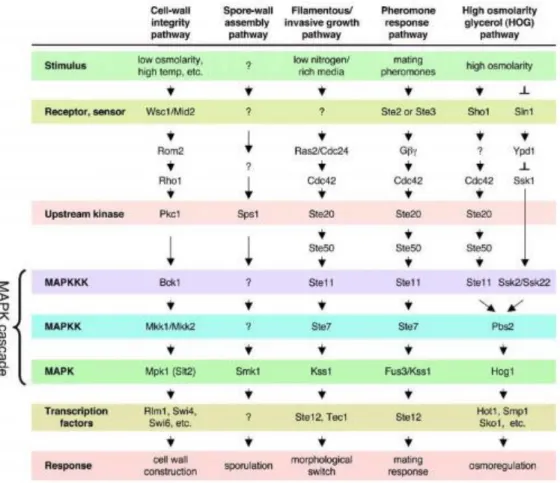
The yeast genome was the first eukaryotic genome to be sequenced (1) and it has allowed pioneer genome scale screening methods, including microarrays (19), two-hybrid analysis (20) and the use of deletion and overexpression libraries (21). Moreover, advances in yeast technology have stimulated the use of this model organism for the creation of high throughput screening platforms for new biologically active compounds, namely through haplo-insufficiency and synthetic lethality screening, or fitness profiling (10). Generally speaking, yeast is considered an excellent model for understanding cellular and molecular processes underlying many diseases. Yeasts harbor well conserved pathways, like TOR, PKC, Calcineurin, stress responsive, secretory, protein sorting pathways and the RAS/cAMP/PKA (22). The MAPK cascades have as principal function regulate transcription factors by MAPK-mediated phosphorylation. Presently, the budding yeast S. cerevisiae has five recognized MAPK pathways, the mating-pheromone response (23), the filamentation-invasion pathway (24), the high osmolarity glycerol (HOG) stress response (25), and the cell integrity pathway (26) (Fig. 1). All of them are operate in vegetative cells during sporulation and regulates the correspondent developmental process (27).
5
Figure 1. MAPK pathways in yeast S. cerevisiae. Withdrawn from (28).
The majority of the cancer-causing mutations were discovered in non-human species, such as yeast, before their role in human cancer was realized. Many of the genes that are frequently altered in tumors have structural or functional orthologues in model genetic systems, including the yeast S. cerevisiae (29). Actually, yeast presents a considerable degree of homology to the human proteome (30). For example, one homology particularly relevant for this work is the one between the oncogenes of the RAS family in human and the two RAS genes RAS1 and RAS2 in yeast (31). Hartwell won 2001 Nobel Prize in Physiology or Medicine for identified in yeast more than 100 genes involved in cell cycle control checkpoints, generally known as the cdc genes (from cell division cycle). The same genes that control the cell cycle in baker's yeast, identically control cell cycle progression in human cells and malfunction in tumor cells (7, 32).
6
1.2 Yeast as a Cell Aging Model
In the last years, the yeast S. cerevisiae has been used as a model to study a range of factors affecting cellular aging, as well as genes involved in pathways controlling life span (33). In view of the specificities of life cycle of yeasts, two types of ageing processes have been identified and can be studied separately: replicative life span (RLS) and chronological life span (CLS). The number of times that a single mother cell, before senescence, originates daughter cells was defined as RLS. On the other hand, the length of time that yeast in non-dividing phase remains viable defined the CLS (34). Several studies using yeast as a model reveled a relation between the longevity and availability of nutrients, thus is now know that the calorie restriction increase the RLS as well as the CLS (34, 35). In others eukaryotic model organisms (worms, flies, zebra fish) the reduction of growth hormones factors promotes longevity as improves overall health by decreasing the probability of developing diseases of diverse types, like cancer, heart attack and diabetes, all related with aging (36).
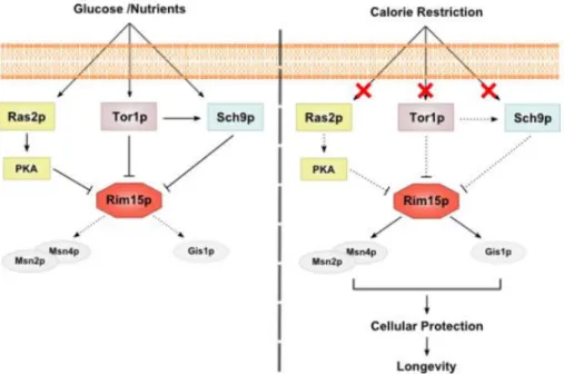
To date, three signaling pathways have been described as regulators of life span: RAS/cAMP/PKA, TOR and Sch9 (37-39) (Fig. 2). In yeast, these pathways are regulated by the availability of nutrients, being activated in presence of glucose and others nutrients inducing cells to proliferate. In opposition, in conditions of nutrient exhaustion, the reduction of signaling of these pathways arrests cell cycle and cells enter a quiescent state (40). Accordingly, the deletions of the genes RAS2, SCH9 and TOR1, as well as the inactivation of other proteins of TOR pathway, increase the yeast CLS and promotes stress resistance (41). The association between these two types of phenotype suggests that increasing cellular protection against damage, and concomitantly increasing the cell repair, can be a strategy to retard aging (40). On the other hand, Tor1p forms a complex with other proteins known as TORC1, which phosphorylates and consequently promotes the activation of the serine threonine kinase Sch9p (42). The deletion of SCH9 triples life span and increases resistance to oxidative and temperature stress (43). The role of Tor1p on longevity could therefore be due to activation of Sch9p (39). Additionally, also mutations decreasing the activity of the RAS/cAMP/PKA pathway extend longevity and increase oxidative stress resistance. This occurs because general stress responsive transcription factors Msn2/Msn4 are activated and induce the transcription of SOD2 the mitochondrial antioxidant enzyme
7
superoxide dismutase (43) and catalase levels, consequently promoting the decrease in oxidative stress and cellular damage (44). Accordingly, growth signaling promotes chronological aging by inducing superoxide anions that inhibit quiescence (45).
Figure 2. RAS/PKA, TOR and Sch9 pathways regulators of life span in S. cerevisiae. Glucose and others
nutrients activate the three pathways which promote the repression of Rim15p and consequently the down regulation of dependent stress resistance system Msn2p/Msn4p and Gis1p. In condition of nutrients restriction the down regulation of RAS/PKA, TOR and Sch9 pathways promotes de activation of Rim15p as well as the protection system Msn2p/Msn4p and Gisp. Withdrawn from (46).
The extension of CLS promoted by the deletions of RAS2, TOR1 and SCH9 or by nutrient restriction is dependent on the activity of a serine/threonine kinase, Rim15. Its deletion causes the reversion of the CLS extension phenotype observed on any of the three mentioned mutants. This suggests that the aging pathways controlled by Sch9p, Tor1p, and Ras2p converge on the protein Rim15p (41), which major role is the activation of the above mentioned stress resistance transcription factors Msn2p, Msn4p and Gis1p (44).
8
1.3 RAS/cAMP/PKA Pathway
The RAS/cAMP/PKA pathway regulates of other processes besides chronological life span. These include cell cycle (47), the polarity of actin cytoskeleton (48), spore morphogenesis (49), the activity of the general amino acid permease Gap1p (50) and DNA damage checkpoint (51). The genome of S. cerevisiae has two RAS genes, RAS1 and RAS2, this the latter more expressed than the former (31). Ras1p and Ras2p are small GTPases with respectively 309 and 322 amino acid residues, which N-terminal portions have high homology to the mammalian Ras proteins, namely KRAS (see section 1.7 of Chapter 1). This region contains G1 to G5 boxes, short stretches of amino acids that are involved in the recognition of guanine nucleotide and phosphate (52). Conversely, it is in the C-terminal that yeast Ras proteins diverge from mammalian Ras. The sequence close to the C-terminus including the 4 terminal amino acids that constitute the CAAX motif (C is cysteine, A is aliphatic amino acid, and X is the C-terminal amino acid) is important for post-translational modifications that facilitate their association with the membrane (53).
The RAS genes are essential for growth, so ∆ras1∆ras2 mutants are nonviable (54, 55). RAS1 is repressed when cells are grown on non-fermentable carbon sources like as glycerol and pyruvate. Therefore the ∆ras2 mutants should not grow on a non-fermentable carbon source, because in those conditions the strain is defective for both Ras1p and Ras2p. Cells with a temperature sensitive RAS2 mutation or ∆ras1 deletion are blocked in the G1 phase of the cell cycle and accumulate as unbudded cells at nonpermissive temperatures (54). Mutations in RAS2 promote accumulation of carbohydrates and increase the sporulation. On the other hand, yeast cells expressing an activating mutant of Ras2p, Ras2val19 exhibit decreased sporulation ability as detected by a reduced glycogen storage level, and are sensitivity to heat shock and nutrient starvation. Also, it is known that the amount of cAMP inside the cell is decreased in the ∆ras mutants, and increased in the activated mutant expressing Ras2val19 (54). Ras1p and Ras2p activate the adenylate cyclase Cyr1p (55) which is associated with a protein called CAP (cyclase-associated protein) promoting the production of cAMP. cAMP binds with the Bcy1 protein that induces its dissociation from the PKA catalytic subunits (encoded by TPK1, TPK2 and TPK3) and consequently activate PKA (Fig. 3). Subsequently, the phosphorylation of several substrates leads to the control of a large
9
variety of functions including cell cycle progression (47). The synthesis of cAMP is also regulated by the Gα protein called Gpa2p that is activated by glucose (56). The activation of PKA pathway enhances activities related with proliferation. The inactivation of cAMP is regulated by Pde1 and Pde2 phosphodiesterases, these enzymes act as antagonists in yeast signaling as well as represents the main control of feedback in PKA pathway. This regulation decreases rapidly the pathway activity. Accordingly, yeast strains harboring mutations in which this type of feedback is inactive, may accumulate high quantities of cAMP (57).
Yeast Ras1p and Ras2p are inhibited by two Ira proteins (Ira1p and Ira2p) (58). They have two very similar genes, IRA1 and IRA2. A region of approximately 360 amino acids called GAP domain is responsible for the intrinsic activation of GTPase activity from Ras (59). Ira1p and Ira2p have similar functions, consequently mutations in IRA1 and IRA2 result in similar phenotypes, and the double mutant has more pronounced phenotypes (60). Apparently, Ira proteins are regulated by Kelch proteins Gpb1p and Gpb2p that bind to a C-terminal domain of Ira1p or Ira2p (61). Gpb is a G mimic that does a protein complex with Gpa2p (62). Gpb1p also has been identified as a binding partner of Ira2p that regulates negatively Ira2p by promoting its ubiquitin-dependent proteolysis (63). On the other hand, Gpb2p regulates positively Ira2p (64). Other important gene in RAS signaling is the CDC25 that encodes an activator of Ras1p and Ras2p, which acts as a GEF (Guanine nucleotide Exchange Factor) that facilitate the exchange of bound GDP with GTP (65, 66). CDC25 is also reported as a gene that is essential for cAMP production. The Sdc25p was been reported to also contain a GEF domain (67). Ras proteins are synthesized in the cytoplasm with a process very similar to the mammalian RAS. The removal of methionine at the N-terminus is the first step in the synthesis, which probably occurs co-translationally. The C-terminal modifications is the next step that include farnesylation, deletion of C-terminal 3 amino acids, carboxyl methylation and, finally, addition of palmitic acid (60).
10
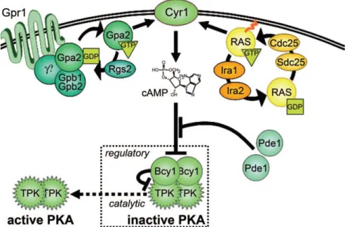
Figure 3. RAS signaling pathway in S. cerevisiae. Ras and Gpa2p (GTP bound G proteins) bind to
adenylate cyclase (Cyr1p) and promote its production of cAMP. Cdc25p and Sdc25p (Ras GEFs) and Ira1p and Ira2p (Ras GAPs) are represented in Ras-Cyr1 complex because they regulate adnylate cyclase by controlling the Ras switch. Gpr1p, a member of the G protein coupled receptor, acts upstream of Gpa2p. Gpa2p was very similar with the mammalian G subunits of heterotrimeric G proteins. Phosphodiesterases (Pde1p and Pde2p) antagonize the signaling via enzymatic inactivation of cAMP. The PKA tetramer is the regulatory target of cAMP. The regulatory Bcy1p subunits keep PKA in an inactive state. cAMP activates the catalytic subunits by binding to Bcy1p subunits and promoting dissociation of the complex. Withdrawn from (68).
1.4 TOR Pathway
In addition to the RAS/cAMP/PKA signaling pathway, the other major nutrient responsive, growth controlling pathway in yeast is the TOR (Fig. 4). Tor (Target of rapamycin) serine/threonine kinases belong to the phosphatidylinositol-3 kinase (PI3K) family, and exert their functions in two distinct multiprotein complexes: TOR Complex 1 (TORC1), which controls many aspects of yeast growth and cell proliferation, and TORC2, which regulates cell polarity and actin cytoskeleton organization (69, 70). The main function of TORC1 is to respond to nutritional status, where its major function appears to be the regulation of translation capacity in response to environmental signals by promoting ribosome biogenesis, amino acid availability, and translation efficiency. Inhibition of TORC1 by rapamycin mimics nutrient starvation and causes G1 arrest, inhibition of protein synthesis, glycogen accumulation, induction of autophagy and entry into quiescence (69, 70). TOR also controls other aspects of ribosome biogenesis,
11
such as the Pol I- and Pol III-dependent transcription of the rDNA and tRNA genes via phosphorylation of dedicated transcription factors (71). Tor1p itself may activate rDNA transcription in rich nutrient conditions by entering the nucleus and binding directly to promoters (72), however, in other studies, Tor1p has been localized to internal membrane structures but not the nucleus (73, 74). TORC1 is also intimately implicated in vesicular trafficking (75). On the other hand, TORC2 signaling is rapamycin insensitive and it is required for the organization of the actin cytoskeleton. Upstream regulators of TORC2 are not known yet (76).
Rapamycin and nitrogen starvation treatment shows very similar responses in S. cerevisiae, suggesting that TORC1 is regulated by the availability of nitrogen source (77). The control of nitrogen metabolism involves the regulation of PP2A and the PP2A-like phosphatase, Sit4p. Yeast cells can adapt the metabolism to the nitrogen sources through the nitrogen catabolite repression pathway (NCR) also known as the nitrogen discrimination pathway (NDP) (78). Two activators, Gln3 and Gat1, and two repressors Dal80 and Gzf3 are the transcription factors that are involved in the regulation of selective use of the nitrogen via NCR (79). Under rich nitrogen sources, Gln3 is phosphorylated and sequestered in the cytoplasm. On the other hand, rapamycin treatment or poor nitrogen sources rapidly triggers the dephosphorylation of Gln3 in a Tap42-phosphatase-dependent manner. The Gln3 enters into the nucleus activating NCR genes (70, 80).
Many functional interactions between TOR and the RAS/cAMP/PKA pathway have been showed (69). It was demonstrated that the activation of PKA signaling pathway confers resistance to rapamycin. So, the activation of the PKA pathway prevents several rapamycin-induced responses. It is also known that TOR controls the subcellular localization of both PKA catalytic subunit Tpk1p and the Ras/cAMP signaling-related kinase Yak1p. However, the detailed relationship between the TOR and RAS/PKA networks is still not understood. Several possibilities have been suggested. On one hand, it was proposed that the TOR and PKA signaling cascades independently coordinated the expression of several genes. On the other hand, it has been proposed that TOR may work upstream of Rasp to regulate PKA activity, thus the RAS/PKA pathway can be a novel TOR effector branch (69, 70, 81).
12
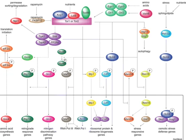
Figure 4. The TOR pathway in S. cerevisiae. The activation of TORC1 by nutrients results in the
stimulation of protein synthesis and the inhibition of stress response genes, autophagy and several pathways that allow growth on poor nitrogen sources. These processes are regulated by the rapamycin sensitive TORC1 complex via the Tap42-Sit4/PPA2c or the Sch9 branch. Withdrawn from (70).
1.5 Sch9 Pathway
Like PKA and TOR, the less well-known Sch9 pathway plays a role in nutrient-mediated signaling in yeast (70). In parallel with the PKA pathway, Sch9p is phosphorylated by TORC1, regulating many of the TORC1 processes. However, Sch9p also acts independently of TORC1, promoting adaptation to stress (70). The main functions of Sch9p are regulation of cell size, activation of ribosomal biogenesis (82), action as a negative regulator of both CLS and RLS and regulation of mitochondrial respiration (37, 83). It was demonstrated that the deletion of SCH9 up-regulates electron transport chain which is associated with an increase in mitochondrial respiration (84). Additionally, it has also been shown that yeast Sch9 is an important component of a network that controls genes involved in a metabolic switch from the TCA cycle and respiration to glycolysis and glycerol biosynthesis. During chronological aging, the
13
Δsch9 exhausts extracellular ethanol and reduces stored lipids, but synthesizes and releases glycerol, suggesting in this way that glycerol production enhances life span (85).
1.6 Gup protein in Saccharomyces cerevisiae
Gup1p and its close homologue Gup2p are members of the membrane-bound O-acetyltransferase (MBOAT) superfamily (86-88). Gup1p was firstly described in Saccharomyces cerevisiae as involved in glycerol metabolism and transport and accordingly included in the major facilitator superfamily (88). Nevertheless, Gup1p is now well known for other aspects of cell physiology that do not relate directly to glycerol active transport, which protein was identified as the Stl1p member of the HXT family of hexose transporters (89). The actual influence of Gup1p on Stl1p activity was found to be indirect through the influence of Pma1 H+ATPase miss localization and consequent defective active transport-driving proton motive force (90). Gup1p is localized in the plasma membrane, more precisely oriented across the membrane plasmatic where the N-terminus is located in periplasmic space, and the C-terminus located intracellularly (88, 91). However, it also co-localizes with cytochrome c oxidase from mitochondria and with NADPH-cytochrome c redutase from the endoplasmatic reticulum (88). These several sub-cellular localizations suggest complex regulation and roles. Gup1p was associated with the integrity and biogenesis of cell wall and plasma membrane (90, 92), and relatedly, the deletion of GUP1 impaired growth under anaerobic conditions and sterol uptake (93). Additionally, this deletion also induced phenotypes on cytoskeleton polarization (94) and bud site selection (95), secretory and endocytic pathway (96), as well as telomere length (97). At the level of cellular morphology, ∆gup1 presents aberrant vacuole morphology (96), while in C. albicans it induces the absence of hyphae formation and consequently defective invasive growth/biofilm formation (98). The extracellular matrix (ECM) of S. cerevisiae is also affected by GUP genes deletion, both at the level of protein and sugar fractions. Many proteins involved in cellular arrangement, carbon metabolism, cell defense and protein fate are not present in S. cerevisiae ECM from ∆gup1 mutant (99). Moreover, also the sugar fractions from S. cerevisiae and C. albicans differ (100). Finally, Tulha et al., (101) reveled the sensitivity of ∆gup1 cells to acetic acid, leading to cell death. This
14
displayed non-apoptotic characteristics and seemed to undergo instead a necrotic death process, ∆gup1 cells presenting a reduced chronological life span. The deletion of GUP1 is further associated with the resistance to complex chemicals like ergosterol synthesis inhibitors, which indicated an interference of Gup1p on sphingolipid and ergosterol synthesis (90), and conversely with the increased sensitivity to sphingolipid synthesis inhibitors, which, together with other evidences, suggested the involvement of Gup1p on the glycosylphosphatidylinositol (GPI) remodeling system (90, 102). Additionally, it was also involved on the resistance to the anti-cancer drug Imatinib (103), together with proteins that regulate the vacuolar pH. Imatinib, marketed as Glivec/Gleevec® by Novartis is a tyrosine kinase inhibitor specific for cancerous cells, namely some types of leukemia (104). Yeasts do not have recognized tyrosine kinases or tyrosine kinase receptors, though the broad sensitivity of S. cerevisiae to this drug (103) suggests otherwise.
Gup1p multiple localizations, and numerous associated processes and phenotypes implies a crucial role for this protein in cellular survival and successful progression through cell cycle. GUP1/2 genes have counterparts in higher Eukaryotes, including mammalians. Abe and co-workers (105) described the mousse homologue of GUP1 as a negative regulator for N-terminal palmitoylation of sonic hedgehog (SHH) protein (Fig. 5). This protein is responsible for the control of morphogenesis, patterning and differentiation during embryogenesis, as well as cellular morphology and proliferation during that process and wound healing. Accordingly, the mammalian Gup1 protein was named Hedgehog acyltransferase-like protein (HHATL) while Gup2, based on amino acid sequence homology was named as Hedgehog acyltransferase protein (HHAT), and these two proteins supposedly exert opposite roles in hedgehog extracellular signal activation prior to export into the outer space. These roles are in accordance with the above-mentioned functions in yeast, suggesting the putative existence of SHH-like pathway in yeast (105).Some evidences suggest the role of SHH pathway in tumor development, because an existence of high expression levels of this protein in neuroblastoma cell lines. When SHH protein is inhibited it promotes apoptosis and stopped proliferation (106). The above-described resistance to an oncologic drug, such as Imatinib of the GUP1 deleted strains concurs. For the time being, no relation was found or searched for that matter between the Gup related processes and phenotypes and the RAS/cAMP/PKA’s above described. Yet, in view of
15
the data available, it is predictable that this relation exists. Importantly, as referred for the hyperactivation of the RAS pathway (107), ∆gup1 is also resistant to rapamycin (108).
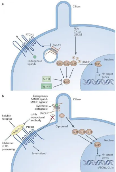
Figure 5. The vertebrate Sonic Hedgehog signaling pathway in the absence or presence of Hh ligands.
In absence of Hh (a),PTCH1, a 12-transmembrane domain protein, is located on the plasma membrane, and the protein GPCR-like receptor Smoothened (SMOH) is located in the membrane of intracellular endosomes. It is proposed that an intracellular small molecule that acts as an agonist for SMOH is transported outside the cell by PTCH1 so that it is not able to bind to SMOH. Under these circumstances, different kinases phosphorylate GLI2/3, creating a repressor form of this transcription factor. Iguana and SUFU prevent the active form of GLI from transactivating Hh-responsive genes in a manner that is still not completely understood.
In presence of Hh ligand (b), PTCH1 is internalized so that it can no longer transport the endogenous agonist molecules outwards. This allows them to accumulate intracellularly and activate SMOH, which itself translocates to the plasma membrane, apparently concentrating in cilia in at least some types of cells. Culminating in the appearance of activator forms of GLI that then regulate the expression of Hh
16
target genes. The known synthetic small-molecule SMOH agonists and antagonists bind to the same site as the putative endogenous ligand. Withdrawn from (109).
1.7 The Ras family of small GTPases
The Ras is a component of the broad family of small GTPases. The Ras genes are transforming oncogenes that have primarily been recognized as murine sarcoma viruses by Jennifer Harvey (Harvey-Ras or HRas) and Werner Kristen (Kristen-Ras or KRas) in 1960 (110, 111). Subsequent studies led to the identification of a third human Ras gene, designated as NRAS in human neuroblastoma cells. So, the three human Ras proteins are designated as HRas, KRas and NRas, which regulate intracellular signaling pathways involved in important cellular processes such as proliferation, cell polarity, differentiation, migration, adhesion, apoptosis and cytoskeletal dynamism (112, 113).
Ras proteins have as principal function the conversion of extracellular stimuli into intracellular signaling cascades, which eventually evoke changes in cellular activities. Thus, in normal mammalian cells, Ras proteins demonstrated functions as molecular switches for critical changes in cellular activities, namely cell proliferation and survival, and their proper regulation is indispensable to maintain the homeostasis of cells. On the other hand, uncontrolled activity of the Ras proteins, or the molecular components of their downstream pathways, can result in cancer or other diseases (113). Approximately 30% of human tumors are estimated to harbor activating mutations in one of the three Ras isoforms. KRAS is most frequently mutated, its mutation rate in all tumors being estimated to lie between 25 and 30%. KRAS mutation is especially frequent in colorectal carcinoma (35–45%), non-small cell lung cancer (16–40%) and pancreatic ductal carcinoma (69–95%) (114). In contrast, activating mutations of NRAS and HRAS are less common (8% and 3%, respectively) (115). The activating oncogenic mutations commonly occur in the GTPase catalytic domains, in codons 12, 13 and 61 (116). All these activating mutations render Ras proteins resistant to GTP hydrolysis, and consequent Ras inactivation stimulated by GTPase activating proteins (GAPs). These constitutively activated oncogenic Ras mutant proteins, therefore, initiate intracellular signaling cascades without the input of extracellular stimuli, resulting in uncontrolled cell proliferation and abnormal cell survival (113).
17
Ras activates several pathways, including the RAF-MEK-ERK/MAPK cascade, which transmits signals downstream and results in the transcription of genes involved in controlling several cellular mechanisms (117). Ras proteins are anchored in the cytoplasmic membrane by carboxylterminal farnesylation but, in some cases, the Ras proteins are bound by Ras-escort proteins which include galectin-1 and galectin-3 that have strong binding affinity to GTP-HRas and GTP-KRas, respectively (117, 118). Ras-escort proteins stabilize the Ras proteins in the GTP-bound state. Disruption of the interaction between these escort proteins and Ras has been exploited as a strategy to modulate aberrant Ras signaling (119). Ras communicates external cellular signals to the nucleus, and its altered activation leads to inappropriate cellular activities including enhanced cell growth, differentiation and survival of the cells (120, 121). The RAS-RAF-MEK-ERK pathway is activated by several known growth factors and cytokines that act through receptor tyrosine kinase signals and by activating mutations in the RAS and RAF genes (120).
The Ras intrinsic GTPase activity, is to hydrolyze the GTP into GDP (122). Ras is therefore a single GTPase molecule that like the other G proteins act as molecular switches and timers that cycle from inactive GDP-bound to active GTP-bound states (123). In normal quiescent cells, Ras is bound to GDP and is inactive (off state), while upon extracellular stimuli, Ras bind to GTP (on state), which has an extra phosphate group than GDP. This extra phosphate holds the two switch regions in a “loaded-spring” configuration. Upon the release of this phosphate, the switch regions relax leading to conformational modifications and return to the inactivate state (Fig. 6). Therefore, a cycling switching between the active/inactive GDP-bound forms controls the activation/inactivation of Ras and several other small G proteins. The cyclic process of GDP/GTP is facilitated by guanine GEFs and the GTPase activating proteins (GAPs) (122).
18
Figure 6. GTPase signaling. GTPase is off when bound to GDP, then a GEF removes GDP and allows GTP to bind to the GTPase, turning it on. All GTPases can hydrolyze GTP to GDP and turn themselves off, though GAPs accelerate this process. Withdrawn from (124).
Normally, ligand binding to receptor tyrosine kinases (RTK) induces dimerization of the receptor and autophosphorylation of specific tyrosine residues in the C-terminal region. This generates binding sites for adaptor proteins like the growth factor receptor-bound protein 2 (GRB2), that recruit the GEF Sos at the plasma membrane, and in turn activates the membrane bound Ras by catalyzing the conversion of GDP into GTP. In its GTP bound conformation, Ras combines with Raf and mobilizes the inactive protein from the cytoplasm recruiting the Raf kinases to the plasma membrane (112, 125). Once the Ras-Raf complex is translocated to the cell membrane, Ras activates the serine/threonine kinase function of Raf isoforms. Upon activation of Ras, Raf acts as a MAP kinase kinase kinase (MAPKKK) to activate MEK1 and MEK2, which, in turn, catalyze the activation of the effector ERK1 and ERK2 kinases, and their translocation into the nucleus. Once activated ERK1/ERK2 broadly phosphorylates several nuclear and cytoplasmic effector proteins involved in diverse cellular responses, such as proliferation, survival and differentiation (Fig. 7) (126, 127).
Although RAF can also be activated by RAS-independent activators (128). Some data have clearly shown that Ras can activate other downstream signaling pathways including phosphatidylinositol 3-kinase (PI3K) and Rac and Rho proteins, associated with the regulation of the cytoskeleton and invasiveness of tumor cells (129).
19
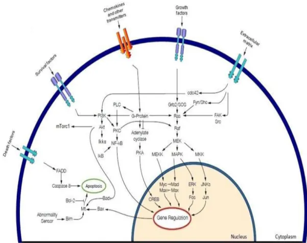
Figure 7. MAPK cascade activation and potential cross talk signals. In the MAPK cascade the growth
factors binding and consequently promotes activation of tyrosine kinase receptors, the activation of the RAS GTPase promotes the kinase activity of the RAF serine/threonine protein kinases. Activated RAF phosphorylates MEK in the cytoplasm, which in turn phosphorylates ERKs that translocates to the nucleus where they phosphorylate and regulate various nuclear and cytoplasmic substrates involved in diverse cellular responses, such as cell proliferation, survival, differentiation, motility, and angiogenesis. RAS may cross-talk with different pathways, such as PI3K. Withdrawn from (112).
The embryonic lethality of KRas knockout mice illustrated the importance of KRas expression during development as a result of liver defects and anemia. In opposition, mice with HRAS or NRAS knockouts are completely viable (130). In another study the expression of oncogenic HRas or KRas under tissue-specific promoters induces various types of malignancies in multiple transgenic mouse models (131).
20
1.8 Metabolic Similarities between Cancer Cells and Yeast
It was supposed that cancer cells suppress mitochondrial metabolism. The early discoveries from Otto Warburg pointed out that cancer cells display a decreased respiration along with an enhanced lactate production, suggesting that they depend mainly on fermentative metabolism for ATP generation (132). The spite of the decrease in energy yield as a consequence of the glycolytic phenotype seems to allow an increase in cell proliferation and be applicable to other fast growing cells (133). In this case, the repression of oxidative metabolism occurs even in the presence of oxygen, this metabolic phenomenon is known as “aerobic glycolysis” or the “Warburg effect”. Moreover, it has been showed that vary cancer cells can reversibly switch between fermentation and oxidative metabolism, depending on the absence or the presence of glucose and the environmental conditions (134, 135). More recently, it was proposed that the “glycolytic” cells could establish a metabolic symbiosis with the “oxidative” ones through lactate shuttling (136). A well defined feature of some cancer cells is the glucose-induced suppression of respiration and oxidative phosphorylation (137, 138). This is a reversible event that is called as “Crabtree effect”. This event might represent an advantage of cancer cells in vivo, as it would allow them to adapt their metabolism to the rather heterogeneous microenvironments in malignant solid growths (139).
The yeast S. cerevisiae is a respiro-fermentative organism, moreover it is a Crabtree positive yeast because upon glucose addition, respiration is inhibited despite the presence of oxygen (140, 141). When glucose amount is high, the yeast uses as main metabolic pathway fermentation, and when this carbon source becomes scarce it can switch to oxidative metabolism (142). In relation to energy metabolism, there are similarities between the glucose-induced repression of oxidative metabolism of yeast and the “aerobic glycolysis” of tumor cells. In both cells, the downregulation of oxidative metabolism is observed with an enhanced fermentation despite the presence of oxygen. Additionally, S. cerevisiae shares with cancer cells the same metabolic features that are identified as the main causes of the above-mentioned Warburg effect. For example, like cancer cells, yeasts overexpress glycolysis enzymes in response to glucose (143, 144). Moreover, the activity and expression pattern of the glycolysis key enzymes, such as hexokinase, phosphofructokinase and pyruvate kinase, are also modified in yeast (144, 145).
21
Although, yeast lacks the genetic defects identified in cancer cells, S. cerevisiae has homologues with genes related with cancer such as p53, cyclin D and Ras (29). Therefore, an interesting approach would be to use “tumourized yeasts” through the introduction of muted genes related with cancer and apply this as a model for anti-cancer drug screening and for metabolic studies.
1.9 The Role of Cell Surface Receptors
Cell signalling requires not only extracellular signal molecules, but also a set of receptor proteins in each cell that enable it to bind and respond to the signal molecules in a characteristic way. These cell surface receptor proteins act as signal transducers. They convert an extracellular ligand-binding event into intracellular signals that alter the behaviour of the target cell (146, 147). The extracellular signal molecules often act at very low concentrations and the receptors that recognize them usually bind them with high affinity. In most cases, the receptors are transmembrane proteins on the target cell surface. When these proteins bind an extracellular signal molecule, they become activated and generate various intracellular signals. In other cases, the receptor proteins are inside the target cell, and the signal molecule needs to enter the cell to bind to them, this process requires that the signal molecule be sufficiently small and hydrophobic to diffuse across the target cell’s plasma membrane (148). This knowledge is common to high and low Eukaryotes. Nevertheless, the presently recognized players at the level of signal reception/sensing are quite different in both types of organisms.
In higher Eukaryotes, the RTKs are a large superfamily of receptors with function as the receptors for a wide array of growth factors, including epidermal growth factor (EGF), nerve growth factor (NGF), platelet derived growth factor (PDGF), vascular endothelial growth factor (VEGF), fibroblast growth factor (FGF), insulin and the insulin-like growth factors (IGF), and the ephrins and angiopoietins (149). RTKs are essential components of cellular signalling pathways that are activated during embryonic development and adult homeostasis. Because of their roles as growth factor receptors, many RTKs have been implicated in the onset or progression of various cancers, either through receptor gain-of-function mutations or through receptor/ligand overexpression (150). Consequently, cell surface receptors are essentials to the mechanism of many chemical toxicants and serve as targets for the development of
22
drugs (149, 150). Growth factors modulate signaling pathways, which control cell proliferation and death in both normal and malignant cells. The EGF was one of the first growth factors to be discovered and is the prototype of a large family of closely related growth factors, which includes TGF, amphiregulin, heparin binding EGF, and betacellulin. Among these growth factors, TGF has been identified as a key modulator in the process of cell proliferation in both normal and malignant epithelial cells. TGF binds to the receptor, the epidermal growth factor receptor (EGFR), which promotes the activation of the EGFR tyrosine kinase enzymatic activity that triggers the intracellular signaling pathway (151). The EGFR is part of a subfamily of four closely related receptors: EGFR (or ErbB-1), HER-2/neu (ErbB-2), HER-3 (ErbB-3), and HER-4 (ErbB-4). The receptors exist as inactive monomers, which dimerize after ligand activation. This causes homodimerization or heterodimerization between EGFR and another member of the Erb receptor family. After ligand binding, the tyrosine kinase intracellular domain of the receptor is activated, with autophosphorylation of the intracellular domain, which initiates a cascade of intracellular events (152, 153). The signaling pathway involves activation of Ras and mitogen activated protein kinase, which activates several nuclear proteins, including cyclin D1, a protein required for cell cycle progression from G1 to S phase (154). EGFR signaling is not only essential for cell proliferation. Several studies have demonstrated that EGFR signaling also mediates other processes that are crucial to cancer progression, including angiogenesis, metastatic spread and the inhibition of apoptosis (153-156). Activation of the TGF-EGFR autocrine growth pathway in cancer cells can be attributed to several mechanisms, such as overexpression of the EGFR, increased concentration of ligand, decreased phosphatase activity, decreased receptor turnover, and the presence of aberrant receptors, including EGFR gene alterations. In this context, the most common EGFR mutant found in human cancer is EGFRvIII (157).
TGF and/or EGFR are overexpressed in many different solid human cancers, including breast, head and neck, gastric, prostate, ovarian, colorectal carcinomas, and glioblastomas, in which it is generally associated with advanced disease and poor prognosis (156, 158, 159). Human EGFR gene locates at chromosome 7p11-13 and the mature protein is synthesized from a 1,210 residues polypeptide precursor. This originates a 170 kDa protein containing approximately 20% of carbohydrate of its molecular mass and is heavily N-glycosylated (160-163). Glycosylation is important in
23
case of protein-protein interactions that occur between protein ligand and their receptors, because it plays a role in determining protein structure and known to affect the three-dimensional configuration of proteins (164).
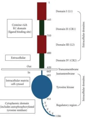
Figure 8. Basic structure of EGFR displaying the relevant domains. (1) The extracellular domains:
domain I/L1; domain II/CR1; domain III/L2; domain IV/CR2. (2) Transmembrane domains. (3) The intracellular domains: juxtamembrane domain; tyrosine kinase domain; regulatory region domain. The phosphorylation of several substrates by the tyrosine kinase domain of the EGFR receptor is responsible for activating of various signaling cascades. Withdrawn from (163).
Like all RTKs EGFR is characterized by three main domains. The extracellular domain of the mature receptor contains 621 amino acids, followed by a single transmembrane domain and a juxtamembrane domain (Fig. 8) (160, 162). Crystallographic studies of the EGFR extracellular domain complexed to its ligands have shown that the domains I, II and III form a ligand-binding pocket (165, 166). In the absence of ligand, EGFR exist as monomers on the cell surface. Binding of ligand to EGFR leads to the formation of receptor homo and heterodimers, depending on whether EGFR dimerizes with another EGFR or with other ErbB family members, respectively (167). EGFR dimerization is entirely receptor-mediated, with no contacts between the two growth factor molecules in the dimeric complex (165). By binding simultaneously
24
to two sites (within domains I and III) in the extracellular region of the receptor, the growth factor alters the special arrangement of the domains (as shown schematically in Fig. 9) (166). Phosphorylation of the EGFR activation loop in contrast to other kinases is not necessary for its activation (168). The EGFR kinase is activated by an asymmetric dimer in which the C-terminal lobes of two-kinase domain bind with each other in a manner analogous to cyclin in activated CDK/cyclin complexes. Thus, ligand binding brings two receptor monomers together and allows for the dimerization and subsequent activation of the kinase domain (169). Ligand induced EGFR dimerization leads to autophosphorylation of several key tyrosine residues in the cytoplasmic domain of each receptor monomer (170). These phosphorylated tyrosine residues then serve as binding sites for a number of adapter and signaling molecules leading to the activation of several intracellular signaling pathways downstream of the receptor. Some of the best characterized EGFR effector pathways are the RAS-RAF-MEK-ERK, PI3K/Akt, JAK/STAT and the PLC -PKC pathways, which upon activation lead to cell proliferation, motility and survival (Fig. 10) (170, 171).
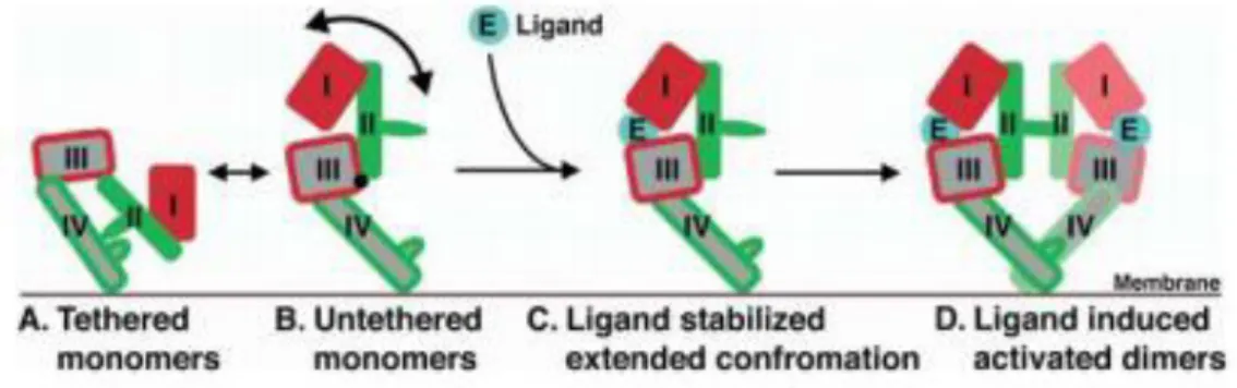
Figure 9. Mechanism of ligand-induced EGFR dimerization. About 95% of the unliganded EGFR exists
in a compact auto-inhibited or tethered conformation, in which domains II and IV form an intra-molecular interaction or tether (A). In 5% of the unliganded molecules, this tether is broken, and the soluble extracellular region of EGFR (sEGFR) can adopt a range of untethered conformations (B). Ligand binds preferentially to untethered molecules, and interacts simultaneously with domains I and III, stabilizing the particular extended form in which domain II is exposed and the receptor can dimerize (C). Dimerizations entirely receptor mediated and dominated by domain II interactions (D). Withdrawn from (166).
The Ras/extracellular signal regulated kinase (ERK) pathway is a critically important route that regulates cell proliferation and survival in yeasts (see above) as in
25
mammalian cells (see above) (172). In these last, GRB2 is an SH2/SH3 domain containing protein that binds EGFR either directly or through the association with the adaptor molecule Shc, and acts as a common adapter protein in a majority of growth factor related signaling events (173, 174).
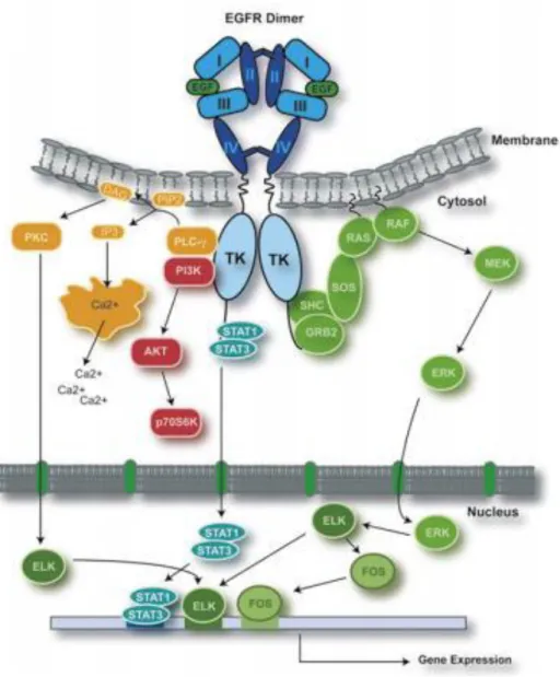
Figure 10. EGFR signaling. Binding of ligand to EGFR leads to receptor dimerization,
autophosphorylation and activation of several downstream signaling pathways. Only selected pathways and transcription factors are presented. Withdrawn from (161).
The PI3K/Akt signaling pathway also affects many cellular processes including cell proliferation, apoptosis and invasion (175, 176). PI3K is recruited to the membrane by directly binding to phosphotyrosine consensus residues of growth factor receptors or
26
adaptors through one or both SH2 domains in the adaptor subunit (177). This leads to allosteric activation of the catalytic subunit. Activation results in the production of the second messenger phosphatidylinositol-3,4,5-trisphosphate (PIP3). The lipid product of PI3K, PIP3, recruits a subset of signaling proteins with pleckstrin homology (PH) domains to the membrane, including PDK1 and Akt. Once activated, Akt mediates the activation and inhibition of several targets, resulting in cellular survival, growth and proliferation (178). The interlinked Ras/MAPK and PI3K/Akt signaling pathways play an important role in tumourigenesis via phosphorylation of various proteins and transcription factors. Furthermore, mutation in KRAS, BRAF, or PIK3CA results in continuous activation of the downstream Ras/MAPK or PI3K pathways, regardless of whether the EGFR is activated or pharmacologically blocked (179-181). EGFR ligands are not only responsible for stimulation of pathways that positively regulate EGFR, but also stimulate pathways that negatively regulate the EGFR coupling to malignant phenotypes and this balance between these positive and negative regulators of EGFR coupling to malignant phenotypes may be altered in tumor cells (169). Generally, 1x10⁵ EGFR per cell are expressed by normal cells, but tumor cells can express more than 2x10⁶ receptors per cell (182). It was reported that the hypoxic microenvironment of tumors can also induce overexpression of EGFR by increasing EGFR mRNA translation, since it was considered that receptor overexpression commonly develops due to gene amplification (183). Further the EGFR overexpression can result in high levels of autocrine signaling (184), autocrine production of TGF-α or EGF reduces the chances of cancer survival (185).
Inactivation of the EGFR can be mediated either by receptor dephosphorylation by phosphotyrosine phosphatases or receptor downregulation. Receptor downregulation is the most prominent regulator of EGFR signal attenuation and involves the internalization and subsequent degradation of the activated receptor in the lysosomes (161, 186).
27
1.9.1 EGFR as a Therapeutic Target
A large body of experimental and clinical work supports the view that the EGFR is a relevant target for cancer therapy. Two therapeutic approaches have been shown most promising and are currently being used to inhibit the EGFR in clinical studies: (a) monoclonal antibodies (MAbs) like Cetuximab (Erbitux®) used in colorectal cancer (CRC) therapy (187, 188), and (b) small molecule inhibitors of the EGFR tyrosine kinase enzymatic activity (TKIs) like Imatinib (Gleevec®) used in leukemia therapy (104, 169). Small-molecule TKIs compete reversibly with adenosine 5’triphosphate to bind to the intracellular catalytic domain of EGFR tyrosine kinase and inhibit the EGFR autophosphorylation and downstream signaling (169). MAbs are generally directed at the external domain of the EGFR to block ligand binding and receptor activation (165, 169). Cetuximab (Ctx) was approved by the FDA in 2004 for squamous cell carcinoma of the head and neck and advanced stage of CRC overexpressing EGFR (189). Ctx is a 152 KDa chimeric monoclonal antibody of the immunoglobulin G1 subclass produced in mammalian cell culture by mouse myeloma cells. It was constructed by attaching the variable regions of the murine monoclonal antibody M225 against EGFR to constant regions of the human IgG1. It has two identical heavy chains consisting of 449 amino acids each and two light chains of 214 amino acids each (Fig. 11) (190, 191).
Cetuximab has a 5-10 fold higher affinity for EGFR than the native ligand, resulting in inhibition of the receptor function (192, 193). It is also able to mediate antibody dependent cell mediated cytotoxicity (194), and receptor downregulation leading to a mitigation of EGFR activity that does not affect other HER family receptors (195). Ctx induces inhibition of EGFR signaling, prevents heterodimerization and leads to downregulation of downstream targets (Fig. 12) (166). It avoids several cell signaling pathways, including the Ras–Raf–MAPK, PI3K/Akt, PKC, STAT and SRC, all of which play important roles in tumor cell proliferation, invasion and inhibition of apoptosis (196). Further, Ctx blocks cell cycle progression by inducing G1 arrest (197-200) as well as the transport of EGFR into the nucleus (201), and also has the potential to kill targets cells by mediating antibody-dependent cell-mediated cytotoxicity (194).