E-Mail [email protected]
Original Paper
Neurosignals 2013;21:1–13 DOI: 10.1159/000332811
Differential Contribution of the Guanylyl
Cyclase-Cyclic GMP-Protein Kinase G Pathway to
the Proliferation of Neural Stem Cells Stimulated
by Nitric Oxide
Bruno P. Carreira
a
Maria Inês Morte
a
Ana Sofia Lourenço
a
Ana Isabel Santos
a
Ângela Inácio
a
António F. Ambrósio
a, b
Caetana M. Carvalho
a
Inês M. Araújo
a, c
a Centre for Neuroscience and Cell Biology, Neuroendocrinology and Neurogenesis Group and b Centre of Ophthalmology and Vision Sciences, IBILI, Faculty of Medicine, University of Coimbra, Coimbra , and c Department of Biomedical Sciences and Medicine, University of Algarve, Faro , Portugal
MAP kinase pathway. The selective inhibitor of PKG, KT5823, prevented the proliferative effect induced by NO at 24 h but not at 6 h. In conclusion, the proliferative effect of NO is ini-tially mediated by the ERK/MAPK pathway, and at later stag-es by the GC/cGMP/PKG pathway. Thus, our work shows that NO induces neural stem cell proliferation by targeting these two pathways in a biphasic manner.
Copyright © 2012 S. Karger AG, Basel
Introduction
An injury to the central nervous system often results
in neuroinflammation, which is characterized by the
activation of resident microglial cells, astrocytes, and
eventually by infiltrating peripheral macrophages and
lymphocytes, recently reviewed by Whitney et al. [1] . It
is now well established that inflammation affects the
formation of new cells following brain injury [2–4] . An
acute brain insult, such as stroke, seizures, or trauma,
often elicits a proliferative response from the
neurogen-ic nneurogen-iches [5–9] . New cells are attracted to the lesioned
areas in order to replace the lost neural cells [10] . Factors
Key Words
Nitric oxide ⴢ Neural stem cells ⴢ cyclic GMP ⴢ
cGMP-dependent kinase ⴢ Proliferation ⴢ Neurogenesis ⴢ Guanylyl cyclase
Abstract
Nitric oxide (NO) is an important inflammatory mediator in-volved in the initial boost in the proliferation of neural stem cells following brain injury. However, the mechanisms un-derlying the proliferative effect of NO are still unclear. The aim of this work was to investigate whether cyclic GMP (cGMP) and the cGMP-dependent kinase (PKG) are involved in the proliferative effect triggered by NO in neural stem cells. For this purpose, cultures of neural stem cells isolated from the mouse subventricular zone (SVZ) were used. We observed that long-term exposure to the NO donor (24 h), NOC-18, increased the proliferation of SVZ cells in a cGMP-dependent manner, since the guanylate cyclase inhibitor, ODQ, prevented cell proliferation. Similarly to NOC-18, the cGMP analogue, 8-Br-cGMP, also increased cell proliferation. Interestingly, shorter exposures to NO (6 h) increased cell proliferation in a cGMP-independent manner via the ERK/
Received: April 19, 2011
Accepted after revision: August 30, 2011 Published online: February 23, 2012
Inês M. Araújo, PhD
Regenerative Medicine Program, Department of Biomedical Sciences and Medicine University of Algarve, Gambelas Campus, Edifício 7, 3º piso
© 2012 S. Karger AG, Basel 1424–862X/13/0212–0001$38.00/0
produced and released by immune cells during the
neu-roinflammatory response affect the proliferation,
mi-gration, and differentiation of neuronal precursors
fol-lowing a lesion [11, 12] . Microglial cells can produce a
wide range of pro- and anti-inflammatory mediators,
which can differentially affect the different steps of
neu-rogenesis [2, 3, 13] . Among such factors, nitric oxide
(NO) is of particular interest given its notable features
such as short half-life, diffusivity, and signaling
proper-ties [1] .
NO is a free radical produced by NO synthases and a
pleiotropic messenger in several biological processes [14] .
Recent studies have shown the involvement of NO on
neurogenesis, either in basal conditions or following an
injury. Particularly, NO was described as a physiological
inhibitor of neurogenesis in mice [15–18] . Other studies
demonstrated that NO is important for the initial
in-crease in proliferation of neural stem cells in the
neuro-genic niches following stroke [19] or seizures [20] . We
re-cently showed that the NO donor 2,2
ⴕ
-(hydroxynitro-sohydrazino)bis-ethanamine (NOC-18) can stimulate
proliferation of isolated neural stem cells in culture when
used in low doses (1–10
M) [20] . Nevertheless, the
mech-anisms underlying the proliferative effect of NO are not
yet identified.
The main biological target of NO is the
heme-con-taining enzyme guanylyl cyclase. Guanylyl cyclase
cata-lyzes the conversion of GTP to cyclic GMP (cGMP),
which in turn can act on further downstream targets
such as the dependent kinase (PKG) and
cGMP-gated channels. In a variety of cell types, NO and cGMP
have been described as important effectors in several
cellular processes, including survival, differentiation,
growth, axon guidance or migration, through the
activa-tion of different downstream signaling cascades [21–23] .
Neurogenesis is positively affected by cGMP and
cGMP-sparing agents such as sildenafil or tadalafil [21, 24–26] .
However, it remains to be established whether cGMP
and PKG are responsible for the proliferative effect of NO
when applied to neural stem cells. Within this scenario,
we investigated the role of the guanylyl
cyclase-cGMP-PKG pathway in the proliferative effect of NO. We
ob-served that cGMP analogues mimic the effect of NO in
increasing cell proliferation. Moreover, blocking
guany-lyl cyclase or PKG prevented the proliferative effect of
NO, thus pointing to the involvement of the NO-cGMP
signaling pathway in the enhancement of neural stem
cell proliferation.
Materials and Methods Materials
Dulbecco’s modified Eagle’s medium:F-12 nutrient mixture (D-MEM/F-12, with GlutaMAX TM -I), B27 supplement,
trypsin-EDTA solution (0.05% trypsin, 1 m M EDTA in Hanks balanced salt solution, antibiotic (10,000 units/ml penicillin, 10 mg/ml streptomycin), epidermal growth factor, basic fibroblast growth factor and Click-iT 쏐 EdU Alexa Fluor 쏐 488 Flow Cytometry As-say Kit were purchased from Invitrogen (Paisley, UK). 5-Bromo-2 ⴕ -deoxyuridine (BrdU), 8-bromoguanosine 3 ⴕ ,5 ⴕ -cyclic mono-phosphate (8-Br-cGMP), phenylmethylsufonyl fluoride, dithio-threitol, orthovanadate, chymostatin, leuptin, antiparin, pepstatin A, trypan blue and 1,4-diamino-2,3-dicyano-1,4-bis( o -amino-phenylmercapto)butadiene monoethanolate (U0126) were pur-chased from Sigma Chemical (St. Louis, Mo., USA). KT5823 was purchased from Alomone Labs (Jerusalem, Israel). 1H-[1,2,4] oxadiozolo[4,3-a]quinoxalin-1-one (ODQ) and zaprinast were obtained from Tocris Bioscience (Bristol, UK). Mouse anti-Sox-2 was purchased from R&D Systems (Minneapolis, Minn., USA) and rabbit anti-nestin from Santa Cruz Biotechnology (Santa Cruz, Calif., USA). NOC-18 was obtained from Alexis Biochemi-cals (San Diego, Calif., USA). A DAKO fluorescence mounting medium was purchased from DakoCytomation (Glostrup, Den-mark). Rat anti-mouse BrdU was obtained from Oxford Biotech-nology (Oxford, UK). Hoechst 33342 dye, anti-rat IgG conjugated with Alexa Fluor 594, anti-mouse IgG conjugated with Alexa Fluor 594 and anti-rabbit IgG conjugated with Alexa Fluor 488 secondary antibodies were purchased from Molecular Probes (Leiden, The Netherlands). Polyvinylidene difluoride mem-branes, enhanced chemifluorescence reagent, cGMP BioTRAK, alkaline phosphatase-linked anti-rabbit and anti-mouse second-ary antibodies were obtained from Amersham Pharmacia Biotech (Little Chalfont, UK). Monoclonal mouse anti-p44/42 MAPK (ERK1/2) and rabbit anti-phospho-ERK1/2 antibodies were ob-tained from Cell Signaling Technology (Danvers, Mass., USA). Other reagents used in immunoblotting experiments were pur-chased from BioRad (Hercules, Calif., USA).
Animals
C57BL/6J mice were obtained from Charles River (Barcelona, Spain) and kept with food and water ad libitum in a 12-hour dark:light cycle. All experiments were performed in accordance with the NIH and the European (86/609/EEC) guidelines for the care and use of laboratory animals.
Subventricular Zone Neural Stem Cell Cultures
Neural stem cell cultures were obtained from the subventricular zone (SVZ) of postnatal day 0–3 C57BL/6J mice, as previously de-scribed [20] . Briefly, the brains were removed from the skull follow-ing decapitation, and placed in a dissection medium composed of Ca 2+ - and Mg 2+ -free Hanks balanced salt solution (137 m M NaCl,
5.36 m M KCl, 0.44 m M KH 2 PO 4 , 0.34 m M Na 2 PO 4 ⴢ 2H 2 O, 4.16 m M
NaHCO 3 , 5 m M glucose, 1 m M sodium pyruvate, 10 m M HEPES,
pH 7.4), supplemented with 0.25% gentamicin. After removing the enveloping meninges, the cerebellum was discarded and the whole cortex tissue was cut in 450- m-thick coronal sections using a MclLwain tissue chopper (The Mickle Laboratory Engineering Co. Ltd., Guildford, UK). The fragments of SVZ encompassing both ependymal and subependymal layers were digested in 0.025%
tryp-sin and 0.265 m M EDTA for 20 min at 37 ° C, and dissociated by
gentle trituration with a pipette tip. Following dissociation, the cells were resuspended in fresh D-MEM/F-12 with GlutaMAX-I, supplemented with 1% B27, 1% antibiotic (10,000 units/ml penicil-lin, 10 mg/ml streptomycin), 10 ng/ml epidermal growth factor and 10 ng/ml basic fibroblast growth factor. Cells were then plated on uncoated Petri dishes at a density of 3,000 cells/cm 2 . The SVZ stem
cells were grown as floating neurospheres in a 95% air/5% CO 2
hu-midified atmosphere at 37 ° C. Seven days following plating, the
pri-mary neurospheres were harvested, centrifuged and dissociated as single cells. Cells were then replated as above and allowed to grow as secondary neurospheres. Neurospheres with 2–4 passages were collected and plated for 5 days on glass coverslips, 16 mm in diam-eter, for immunocytochemistry assays, or on 12-well plates, coated with poly- L -lysine, in a serum-free medium, without growth fac-tors, for preparation of lysates or flow cytometry assays.
Characterization of SVZ Primary Cultures
SVZ-derived neural stem cells plated on poly- L -lysine-coated coverslips for 5 days (in a minimum of 3 independent experi-ments) were fixed with 4% paraformaldehyde/4% sucrose in phos-phate-buffered saline (PBS, 0.1 M ). The cells were then permeabi-lized with 1% Triton X-100 for 5 min. Nonspecific binding was blocked with 3% BSA in 0.2% Tween 20 in PBS (PBS-T) for 1 h, and cells were labeled with the primary antibodies mouse anti-Sox-2 (1: 100) and rabbit anti-nestin (1: 100) for 90 min at room temperature. The cells were then exposed to secondary antibodies anti-mouse and anti-rabbit IgGs conjugated with Alexa Fluor 594 and 488 (1: 200), respectively, for 1 h at room temperature. Nuclei were stained with Hoechst 33342 (1 g/ml) for 3 min. Coverslips were mounted on glass slides using a DAKO fluorescence mount-ing medium. Images were acquired in a laser scannmount-ing micro-scope LSM 510 META (Zeiss, Jena, Germany).
Experimental Treatments in SVZ Cell Cultures
SVZ-derived neural stem cells were exposed to the NO donor NOC-18 (10 M ) or to the cGMP analogue 8-Br-cGMP (20 M ) for different periods of time, as indicated in detail in the figure legends and in the text. The guanylyl cyclase inhibitor ODQ (50 M ), the protein kinase G (PKG) inhibitor KT5823 (1 M ) and the MEK1/2 inhibitor U0126 (1 M ) were added 30 min before NOC-18 or 8-Br-cGMP and kept throughout the incubation period.
Decomposed NOC-18 was prepared by diluting a 10 m M NOC-18 stock solution (prepared in 0.01 M NaOH, which keeps NOC-18 from releasing NO in alkaline solutions) to 1 m M NOC-18 in a culture medium (pH 7.0, since NOC-18 releases NO at this pH), which was kept for 72 h at room temperature on a lab bench, un-protected from light. This solution was used to treat SVZ cultures in a final concentration of 10 M decomposed NOC-18, and ex-periments were run with fresh NOC-18.
Determination of cGMP Levels
The cGMP levels in SVZ-derived neural stem cell cultures were determined after exposure to drugs for 6 or 24 h using a cGMP enzyme immunoassay system (Amersham). All experi-ments were carried out in duplicate and in the presence of zapri-nast (50 M ), a phosphodiesterase inhibitor, as previously de-scribed [27] . Cell lysis and cGMP measurement were performed according to the manufacturer’s instructions. The results are ex-pressed as femtomoles per million cells.
Detection of Cell Proliferation and Cell Cycle Analysis by Flow Cytometry
SVZ cell proliferation was assessed by the incorporation of 5-ethynyl-2 ⴕ -deoxyuridine (EdU) and detected by flow cytome-try, using the Click-iT EdU Alexa Fluor 488 Flow Cytometry As-say Kit. EdU was added to the SVZ cultures 4 h before fixation. Fixation was performed with 70% ethanol overnight. Detection of EdU incorporation was based on click chemistry, a copper-cata-lyzed reaction between an azide (conjugated to a fluorophore) and an alkyne (EdU). Fixed cells were incubated for 30 min with Alexa Fluor 488 azide and copper sulfate. The cells were then incubated with RNase and the nuclear dye 7-actinomycin D (7-AAD) for 30 min. The cells were analyzed for EdU incorporation and nuclear staining on a FACScalibur using the Cellquest software (Becton Dickinson, San Jose, Calif., USA). Thirty thousand events were acquired per experiment. A minimum of 3 independent experi-ments were analyzed for each condition. The flow cytometer was calibrated with fluorescent standard microbeads (CaliBRITE Beads, BD Biosciences, San Jose, Calif., USA) for accurate instru-ment setting.
Detection of BrdU Incorporation
Cell proliferation was also assessed by the incorporation of the thymidine analogue BrdU, which is incorporated into the DNA of dividing cells during S phase. BrdU (10 M ) was added to the cultures 16 h before fixation [20, 28] . Nuclei that incorporated BrdU in this time window were detected by immunofluorescence as follows: following 20 min fixation with 4% paraformalde-hyde/4% sucrose in PBS, the cells were permeabilized with 1% Triton X-100 for 5 min, and DNA was denaturated by treatment with 1 M HCl for 30 min at 37 ° C. Nonspecific binding was blocked
with 3% BSA in PBS-T for 1 h, and then BrdU-positive cells were labeled with a rat anti-BrdU antibody (1: 50) for 90 min at room temperature. The cells were then incubated with a secondary an-tibody goat anti-rat IgG conjugated with Alexa Fluor 594 (1: 200) for 1 h at room temperature. Nuclei were stained with Hoechst 33342 (1 g/ml) for 3 min. Coverslips were mounted on glass slides using DAKO fluorescence mounting medium Dako Cyto-mation. The cells were visualized in a fluorescence microscope (Axioskop 2 Plus, Zeiss, Jena, Germany) and images were ac-quired with the Axiovision software 4.7. The number of BrdU-positive nuclei was counted in 7–10 randomly selected fields for each coverslip (in a total of approximately 900–1,200 cells per cov-erslip), and the data were expressed as percentages of the total number of living cells. A minimum of 3 independent experiments were analyzed for each condition.
Western Blot Analysis
Cells were lysed in 50 m M Tris-HCl, 10 m M EGTA, 1% Triton X-100 and 2 m M MgCl 2 , supplemented with 100 M
phenylmeth-ylsufonyl fluoride, 1 m M dithiothreitol, 1 g/ml chymostatin, 1 g/ml leupeptin, 1 g/ml antiparin, 5 g/ml pepstatin A, 1 m M sodium orthovanadate, and 50 m M NaF, pH 7.4, at 4 ° C. Protein
concentration was determined by the bicinchoninic acid method (BCA TM Protein Assay kit, Pierce, Rockford, Ill., USA), and the
samples were used for Western blot analysis, after adding 6 ! con-centrated sample buffer (0.5 M Tris, 30% glycerol, 10% sodium dodecyl sulfate, 0.6 M dithiothreitol, 0.012% bromophenol blue) and heating, for 5 min, at 95 ° C. Equal amounts of protein were
sulfate-poly-acrylamide gels, and transferred electrophoretically to polyvi-lydene difluoride membranes. These were then blocked for 1 h at room temperature in Tris-buffered saline (137 m M NaCl, 20 m M Tris-HCl, pH 7.6) containing 0.1% Tween 20 (TBS-T) and 3% BSA. Incubations with primary antibodies (rabbit anti-phospho-ERK1/2 or mouse anti-anti-phospho-ERK1/2, 1: 1,000; Cell Signaling, Danvers, Mass., USA) in TBS-T 1% BSA were performed overnight at 4 ° C.
The membranes were subsequently incubated for 1 h at room tem-perature with alkaline phosphatase-linked secondary antibodies (anti-rabbit or anti-mouse IgG, 1: 20,000) in TBS-T 1% BSA. After extensive washing in TBS-T with 0.5% BSA, immunoreactive bands were visualized on a VersaDoc 3000 Imaging System (Bio-Rad, Hercules, Calif., USA) following incubation of the mem-brane with enhanced chemifluorescence reagent for 5 min. The results are expressed as the percentage of control of phospho-ERK/total ERK ratio.
Statistical Analysis
Data are expressed as means 8 SEM. Statistical significance was determined by using two-tailed t tests or one-factor analysis of variance (ANOVA) as appropriate, followed by post hoc Bon-ferroni’s or Dunnet’s tests, as indicated in the figure legends and in the text. Differences were considered significant when p ! 0.05.
Results
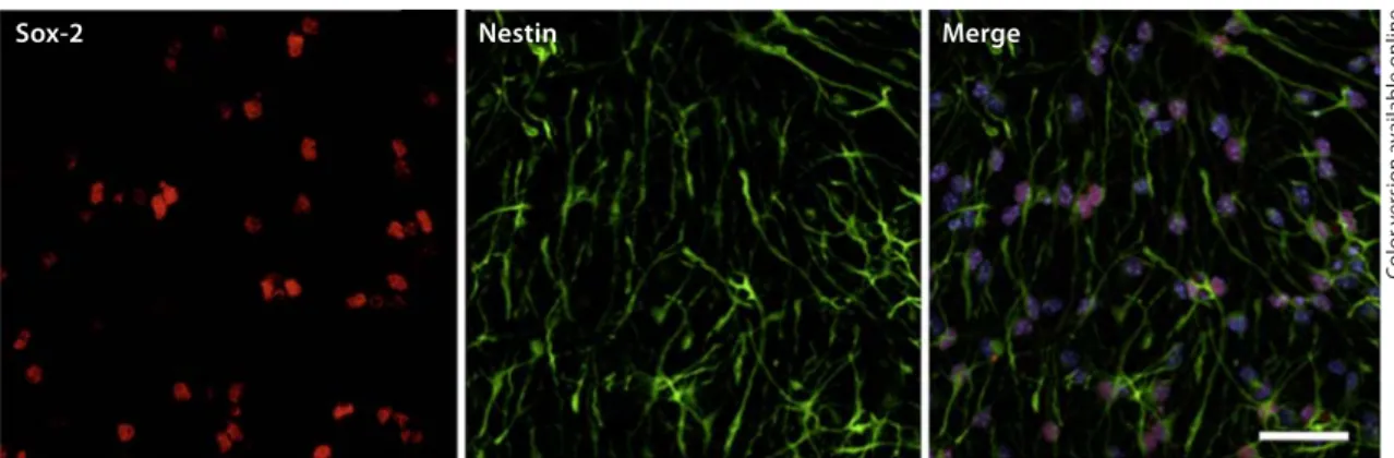
Characterization of SVZ Primary Cultures
Cells isolated from the SVZ were cultured as described
previously (see Materials and Methods), and plated on
poly-
L-lysine-coated coverslips for 5 days. At this stage
the cells were immunoreactive for the transcription
fac-tor Sox-2, essential for the self-renewal of
undifferenti-ated stem cells, and nestin, a neural precursor cell
mark-er. The percentage of double-labeled cells was
approxi-mately 70%, suggesting that the majority of cells remain
undifferentiated before being used for experiments
( fig. 1 ). This abundance of undifferentiated SVZ cells is
similar to that observed in SVZ primary cultures used in
previous studies investigating the effects of NO on cell
proliferation by our group [20] as well as by others [18,
29] .
NO Increases Cell Proliferation via the Guanylyl
Cyclase-cGMP Pathway
To investigate the involvement of cGMP in the
prolif-erative effect of NO, we evaluated the incorporation of
thymidine analogues (EdU or BrdU) by SVZ cell cultures
following treatment with a NO donor (NOC-18). We have
previously shown that treatment with NOC-18 in the
range of 1–10
Mincreases proliferation of SVZ cells, but
whether cGMP is involved in NO-induced neural stem
cell proliferation has not been addressed. We first
inves-tigated, by flow cytometry, the involvement of cGMP in
the proliferative effect of NO after 6 or 24 h following
treatment with NOC-18 (10
M). Exposure to NOC-18 for
6 h increased the incorporation of EdU to 133.4 8 5.12%
of the control (p ! 0.001) ( fig. 2 a). After 24 h of NOC-18
treatment we further increased the EdU incorporation to
165.3 8 10.2% of the control (p ! 0.001) ( fig. 2 b). In
con-trol conditions (untreated cells) the percentage of
EdU-positive cells (percentage of total living cells) was 2.4 8
0.7% at 6 h and 2.2 8 0.9% at 24 h.
The involvement of cGMP in the proliferative effect of
NOC-18 was evaluated using the guanylyl cyclase
inhibi-tor ODQ. Treatment with ODQ prevented NO-induced
EdU incorporation at 24 h (p ! 0.05), compared to SVZ
cells treated with NOC-18 alone ( fig. 2 b), suggesting that
cGMP mediates the effect of NO on cell proliferation for
Sox-2 Nestin Merge
Fig. 1. Characterization of SVZ stem cell cultures. Representative laser scanning confocal images of SVZ cells
labeled against Sox-2 (red) and nestin (green) are shown. Nuclei were labeled with Hoechst 33342 (blue). Scale bar: 45 m. Colors refer to the online version only.
Co lo r v e rs io n av a il a b le o n li n e
f e
NOC-18 D-NOC-18 NOC-18 D-NOC-18
U0126 EdU-positiv e c e lls (% of c o ntr ol) NOC-18
***
+++ 0 50 100 150 200 ODQ EdU-positiv e c e lls (% of c o ntr ol) d 24 h 24 h b 24 h NOC-18***
+++ 0 50 100 150 200 ODQ % Br dU-positiv e c e lls NOC-18***
***
***
+++ 0 5 10 15 EdU-positiv e c e lls (% of c o ntr ol) 0 50 100 150 200 EdU-positiv e c e lls (% of c o ntr ol) 0 50 100 150 200 U0126 EdU-positiv e c e lls (% of c o ntr ol) 6 h NOC-18*
+ 0 50 100 150 200 a ODQ EdU-positiv e c e lls (% of c o ntr ol) 6 h c 6 h g NOC-18***
0 50 100 150 200Control NOC-18 NOC-18 + ODQ ODQ
Co lo r v e rs io n av a il a b le o n li n e 2
Table 1. D etermination of cGMP levels in neural stem cell cul-tures following exposure to NOC-18 for 6 or 24 h
Treatment cGMP levels, fmol/106 cells
6 h Control NOC-18 NOC-18 + ODQ ODQ 23.383.6 257.8870.1* 25.283.5+ 25.882.1 24 h Control NOC-18 NOC-18 + ODQ ODQ NOC-18 + U0126 U0126 52.483.4 266.3816.7** 25.988.7++ 25.2811.9 308.1832.7* 46.3819.3
D ata are expressed as means 8 SEM of 2–4 independent ex-periments. SVZ cells were treated with NOC-18 (10 M) with or without ODQ (50 M), or U0126 (1 M). ODQ and U0126 were added before and kept throughout exposure to NOC-18.
* p < 0.001 or ** p < 0.01, significantly different from control;
+ p < 0.001 or ++ p < 0.01, significantly different from NOC-18,
one-way ANOVA (Bonferroni’s post-test).
the 24-hour period. However, the proliferative effect of
NOC-18 after 6 h of treatment was not significantly
af-fected by ODQ (p 1 0.05; fig. 2 a), suggesting that
mecha-nisms other than guanylyl cyclase and cGMP are
respon-sible for the proliferative effect of NO at 6 h of incubation
with NOC-18. We then investigated whether the
mito-gen-activated kinase ERK1/2 was involved in the initial
proliferative effect of NO at 6 h. Inhibition of ERK1/2
ac-tivation by U0126 indeed prevented the increase in
EdU-positive cells stimulated by NOC-18 at both 6 h (p ! 0.05)
and 24 h (p ! 0.001) of treatment ( fig. 2 c, d).
We confirmed that ODQ prevented cell proliferation
at 24 h following treatment with NOC-18 by evaluating
the incorporation of BrdU by immunocytochemistry and
microscopy analysis. Treatment with NOC-18 alone for
24 h increased the number of BrdU-positive cells from 8.5
8 0.3% of total cells (control) to 13.4 8 0.5% (p ! 0.001),
and ODQ significantly blocked NOC-18-induced
prolif-eration (4.6 8 0.3%, p ! 0.001) compared to NOC-18
alone ( fig. 2 e, f).
Unlike fresh NOC-18, decomposed NOC-18 did not
stimulate proliferation of SVZ cells for either 6 or 24 h of
treatment, as evaluated by EdU incorporation and
detec-tion by flow cytometry ( fig. 2 g).
We also analyzed intracellular cGMP levels in SVZ
cells ( table 1 ). In control conditions, cGMP levels were
23.3 8 3.6 fmol cGMP/10
6cells at 6 h and 52.4 8 3.4 fmol
cGMP/10
6cells at 24 h. NOC-18 alone increased cGMP
levels more than 5-fold for either 6 or 24 h of treatment
(p ! 0.001). ODQ completely blocked NOC-18-induced
cGMP production at both 6 h (p ! 0.001) and 24 h (p !
0.01) of treatment (25.2 8 3.5 and 25.9 8 8.7 fmol
cGMP/10
6cells, respectively) when compared to NOC-18
alone. We further investigated a possible crosstalk of the
ERK1/2 pathway with the production of cGMP at 24 h. A
blockade of MEK1/2 by U0126 had no effect on cGMP
production (24 h) compared to cultures treated with
NOC-18 alone (266.3 8 16.7 fmol cGMP/10
6cells).
Since NO may induce apoptosis in neural stem cells
[30] , cell death was evaluated in the cultures following
treatment with NOC-18. Flow cytometry analysis of
nu-clei stained with 7-AAD, as described in the Materials and
Methods section, showed that NOC-18, alone or in
com-bination with ODQ or U0126, did not significantly affect
cell viability compared to untreated cultures ( table 2 ).
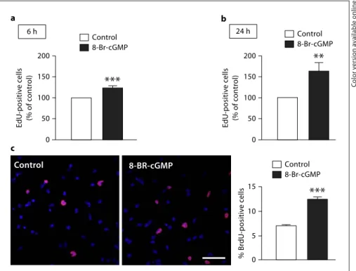
The cGMP Analogue 8-Br-cGMP Mimics the
Proliferative Effect of NOC-18
Since cGMP appears to mediate the proliferative effect
of NO at 24 h, we assessed the proliferative effect of a
Fig. 2. NO increases cell proliferation via the guanylyl
cyclase-cGMP pathway for longer (24 h) but not for shorter (6 h) periods of cell exposure to NO. Cell proliferation following treatment with NOC-18 (10 M ) in the absence or presence of 50 M ODQ for 6 h ( a ) or 24 h ( b ), evaluated by the incorporation of EdU and assessed by flow cytometry. Data are expressed as means 8 SEM of at least 6 independent experiments. One-way ANOVA (Bonfer-roni’s post-test). * * * p ! 0.001, significantly different from con-trol; +++ p ! 0.001, significantly different from NOC-18. EdU
in-corporation in neural stem cells following exposure to NOC-18 (10 M ) in the absence or presence of 1 M U0126, a selective MEK1 and MEK2 inhibitor, for 6 h ( c ) or 24 h ( d ), as assessed by flow cytometry. Data are expressed as means 8 SEM of at least 4 independent experiments. One-way ANOVA (Bonferroni’s post-test). * p ! 0.05 or * * * p ! 0.001, significantly different from con-trol; + p ! 0.05 or +++ p ! 0.001, significantly different from
NOC-18. e Representative images of BrdU (red) incorporation in neural stem cells following exposure to NOC-18 (10 M ) for 24 h in the absence or presence of a guanylyl cyclase inhibitor, ODQ (50 M ). Nuclei are labeled by Hoechst 33342 (blue). Scale bar: 20 m.
f ODQ completely blocks the increase in the number of BrdU-positive cells. Data are expressed as means 8 SEM of at least 4 independent experiments. One-way ANOVA (Bonferroni’s post-test). * * * p ! 0.001, significantly different from control;
+++ p ! 0.001, significantly different from 10 M NOC-18. g
De-composed NOC-18 (D-NOC-18) does not stimulate proliferation of SVZ-derived neural stem cells, unlike fresh NOC-18. Data are expressed as means 8 SEM of at least 4 independent experiments. One-way ANOVA (Dunnet’s post-test). * * * p ! 0.001, significant-ly different from control. Colors refer to the online version onsignificant-ly.
cGMP analogue, 8-Br-cGMP (20
M), by flow cytometry.
We observed a significant increase in EdU incorporation
following 6 and 24 h of treatment with 8-Br-cGMP to
123.6 8 6.1% (p ! 0.001; fig. 3 a) and 162.7 8 20.1%
(p ! 0.01; fig. 3 b) of the control, respectively, compared to
untreated cultures. We further confirmed the 24-hour
observations by evaluating the incorporation of BrdU by
immunocytochemistry and microscopy analysis.
Treat-ment with 8-Br-cGMP for 24 h significantly increased the
number of BrdU-positive cells to 12.4 8 0.5% (p ! 0.01)
compared to control (7.0 8 0.2%; fig. 3 c).
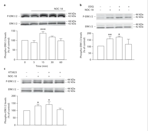
NO-Induced Activation of the Guanylyl Cyclase
Pathway Is Independent of ERK/MAPK Pathway
Activation
To identify the intracellular pathways that mediate the
proliferative effect of NO, we investigated whether the
guanylyl cyclase pathway is involved in the activation of
the ERK/MAPK pathway. We showed that NOC-18 alone
stimulates proliferation of SVZ cultures by activating
ERK1/2 [20] . To evaluate how fast the phosphorylation of
ERK1/2 occurs following exposure to NOC-18, we
ana-lyzed the phospho-ERK1/2:total ERK1/2
immunoreac-tivity ratio at several time points after the stimulus (at 5,
15, 30 and 60 min). The phosphorylation of ERK1/2
tran-siently increased to 138.1 8 8.4% of the control at 15 min
Table 2. Cell viability in neural stem cell cultures following
expo-sure to NOC-18 with or without U0126 or ODQ
Treatment Live cells
6 h Control 10 M NOC-18 10 M NOC-18 + 1 M U0126 1 M U0126 10 M NOC-18 + 50 M ODQ 50 M ODQ 90.981.4% 90.081.7% (n.s.) 90.281.9% (n.s.) 92.681.2% (n.s.) 90.681.7% (n.s.) 90.081.6% (n.s.) 24 h Control 10 M NOC-18 10 M NOC-18 + 1 M U0126 1 M U0126 10 M NOC-18 + 50 M ODQ 50 M ODQ 90.681.2% 91.081.4% (n.s.) 90.181.4% (n.s.) 89.881.8% (n.s.) 91.481.1% (n.s.) 90.582.0% (n.s.) C ell viability was assessed using the nuclear dye 7-AAD, de-tected by flow cytometry.
Data are expressed as means 8 SEM of at least 3 independent experiments.
p > 0.05 (nonsignificant; n.s.), not different from control, one-way ANOVA (Dunnett’s post-test).
c b 24 h % Br dU-positiv e c e lls 0 5 10 15 EdU-positiv e c e lls (% of c o ntr ol) 0 50 100 150 200 EdU-positiv e c e lls (% of c o ntr ol) 0 50 100 150 200 Control 8-Br-cGMP a 6 h Control 8-BR-cGMP
***
***
**
Control 8-Br-cGMP Control 8-Br-cGMPFig. 3. Effect of the cGMP analogue,
8-Br-cGMP, on cell proliferation in SVZ neuro-sphere cultures. EdU incorporation in neural stem cells following exposure to 20
M 8-Br-cGMP for 6 h ( a ) or 24 h ( b ), as assessed by flow cytometry. Two-tailed t test. * * * p ! 0.001 or * * p ! 0.01, signifi-cantly different from control. Data are ex-pressed as means 8 SEM of at least 4 in-dependent experiments. c 8-Br-cGMP (20 M ) mimics the proliferative effect of NO, as determined by BrdU incorporation fol-lowing 24 h of treatment. Representative images of BrdU-positive cells (red) in neu-ral stem cell cultures after exposure to 20 M 8-Br-cGMP, for 24 h, are shown in the panels in c . Nuclei are labeled with Hoechst 33342 (blue). Scale bar: 20 m. The data represent the percentage of BrdU-positive cells and are expressed as means 8 SEM of at least 3 independent experiments. Two-tailed t test. * * * p ! 0.01, significantly different from control. Colors refer to the online version only.
Co lo r v e rs io n av a il a b le o n li n e
early activation of ERK1/2 by exogenous NO, since
KT5823, a PKG inhibitor, did not prevent the increased
phosphorylation of ERK1/2 following exposure to
NOC-18 for 15 min ( fig. 4 c).
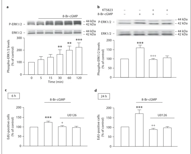
The cGMP Analogue 8-Br-cGMP Activates the
ERK/MAPK Pathway via PKG
Since the cGMP analogue 8-Br-cGMP increased cell
proliferation at 6 and 24 h in a similar manner to NOC-18,
after treatment with 10
MNOC-18 (p ! 0.001; fig. 4 a).
ODQ did not prevent the increase in the phosphorylation
of ERK1/2 triggered by NOC-18 (164.3 8 6.5% over the
control), suggesting the activation of ERK by NO is an
event independent of guanylyl cyclase ( fig. 4 b).
Since cGMP can activate the cGMP-dependent PKG,
the involvement of PKG in the activation of ERK1/2
fol-lowing treatment with NOC-18 was also evaluated. We
observed that PKG does not appear to be involved in the
a c b NOC-18 NOC-18 KT5823 – – + + – + + – – – + + – + + – Phospho -ERK1/2 lev e ls (% of c o ntr ol) NOC-18
**
*
0 50 100 150 200*
*
0 50 100 150 200 Phospho -ERK1/2 lev e ls (% of c o ntr ol) Phospho -ERK1/2 lev e ls (% of c o ntr ol)***
0 0 5 15 30 60 44 kDa 44 kDa 42 kDa 42 kDa 50 100 150 P-ERK1/2 ERK1/2 P-ERK1/2 ERK1/2 44 kDa 44 kDa 42 kDa 42 kDa 44 kDa 44 kDa 42 kDa 42 kDa P-ERK1/2 ERK1/2 ODQ Time (min)Fig. 4. NO activates the ERK/MAPK pathway in a
cGMP-inde-pendent manner. a Time course analysis of the phosphorylation of ERK1/2 following exposure to NOC-18 (10 M ). NOC-18 en-hanced ERK1/2 phosphorylation as early as at 15 min of treat-ment. Data are expressed as means 8 SEM of at least 4 indepen-dent experiments. One-way ANOVA (Bonferroni’s post-test). * * * p ! 0.001, significantly different from control. b Western blot analysis of the involvement of guanylyl cyclase in the phosphory-lation of ERK1/2, in lysates of neural stem cell cultures treated
with NOC-18 for 15 min. No effect of ODQ (50 M ) on ERK1/2 phosphorylation was observed. Data are expressed as means 8 SEM of at least 3 independent experiments. One-way ANOVA (Bonferroni’s post-test). * * p ! 0.01 and * p ! 0.05, significantly different from control. c No effect of the PKG inhibitor (KT5823; 1 M ) on the phosphorylation of ERK1/2 stimulated by exposure to NOC-18 (10 M ) for 15 min. One-way ANOVA (Bonferroni’s post-test). * p ! 0.05, significantly different from control.
we also investigated the ability of 8-Br-cGMP to activate
the ERK pathway. We analyzed the phospho-ERK1/2:
total ERK1/2 immunoreactivity ratio at 5, 15, 30, 60 and
120 min following treatment with 8-Br-cGMP. There was
a steady increase in the levels of phosphorylated ERK1/2
up to 2 h after treatment. At this time point, 20
M8-Br-cGMP induced a two-fold increase in
phospho-ERK1/2:total ERK1/2 immunoreactivity ratio (221.1 8
12.3% of the control), as compared to untreated cultures
( fig. 5 a). Inhibition of PKG with KT5823 prevented
ERK1/2 phosphorylation following exposure to
8-Br-cGMP for 2 h ( fig. 5 b), suggesting that PKG activation by
8-Br-cGMP is important for activation of ERK1/2 and
oc-curs upstream of ERK1/2 phosphorylation. Likewise, the
blockade of the ERK pathway by U0126 prevented EdU
incorporation stimulated by treatment with 8-Br-cGMP
for 6 h (102.9 8 4.8%, p ! 0.05; fig. 5 c) or 24 h (87.0 8
5.8%, p ! 0.01; fig. 5 d).
– – + + – + + –***
+++ + 0 50 100 150 200 Phospho -ERK1/2 lev e ls (% of c o ntr ol) Phospho -ERK1/2 lev e ls (% of c o ntr ol) 0 Time (min) 0 5 15 30 60 120 100 200 300 a b 8-Br-cGMP 8-Br-cGMP 8-Br-cGMP KT5823 P-ERK1/2 ERK1/2 P-ERK1/2 ERK1/2 44 kDa 44 kDa 42 kDa 42 kDa 44 kDa 44 kDa 42 kDa 42 kDa**
**
***
EdU-positiv e c e lls (% of c o ntr ol) 0 50 100 150 200 c 6 h ++ EdU-positiv e c e lls (% of c o ntr ol) 0 50 100 150 200 d 24 h***
8-Br-cGMP U0126 U0126***
Fig. 5. The cGMP analogue 8-Br-cGMP increases the
phosphory-lation of ERK1/2. a Time course analysis of the phosphorylation of ERK1/2 in lysates of neural stem cell cultures upon treatment with 20 M 8-Br-cGMP. Following exposure to 8-Br-cGMP there is a time-dependent increase in the phosphorylation of ERK1/2, followed for up to 120 min. Data are expressed as means 8 SEM of at least 4 independent experiments. One-way ANOVA (Bonfer-roni’s post-test). * * * p ! 0.001 and * * p ! 0.01, significantly differ-ent from control. b KT5823 prevents the phosphorylation of ERK1/2 stimulated by treatment with 8-Br-cGMP for 2 h.
One-way ANOVA (Bonferroni’s post-test). * * * p ! 0.001, significantly different from control; +++ p ! 0.001, significantly different from
8-Br-cGMP. EdU incorporation in neural stem cells following ex-posure to 8-Br-cGMP (20 M ) in the absence or presence of 1 M U0126, for 6 h ( c ) or 24 h ( d ), as assessed by flow cytometry. Data are expressed as means 8 SEM of at least 4 independent experi-ments. One-way ANOVA (Bonferroni’s post-test). * * * p ! 0.001, significantly different from control; + p ! 0.05 or ++ p ! 0.01,
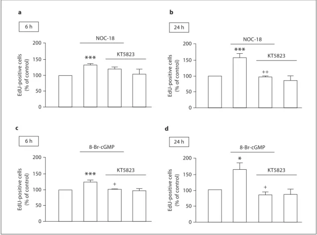
PKG Contributes to Late but Not to Early Proliferation
Induced by NO
The involvement of PKG in the proliferative effect of
NO was studied by flow cytometry by evaluating the
in-corporation of EdU by SVZ cell cultures following
treat-ment with NOC-18 or 8-Br-cGMP. The blockade of PKG
by KT5823 had no effect on early proliferation (6 h)
( fig. 6 a), but significantly prevented the EdU
incorpora-tion induced by NOC-18 treatment at 24 h (97.5 8 15.2%,
p ! 0.01), compared to cultures treated with NOC-18
alone (157.3 8 12.4%; fig. 6 b). Concerning the exposure
of SVZ cells to the cGMP analogue, the inhibition of
PKG prevented the proliferation induced by 8-Br-cGMP
both for 6 h (102.9 8 2.0%, p ! 0.05; fig. 6 c) and 24 h
(83.8 8 8.1%, p ! 0.05; fig. 6 d) compared to 8-Br-cGMP
alone (123.8 8 6.1 and 162.7 8 20.1% for 6 and 24 h,
respectively). We further confirmed by
immunocyto-chemistry the effect of the PKG inhibitor on the
incor-poration of BrdU in cultures treated with NOC-18
( fig. 7 a) or with 8-Br-cGMP ( fig. 7 b). KT5823
significant-ly prevented BrdU incorporation following exposure for
24 h to either NOC-18 (p ! 0.05) or 8-Br-cGMP (p !
0.01).
Flow cytometry analysis of nuclei stained with 7-AAD,
for the treatments described above, showed that KT5823
alone or in combination with NOC-18 or 8-Br-cGMP did
not significantly affect the cell viability of SVZ cells
com-pared to untreated cultures ( table 3 ).
a b c d + 8-Br-cGMP EdU-positiv e c e lls (% of c o ntr ol) 0 50 100 150 200 6 h + EdU-positiv e c e lls (% of c o ntr ol) 0 50 100 150 200 24 h
*
8-Br-cGMP KT5823 KT5823***
NOC-18 EdU-positiv e c e lls (% of c o ntr ol) 0 50 100 150 200 6 h ++ EdU-positiv e c e lls (% of c o ntr ol) 0 50 100 150 200 24 h***
NOC-18 KT5823 KT5823***
Fig. 6. Involvement of the cGMP/PKG signaling pathway in the
proliferation of neural stem cells. Cell proliferation following treatment with NOC-18 (10 M ) in the absence or presence of 1 M KT5823, a selective PKG inhibitor, for 6 h ( a ) or 24 h ( b ), evaluated by incorporation of EdU and assessed by flow cytom-etry. Data are expressed as means 8 SEM of at least 4 independent experiments. One-way ANOVA (Bonferroni’s post-test). * * * p ! 0.001, significantly different from control; ++ p ! 0.001,
signifi-cantly different from NOC-18. EdU incorporation in neural stem cells following exposure to 8-Br-cGMP (20 M ) in the absence or presence of 1 M KT5823, for 6 h ( c ) or 24 h ( d ), as assessed by flow cytometry. Data are expressed as means 8 SEM of at least 4 independent experiments. One-way ANOVA (Bonferroni’s post-test). * p ! 0.05 or * * * p ! 0.001, significantly different from con-trol; + p ! 0.05, significantly different from 8-Br-cGMP.
Discussion
In this work, we show that cGMP and PKG are
in-volved in the late proliferative effect triggered by NO in
neural stem cell cultures, since the inhibition of guanylyl
cyclase or PKG abolishes cell proliferation induced by
NO. Although cGMP and PKG were not involved in the
early activation of ERK1/2, they were mandatory for the
increase in cell proliferation following treatment with the
NO donor NOC-18 for 24 h. Moreover, the cGMP
ana-logue 8-Br-cGMP also increased neural stem cell
prolif-eration. We also found that the crosstalk between the
ERK1/2 pathway and the PKG pathway differs when
neu-ral stem cells are treated directly with the NO donor or
with the cGMP analogue.
Our results demonstrate that NO rapidly and
tran-siently activates ERK1/2 in a cGMP-independent
man-ner, since ODQ did not prevent ERK1/2 phosphorylation.
Also, the blockade of guanylyl cyclase did not prevent the
early proliferative effect of NOC-18 at 6 h. Furthermore,
we have previously shown that phosphorylation and
acti-vation of ERK1/2 is essential for the proliferative effect of
NO, either at the early stages of cell proliferation
follow-ing exposure to NO (as early as 30 min) or for later
end-points (24 h) [20] . These data strongly suggest that the
initial proliferation triggered by NO is dependent on the
activation of the ERK1/2 pathway, does not require
acti-vation of guanylyl cyclase, and is not dependent on cGMP.
This early proliferative effect is also independent of PKG,
which does not contribute to either ERK phosphorylation
or proliferation. Possibly, NO might directly activate
components of the ERK pathway (upstream of ERK1/2)
that are sensitive to NO signaling by S-nitrosylation of
cysteine residues. For instance, p21Ras has a cysteine
res-idue (Cys118) that is sensitive to NO, and when
nitrosyl-ated, p21Ras becomes activated [31, 32] .
Long-term exposure (24 h) to NO increased cell
pro-liferation in a cGMP-dependent manner. The blockade of
b a 8-Br-cGMP ++ % Br dU-positiv e c e lls 0 5 10 15
*
NOC-18 KT5823 + % Br dU-positiv e c e lls 0 5 10 15*
KT5823Fig. 7. The proliferative effect of NO is PKG-dependent. a Effect
of the PKG inhibitor (KT5823; 1 M ) on cell proliferation, upon exposure to NOC-18 (10 M ), for 24 h. One-way ANOVA (Bonfer-roni’s post-test). * p ! 0.05, significantly different from control;
+ p ! 0.05, significantly different from NOC-18. b KT5823
pre-vents cell proliferation stimulated by treatment with 8-Br-cGMP for 24 h. One-way ANOVA (Bonferroni’s post-test). * p ! 0.05, significantly different from control; + p ! 0.05, significantly
dif-ferent from 8-Br-cGMP.
Table 3. Cell viability in neural stem cell cultures following
expo-sure to NOC-18 or 8-Br-cGMP with or without KT5823
Treatment Live cells
6 h Control 10 M NOC-18 10 M NOC-18 + 1 M KT5823 20 M 8-Br-cGMP 20 M 8-Br-cGMP + 1 M KT5823 1 M KT5823 89.781.1% 91.081.9% (n.s.) 89.281.1% (n.s.) 93.081.2% (n.s.) 90.281.2% (n.s.) 92.181.3% (n.s.) 24 h Control 10 M NOC-18 10 M NOC-18 + 1 M KT5823 20 M 8-Br-cGMP 20 M 8-Br-cGMP + 1 M KT5823 1 M KT5823 87.981.9% 91.082.1% (n.s.) 92.182.3% (n.s.) 89.980.8% (n.s.) 90.081.2% (n.s.) 93.581.1% (n.s.) C ell viability was assessed using the nuclear dye 7-AAD, de-tected by flow cytometry.
Data are expressed as means 8 SEM of at least 3 independent experiments.
p > 0.05 (nonsignificant; n.s.), not different from control, one-way ANOVA (Dunnett’s post-test).
References
guanylyl cyclase or PKG completely abolished the
prolif-erative effect of NO at 24 h, suggesting that cGMP and
PKG signaling are essential for the proliferative effect of
NO. Our data strongly suggest that the proliferative effect
of NO is biphasic, i.e. an early increase in proliferation is
dependent on the MAPK pathway and does not rely on
guanylyl cyclase or PKG, and for later stages cGMP and
PKG contribute heavily to the proliferation induced by
NO. It is worth noting that the production of cGMP was
independent of the MAPK pathway, since U0126 did not
prevent the increase in cGMP levels observed following a
24-hour exposure to NOC-18.
The cGMP analogue 8-Br-cGMP also triggered cell
proliferation, and our data show that PKG is important
for this proliferative effect. 8-Br-cGMP also induced
ac-tivation of ERK1/2, which was now sensitive to the
inhi-bition of PKG, suggesting that phosphorylation of ERK1/2
following 8-Br-cGMP application to neural stem cells
re-lies on a mechanism dependent on PKG. It remains to be
established whether PKG directly activates ERK by
phos-phorylation. Furthermore, we observed that blocking
ERK1/2 activation with U0126 prevents proliferation of
neural stem cells following treatment with 8-Br-cGMP,
suggesting that the activation of the MAPK pathway is
responsible for the proliferative effect of the cGMP
ana-logue. PKG is a serine/threonine kinase that is activated
upon binding of cGMP, and NO-induced elevation of the
intracellular levels of cGMP has been reported to
direct-ly regulate the activity of PKG [33–35] . PKG has been
im-plicated in the regulation of gene expression as reviewed
by Madhusoodanan and Murad [23] . Some studies
sug-gest that the cGMP/PKG pathway is involved in the
acti-vation of the MAPK pathway, particularly ERK1/2 [36,
37] , which appears to be the case in our study where
neu-ral stem cells are treated with a cGMP analogue. When
the neural stem cells are treated with a NO donor, we
ob-served that the initial ERK1/2 activation is independent
of cGMP-dependent signaling to initiate the early
prolif-erative events triggered by NO. For longer exposures,
sig-naling via guanylyl cyclase and PKG gains relevance for
the proliferation stimulated by NO.
The GC-cGMP pathway is the main effector pathway
of the biological effects of NO as a second messenger.
Al-though this pathway is expected to participate in the
sig-naling events contributing to the proliferative effect of
NO treatment in neural stem cells, most evidence up to
now was circumstantial. Regarding the formation of
newborn cells from stem cells, it was reported that the
elevation of cGMP levels by PDE5 inhibition promoted
PKG activation, enhancing the proliferation of
mesen-chymal stem cells [38] . Other studies correlated the
eleva-tion of cGMP levels with the enhancement of
neurogen-esis [25, 26, 39] . Understanding how cGMP and its
down-stream target PKG contribute to the proliferative effect of
NO can help in better targeting these systems to design
well-grounded therapies for stimulating endogenous
neurogenesis for brain repair purposes.
Altogether, we show that NO can activate two
inde-pendent pathways in a biphasic manner that act to
in-crease neural stem cell proliferation, initially the ERK/
MAPK pathway, and for later stages the guanylyl/cGMP/
PKG pathway. Although there is no evidence of crosstalk
between these two pathways for the early effect of NO,
this possibility cannot be excluded for NO-induced cell
proliferation at later stages. While the early proliferation
of neural stem cells triggered by NO is independent of
cGMP and PKG, the complete blockade of the
prolifera-tive effect of NO at later stages by inhibition of either
gua-nylyl cyclase, PKG or ERK1/2 suggests a crosstalk
be-tween these different signaling pathways.
Acknowledgments
This work was supported by the Calouste Gulbenkian Foun-dation, L’Oréal, UNESCO, and the Foundation for Science and Technology (FCT, Portugal), COMPETE and FEDER (project PTDC/SAU-NEU/102612/2008). Bruno P. Carreira and Maria Inês Morte were supported by FCT, Portugal (fellowships SFRH/BPD/78901/2011, SFRH/BD/23754/2005 and SFRH/ BD/38127/2007).
1 Whitney NP, Eidem TM, Peng H, Huang Y, Zheng JC: Inflammation mediates varying effects in neurogenesis: relevance to the pathogenesis of brain injury and neurode-generative disorders. J Neurochem 2009; 108: 1343–1359.
2 Monje ML, Toda H, Palmer TD: Inflamma-tory blockade restores adult hippocampal neurogenesis. Science 2003; 302: 1760–1765. 3 Ekdahl CT, Claasen JH, Bonde S, Kokaia Z,
Lindvall O: Inflammation is detrimental for neurogenesis in adult brain. Proc Natl Acad Sci USA 2003; 100: 13632–13637.
4 Ekdahl CT, Kokaia Z, Lindvall O: Brain in-flammation and adult neurogenesis: the dual role of microglia. Neuroscience 2009; 158: 1021–1029.
5 Bengzon J, Kokaia Z, Elmer E, Nanobashvili A, Kokaia M, Lindvall O: Apoptosis and pro-liferation of dentate gyrus neurons after sin-gle and intermittent limbic seizures. Proc Natl Acad Sci USA 1997; 94: 10432–10437.
6 Parent JM, Yu TW, Leibowitz RT, Geschwind DH, Sloviter RS, Lowenstein DH: Dentate granule cell neurogenesis is increased by sei-zures and contributes to aberrant network reorganization in the adult rat hippocampus. J Neurosci 1997; 17: 3727–3738.
7 Parent JM, Lowenstein DH: Seizure-induced neurogenesis: are more new neurons good for an adult brain? Prog Brain Res 2002; 135: 121–131.
8 Arvidsson A, Kokaia Z, Airaksinen MS, Saa-rma M, Lindvall O: Stroke induces wide-spread changes of gene expression for glial cell line-derived neurotrophic factor family receptors in the adult rat brain. Neurosci-ence 2001; 106: 27–41.
9 Arvidsson A, Collin T, Kirik D, Kokaia Z, Lindvall O: Neuronal replacement from en-dogenous precursors in the adult brain after stroke. Nat Med 2002; 8: 963–970.
10 Kernie SG, Parent JM: Forebrain neurogen-esis after focal ischemic and traumatic brain injury. Neurobiol Dis 2010; 37: 267–274. 11 Jakubs K, Bonde S, Iosif RE, Ekdahl CT,
Ko-kaia Z, KoKo-kaia M, Lindvall O: Inflammation regulates functional integration of neurons born in adult brain. J Neurosci 2008; 28: 12477–12488.
12 Thored P, Heldmann U, Gomes-Leal W, Gisler R, Darsalia V, Taneera J, Nygren JM, Jacobsen SE, Ekdahl CT, Kokaia Z, Lindvall O: Long-term accumulation of microglia with proneurogenic phenotype concomitant with persistent neurogenesis in adult sub-ventricular zone after stroke. Glia 2009; 57: 835–849.
13 Hoehn BD, Palmer TD, Steinberg GK: Neu-rogenesis in rats after focal cerebral ischemia is enhanced by indomethacin. Stroke 2005; 36: 2718–2724.
14 Contestabile A, Ciani E: Role of nitric oxide in the regulation of neuronal proliferation, survival and differentiation. Neurochem Int 2004; 45: 903–914.
15 Packer MA, Stasiv Y, Benraiss A, Chmiel-nicki E, Grinberg A, Westphal H, Goldman SA, Enikolopov G: Nitric oxide negatively regulates mammalian adult neurogenesis. Proc Natl Acad Sci USA 2003; 100: 9566– 9571.
16 Moreno-Lopez B, Romero-Grimaldi C, No-val JA, Murillo-Carretero M, Matarredona ER, Estrada C: Nitric oxide is a physiological inhibitor of neurogenesis in the adult mouse subventricular zone and olfactory bulb. J Neurosci 2004; 24: 85–95.
17 Matarredona ER, Murillo-Carretero M, Moreno-Lopez B, Estrada C: Nitric oxide synthesis inhibition increases proliferation of neural precursors isolated from the post-natal mouse subventricular zone. Brain Res 2004; 995: 274–284.
18 Torroglosa A, Murillo-Carretero M, Rome-ro-Grimaldi C, Matarredona ER, Campos-Caro A, Estrada C: Nitric oxide decreases subventricular zone stem cell proliferation by inhibition of epidermal growth factor re-ceptor and phosphoinositide-3-kinase/Akt pathway. Stem Cells 2007; 25: 88–97. 19 Zhu DY, Liu SH, Sun HS, Lu YM: Expression
of inducible nitric oxide synthase after focal cerebral ischemia stimulates neurogenesis in the adult rodent dentate gyrus. J Neurosci 2003; 23: 223–229.
20 Carreira BP, Morte MI, Inacio A, Costa G, Rosmaninho-Salgado J, Agasse F, Carmo A, Couceiro P, Brundin P, Ambrosio AF, Car-valho CM, Araujo IM: Nitric oxide stimu-lates the proliferation of neural stem cells by-passing the epidermal growth factor recep-tor. Stem Cells 2010; 28: 1219–1230. 21 Gomez-Pinedo U, Rodrigo R, Cauli O,
Her-raiz S, Garcia-Verdugo JM, Pellicer B, Pelli-cer A, Felipo V: cGMP modulates stem cells differentiation to neurons in brain in vivo. Neuroscience 2010; 165: 1275–1283.
22 Tegenge MA, Rockel TD, Fritsche E, Bicker G: Nitric oxide stimulates human neural progenitor cell migration via cGMP-mediat-ed signal transduction. Cell Mol Life Sci 2011; 68: 2089–2099.
23 Madhusoodanan KS, Murad F: No-cGMP sig-naling and regenerative medicine involving stem cells. Neurochem Res 2007; 32: 681–694. 24 Zhang R, Wang Y, Zhang L, Zhang Z, Tsang
W, Lu M, Chopp M: Sildenafil (Viagra) in-duces neurogenesis and promotes functional recovery after stroke in rats. Stroke 2002; 33: 2675–2680.
25 Zhang L, Zhang Z, Zhang RL, Cui Y, LaPointe MC, Silver B, Chopp M: Tadalafil, a long-acting type 5 phosphodiesterase iso-enzyme inhibitor, improves neurological functional recovery in a rat model of embol-ic stroke. Brain Res 2006; 1118: 192–198. 26 Wang L, Gang Zhang Z, Lan Zhang R,
Chopp M: Activation of the PI3-K/Akt path-way mediates cGMP enhanced-neurogenesis in the adult progenitor cells derived from the subventricular zone. J Cereb Blood Flow Metab 2005; 25: 1150–1158.
27 Araujo IM, Ambrosio AF, Leal EC, Santos PF, Carvalho AP, Carvalho CM: Neuronal nitric oxide synthase proteolysis limits the involvement of nitric oxide in kainate-in-duced neurotoxicity in hippocampal neu-rons. J Neurochem 2003; 85: 791–800.
28 Alvaro AR, Martins J, Araujo IM, Ros-maninho-Salgado J, Ambrosio AF, Cavadas C: Neuropeptide Y stimulates retinal neural cell proliferation – involvement of nitric ox-ide. J Neurochem 2008; 105: 2501–2510. 29 Covacu R, Danilov AI, Rasmussen BS,
Hal-len K, Moe MC, Lobell A, Johansson CB, Svensson MA, Olsson T, Brundin L: Nitric oxide exposure diverts neural stem cell fate from neurogenesis towards astrogliogenesis. Stem Cells 2006; 24: 2792–2800.
30 Canals S, Casarejos MJ, Rodriguez-Martin E, de Bernardo S, Mena MA: Neurotrophic and neurotoxic effects of nitric oxide on fetal midbrain cultures. J Neurochem 2001; 76: 56–68.
31 Lander HM, Hajjar DP, Hempstead BL, Mir-za UA, Chait BT, Campbell S, Quilliam LA: A molecular redox switch on p21(ras). Struc-tural basis for the nitric oxide-p21(ras) inter-action. J Biol Chem 1997; 272: 4323–4326. 32 Deora AA, Hajjar DP, Lander HM:
Recruit-ment and activation of Raf-1 kinase by nitric oxide-activated Ras. Biochemistry 2000; 39: 9901–9908.
33 Fiscus RR, Rapoport RM, Murad F: Endo-thelium-dependent and nitrovasodilator-in-duced activation of cyclic GMP-dependent protein kinase in rat aorta. J Cyclic Nucleo-tide Protein Phosphor Res 1983; 9: 415–425. 34 Fiscus RR: Involvement of cyclic GMP and
protein kinase G in the regulation of apopto-sis and survival in neural cells. Neurosignals 2002; 11: 175–190.
35 Forstermann U, Gorsky LD, Pollock JS, Schmidt HH, Heller M, Murad F: Regional distribution of EDRF/NO-synthesizing enzyme(s) in rat brain. Biochem Biophys Res Commun 1990; 168: 727–732.
36 Zaragoza C, Balbin M, Lopez-Otin C, Lamas S: Nitric oxide regulates matrix metallopro-tease-13 expression and activity in endothe-lium. Kidney Int 2002; 61: 804–808. 37 Ota KT, Pierre VJ, Ploski JE, Queen K, Schafe
GE: The NO-cGMP-PKG signaling pathway regulates synaptic plasticity and fear memo-ry consolidation in the lateral amygdala via activation of ERK/MAP kinase. Learn Mem 2008; 15: 792–805.
38 Haider HK, Lee YJ, Jiang S, Ahmad RP, Ryon MD, Ashraf M: Phosphodiestrase inhibition with tadalafil provides longer and sustained protection of stem cells. Am J Physiol Heart Circ Physiol 2010; 299:H1395–H1404. 39 Zhang RL, Zhang Z, Zhang L, Wang Y,
Zhang C, Chopp M: Delayed treatment with sildenafil enhances neurogenesis and im-proves functional recovery in aged rats after focal cerebral ischemia. J Neurosci Res 2006; 83: 1213–1219.