Outubro de 2011
Universidade do Minho
Escola de Engenharia
Diana Filipa Barros Alves
Influence of a Quaternary Ammonium
Compound on the Cell Structure of
Bacteria using Atomic Force
Microscopy
UMinho|20
11
Diana Filipa Barr
os Alves
Influence of a Quaternar
y Ammonium Compound on t
he Cell Structure of Bacteria using Atomic F
Dissertação de Mestrado
Mestrado Integrado em Engenharia Biomédica
Ramo de Engenharia Clínica
Trabalho realizado sob a orientação da
Professora Doutora Lígia Marona Rodrigues
e da
Professora Doutora Henny C. van der Mei
Outubro de 2011
Diana Filipa Barros Alves
Influence of a Quaternary Ammonium
Compound on the Cell Structure of
Bacteria using Atomic Force
É AUTORIZADA A REPRODUÇÃO PARCIAL DESTA DISSERTAÇÃO APENAS PARA EFEITOS
DE INVESTIGAÇÃO, MEDIANTE DECLARAÇÃO ESCRITA DO INTERESSADO, QUE A TAL SE
COMPROMETE;
Universidade do Minho, ___/___/______
Influence of a Quaternary Ammonium Compound on the Cell Structure of Bacteria using AFM
A
A
C
C
K
K
N
N
O
O
W
W
L
L
E
E
D
D
G
G
E
E
M
M
E
E
N
N
T
T
S
S
At the end of a short but extremely rewarding journey I cannot forget a number of people that somehow contributed for the development of my master thesis.
First of all, I would like to express my gratitude to Prof. Dr. Henk Busscher and Prof. Dr. Henny van der Mei for the opportunity to develop my experimental work at the Biomedical Engineering Department (BME) of University Medical Centre of Groningen (the Netherlands) where I was introduced to the world of research. To Prof. Dr. Henny van der Mei I would like to thank for all the wise advices and constant challenges during all the process and for remind me that in research all kind of results are indeed results. I also would like to thank Prof. Dr. Lígia Rodrigues that, even far, was a constant presence in the development of the present thesis. Her guidance, incentive and motivation from the beginning were essential for the success of the work developed. I would like to thank Prof. Dr. Miguel Gama as the coordinator of Clinical Engineering specialization of the Integrated Master in Biomedical Engineering and for the incentive provided in a moment of indecision.
A special thanks to Mihaela Crismaru, a PhD student from BME, who shared her experience with me and gave me the opportunity to integrate her project. I would like to thank for all the helpful suggestions, the insightful tips, the optimism and the friendship. I also would like to thank Joop de Vries for teaching me how to use such a complex technique as AFM and for all the assistance provided afterwards. To Betsy, I would like to thank for teaching me how the laboratory works and everything about bacterial culture. As well, I would like to thank all the professors, PhD students and technicians from BME who made me feel accepted in their group.
I would like to thank all the good friends I made in Groningen that were essential for such a nice stay. A special thanks to my roommate Catarina Leirós, whose friendship and support helped me to face all the personal and professional challenges, as well to a good friend from BME, Theerthankar Das, whose life will always be an example and inspiration from me.
To my best friends, Joana, Graça, Sara, Nelson, Rita and Águeda thank you for the friendship and support all over these years. To a special friend, Ângela Prego, who has
iv
Influence of a Quaternary Ammonium Compound on the Cell Structure of Bacteria using AFM
been a real sister, thank you for the unconditional friendship and for always having the right words.
Finally, I cannot forget to thank my family. To my parents, I would like to thank for all the support and unconditional love. Thank you for being the best parents in the world! To my little sister (yes, you will be always little to me) I would like to thank all the patience and support.
“Whatever you do will be insignificant, but it is very important that you do it”.
Influence of a Quaternary Ammonium Compound on the Cell Structure of Bacteria using AFM
A
A
B
B
S
S
T
T
R
R
A
A
C
C
T
T
Bacterial adhesion and subsequent biofilm formation remains a serious concern, especially in clinical applications, since bacteria associated with biofilms are more resistant to antibiotic treatment and to the host immune system. A potential approach to deal with this drawback may rely on the use of antimicrobial coatings comprising quaternary ammonium compounds (QACs).
The main purpose of the present thesis was to study the efficacy of a QAC against staphylococci when compared with two other antimicrobial compounds, an antibiotic (Gentamicin) and an antimicrobial peptide (Gramicidin S) using atomic force microscopy (AFM). After assessing the antimicrobial activity of the compounds against planktonic cultures by determining their minimal inhibitory (MIC) and minimal bactericidal concentrations (MBC), adhering staphylococcal cells were exposed to the antimicrobial compounds and their cell surfaces analyzed with AFM. The number of bacteria removed by the AFM tip was determined and taken as an indication of cell surface damage. The antimicrobial action of the compounds on staphylococcal biofilms was evaluated by the determination of the metabolic activity of biofilm through an 3-(4,5-dimethylthiazol-2-yl)-2,5-diphenyl-tetrazolium bromide (MTT) assay. Finally, in order to get some insights about QAC mode of action against bacterial cells, its antimicrobial activity in planktonic cultures, as well as their effects on adhering staphylococcal cells by AFM were performed in the presence of calcium ions.
All the antimicrobial compounds proved to exhibit antimicrobial activity against staphylococcal planktonic cultures. AFM measurements allowed the analysis in situ of the bacterial cells behaviour when exposed to antimicrobial compounds. Bacterial cell surface wrinkled upon exposure to the antimicrobials until bacteria disappear from the surface. Gentamicin yielded faster wrinkling and removal of bacteria from the surface as compared to QAC and Gramicidin S which yielded similar results. As the staphylococci outside the continuously scanned area did not seem affected by the compounds it was suggested that the pressure of the AFM tip assisted the incorporation of antimicrobials in the membrane, enhancing their bactericidal efficacy. Staphylococci biofilm proved to be susceptible to all the antimicrobials, with QAC being the most effective one, causing a complete loss of metabolic activity at 2xMBC. The antimicrobial action of the QAC was compromised by the presence of calcium ions both in planktonic cultures and in adhering staphylococci which suggests that its mode of action relies on its ability to exchange with calcium ions that are responsible for membrane stability.
Influence of a Quaternary Ammonium Compound on the Cell Structure of Bacteria using AFM
R
R
E
E
S
S
U
U
M
M
O
O
A adesão bacteriana e consequente formação de biofilmes constitui um problema sério, sobretudo na área clínica, visto que as bactérias associadas em biofilmes são menos susceptíveis à terapia antibiótica e à acção do sistema imunitário. Uma potencial estratégia para lidar com este problema consiste no uso de revestimentos antimicrobianos constituídos por compostos quaternários de amónio (QACs).
A presente tese teve como principal objectivo o estudo da eficácia de um QAC sobre uma estirpe bacteriana do género estafilococos quando comparado com outros dois compostos antimicrobianos, um antibiótico (Gentamicina) e um péptido antimicrobiano (Gramicidina S). Depois de avaliada a actividade antimicrobiana dos compostos em culturas planctónicas através da determinação da concentração mínima inibitória (MIC) e da concentração mínima bactericida (MBC), as bactérias aderidas a uma superfície foram expostas aos compostos e a sua superfície celular foi analisada por AFM. O número de bactérias removidas pela ponta do AFM foi determinado e considerado como uma indicação dos danos causados na superfície celular. A susceptibilidade dos biofilmes aos compostos foi avaliada pela determinação da actividade metabólica do biofilme através de um ensaio de 3-(4,5-dimetiltiazol-2-il)-2-5 difeniltatrazólio de brometo (MTT). Finalmente, com o intuito de investigar o modo de acção do QAC sobre as células bacterianas, a sua actividade antimicrobiana, bem como os seus efeitos nas células aderidas e analisados por AFM foram realizados na presença de iões de cálcio. Todos os compostos exibiram actividade antimicrobiana no estado planctónico. O efeito dos compostos sobre as células bacterianas foi analisado in situ com o AFM. A superfície celular apresentou-se mais enrugada depois de exposta aos compostos até as bactérias serem removidas da superfície. A Gentamicina resultou numa remoção mais rápida que o QAC e a Gramicidina S que exibiram resultados semelhantes. Como as bactérias localizadas fora da área continuamente analisada pelo AFM não foram igualmente influenciadas pelos compostos foi colocada a hipótese de que a pressão da ponta do AFM auxiliou a incorporação dos compostos na membrana, melhorando assim a sua eficácia bactericida. O biofilme formado pela estirpe estudada na presente tese apresentou-se susceptível a todos os compostos antimicrobianos, sendo o QAC o mais eficaz ao provocar uma redução completa da actividade metabólica do biofilme a 2xMBC. A acção antimicrobiana do QAC foi comprometida pela presença de iões de cálcio o que sugere que o seu modo de acção depende da sua capacidade de substituir estes iões na membrana.
Influence of a Quaternary Ammonium Compound on the Cell Structure of Bacteria using AFM
T
T
A
A
B
B
L
L
E
E
O
O
F
F
C
C
O
O
N
N
T
T
E
E
N
N
T
T
S
S
ACKNOWLEDGEMENTS iii ABSTRACT v RESUMO vii TABLE OF CONTENTS ix LIST OF FIGURES xi LIST OF TABLES xv ABBREVIATIONS xviiSCOPE AND AIMS xix
CHAPTER 1| GENERAL INTRODUCTION 1
1.1 Bacterial adhesion and biofilm formation onto biomaterials 3 1.1.1 Biofilms: a survival strategy of microorganisms 5 1.1.2 Coagulase-negative staphylococci as nosocomial pathogens 7 1.2 Treatment and prevention of biomaterial associated infections 9 1.2.1 The role of quaternary ammonium compounds on preventing BAI 10 1.2.1.1 QAC's mechanisms of action 11 1.2.1.2 Antimicrobial and anti-adhesive activities of QACs 13 1.3 Microscopy techniques to study the antimicrobial effects 14 1.3.1 Basic principles of AFM and AFM imaging modes 16 1.3.2 Sample preparation for AFM 18 1.3.3. Applications of AFM in microbiology 19
CHAPTER 2| MATERIALS AND METHODS 23
2.1 Microorganisms and growth conditions 25
2.2 Antimicrobial compounds 25
2.3 Antimicrobial assay 26
2.4 Atomic force microscopy 27
x
Influence of a Quaternary Ammonium Compound on the Cell Structure of Bacteria using AFM
2.6 Fluorescence microscopy 28
2.7 MTT assay 29
2.8 Calcium effect on QAC's mode of action 29
2.9 Statistical analysis 31
CHAPTER 3| RESULTS 33
3.1 Antimicrobial activity 35
3.2 Atomic force microscopy 35
3.3 Fluorescence microscopy 43
3.4 MTT assay 45
3.5 Calcium effect on QAC's mode of action 46
CHAPTER 4| DISCUSSION 49
4.1 Antimicrobial effect on adhering staphylococcal cell wall 52 4.2 Antimicrobial effect on biofilm 54 4.3 Influence of calcium cations on QAC mechanism 55
CHAPTER 5| CONCLUSIONS AND RECOMMENDATIONS 57
5.1 Conclusions 59
5.2 Recommendations 60
Influence of a Quaternary Ammonium Compound on the Cell Structure of Bacteria using AFM
L
L
I
I
S
S
T
T
O
O
F
F
F
F
I
I
G
G
U
U
R
R
E
E
S
S
C
C
HHAAPPTTEERR1
1
Figure 1.1 Schematic model of the steps involved in biofilm formation on a surface: conditioning film formation (1), reversible attachment (2), irreversible attachment (3), maturation (4) and detachment (5).
5
Figure 1.2 Schematic representation of the phases involved in Staphylococcus epidermidis biofilm formation and bacterial factors involved. 8
Figure 1.3 General chemical structure of a surfactant (R represents a functional group and X- represents a counter ion such as Cl-, Br- or NO3
-). 10
Figure 1.4 Schematic representation showing the mechanism of action for QACs. Progressive adsorption of the quaternary head group to acidic phospholipids in the membrane with increasing QAC exposure/concentration leads to decreased fluidity of the bilayers and the creation of hydrophilic voids in the membrane.
12
Figure 1.5 Schematic diagram of AFM work principle. 17 Figure 1.6 Schematic representation of an AFM tip operating in the contact mode (A)
and dynamic mode (B). 18
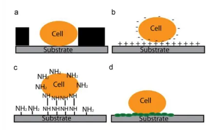
Figure 1.7 Schematic representation of the most commonly immobilization methods for AFM: (a) physical entrapment, (b) attractive electrostatic interactions, (c) covalent binding to amine-functionalized surface by glutaraldehyde and (d) attachment to polyphenolic adhesive proteins.
19
Figure 1.8 Schematic representation of a typical force curve with the different regions of the approach and withdrawal portions. 21
C
C
HHAAPPTTEERR2
2
Figure 2.1 Chemical structure of Ethoquad C/25 (Cocoalkyl methyl (polyoxyethylene) ammonium chloride) (A), Gramicidin S (B) and Gentamicin sulphate (C). 26
C
C
HHAAPPTTEERR3
3
Figure 3.1 AFM Peak Force images of Staphylococcus epidermidis ATCC 14990 after being exposed to 10 mM potassium phosphate buffer (control) for 0 min (A), 103 min (B), 161 min (C) and 300 min (D), to 2 µg/ml Gentamicin sulphate for 0 min (E), 182 min (F), 240 min (G) and 284 min (H), to 110 µg/ml QAC for 0 min (I), 90 min (J), 141 min (K) and 264 min (L) and to 8 µg/ml Gramicidin S for 0 min (M), 170 min (N), 259 min (O) and 274 min (P). Experiments were performed with a 3 nN force.
xii
Influence of a Quaternary Ammonium Compound on the Cell Structure of Bacteria using AFM Figure 3.2 AFM Peak Force images of S. epidermidis ATCC 14990 when exposed to
10 mM potassium phosphate buffer (A, B) and 2 µg/ml Gentamicin sulphate solution (C, D) after a single scan (30 µm x 30 µm) and after multiple scans in the smaller region (10 µm x 10 µm). Experiments were performed with a 3 nN force.
38
Figure 3.3 AFM Peak Force images of S. epidermidis when exposed to 10 mM potassium phosphate buffer (A) and QAC solution for 60 min (B,C). Experiments were performed with a 3 nN force.
39
Figure 3.4 Kaplan-Meier curve expressing the percentage of Staphylococcus epidermidis ATCC 14990 adhering on a substrate surface during exposure to 10 mM
potassium phosphate buffer (control), 2 µg/ml Gentamicin sulphate solution, 110 µg/ml QAC solution and 8 µg/ml Gramicidin S solution while continuously scanning. Experiments were performed in triplicate with a 3 nN force.
39
Figure 3.5 Roughness (nm) of Staphylococcus epidermidis ATCC 14990 adhering on a substrate surface before and after exposure to 10 mM potassium phosphate buffer (control) (A), 110 µg/ml QAC solution (B), 8 µg/ml Gramicidin S solution (C) and 2 µg/ml Gentamicin sulphate solution (D).
41
Figure 3.6 AFM Peak Force images of Staphylococcus epidermidis ATCC 14990 after being exposed to 10 mM potassium phosphate buffer (control) for 0 min (A), 181 min (B) and 274 min (C), to 2 µg/ml Gentamicin sulphate for 0 min (D), 33 min (E) and 55 min (F) and to 110 µg/ml QAC for 0 min (G), 58 min (H) and 72 min (I). Experiments were performed with a 6 nN force.
42
Figure 3.7 Kaplan-Meier curve expressing the percentage of Staphylococcus epidermidis ATCC 14990 adhering on a substrate surface during exposure to 10 mM
potassium phosphate buffer (control), 2 µg/ml Gentamicin sulphate solution and 110 µg/ml QAC solution while continuously scanning. Experiments were performed once with a 6 nN force.
43
Figure 3.8 Fluorescent live-dead images of Staphylococcus epidermidis ATCC 14990 after being exposed for 60 min to 10 mM potassium phosphate buffer as a control (A), to 110 µg/ml QAC solution (B), 8 µg/ml Gramicidin S solution (C) and to 2 µg/ml Gentamicin sulphate solution (D).
44
Figure 3.9 Percentage relative of metabolic activity of Staphylococcus epidermidis ATCC 14990 biofilm after being exposed overnight to QAC, Gentamicin sulphate and Gramicidin S solutions at different concentrations (0.5; 1 and 2xMBC). Results correspond to the average of three independent assays.
45
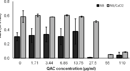
Figure 3.10 Staphylococcus epidermidis ATCC 14990 growth in the absence (control) and in the presence of several concentrations of QAC diluted in nutrient broth (NB) and NB supplemented with 0.1 M CaCl2. Results correspond to the average of two
independent assays.
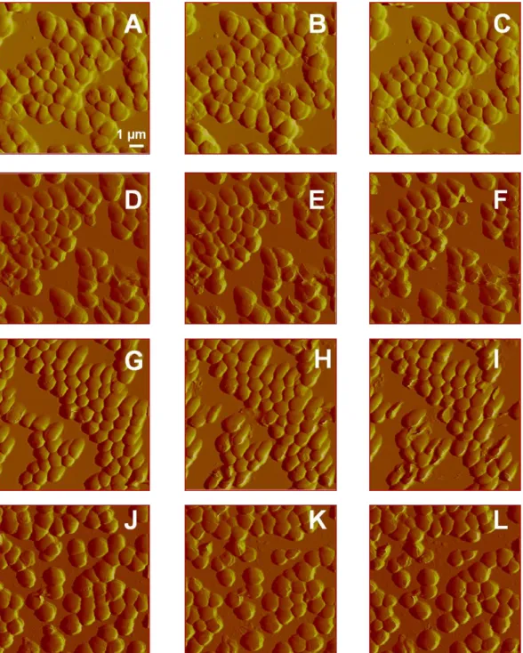
Influence of a Quaternary Ammonium Compound on the Cell Structure of Bacteria using AFM Figure 3.11 AFM Peak Force images of Staphylococcus epidermidis ATCC 14990
after being exposed to demineralized water (control) for 0 min (A), 188 min (B) and 300 min (C), to 0.1 M Cacl2 (control) for 0 min (D), 182 min (E) and 300 min (F), to 110
µg/ml QAC in demineralized water for 0 min (G), 191 min (H) and 300 min (I) and to 110 µg/ml QAC in CaCl2 for 0 min (J), 192 min (K) and 300 min (L). Experiments were
performed with a 3 nN force
47
Figure 3.12 Kaplan-Meier curve expressing the percentage of Staphylococcus epidermidis ATCC 14990 adhering on a substrate surface during exposure to
demineralized water (control), 0.1 M CaCl2 (control), 110 µg/ml QAC in demineralized
water and 110 µg/ml QAC in CaCl2 while continuously scanning. Experiments were
performed in duplicate with a 3nN force.
Influence of a Quaternary Ammonium Compound on the Cell Structure of Bacteria using AFM
L
L
I
I
S
S
T
T
O
O
F
F
T
T
A
A
B
B
L
L
E
E
S
S
C
C
HHAAPPTTEERR1
1
Table 1.1 Incidence and microorganisms most commonly isolated from infections on different biomedical implants and devices. 3 Table 1.2 The major factors contributing to Staphylococcus epidermidis biofilm
formation and their function. 8
Table 1.3 Comparison between the most commonly used techniques used to visualize the effect of antimicrobials on bacteria. 15
C
C
HHAAPPTTEERR3
3
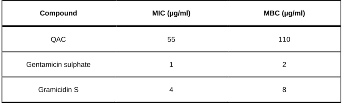
Table 3.1 MIC and MBC values of QAC, Gentamicin sulphate and Gramicidin S obtained for Staphylococcus epidermidis ATCC 14990. Data for individual strains were obtained in triplicate yielding identical results.
Influence of a Quaternary Ammonium Compound on the Cell Structure of Bacteria using AFM
A
A
B
B
B
B
R
R
E
E
V
V
I
I
A
A
T
T
I
I
O
O
N
N
S
S
AFM ATCC
Atomic force microscopy
American type culture collection BAI
CLSM CNC
Biomaterial associated infections Confocal laser scanning microscopy Coagulase-negative staphylococci CTAB Cetyltrimethylammonium bromide EPS
MBC MIC
Extracellular polymeric substances Minimal bactericidal concentration Minimal inhibitory concentration
MTT 3-(4,5-dimethylthiazol-2-yl)-2,5-diphenyl-tetrazolium bromide NB Nutrient broth
PEI Polyethyleneimine
PIA Polysaccharide intercellular adhesion PLL Poly-L-lysine
QAC QAS
Quaternary ammonium compound Quaternary ammonium silane QNM Quantitative nanomechanical SEM Scanning electronic microscopy TEM Transmission electronic microscopy TSB Tryptone soy broth
Influence of a Quaternary Ammonium Compound on the Cell Structure of Bacteria using AFM
S
S
C
C
O
O
P
P
E
E
A
A
N
N
D
D
A
A
I
I
M
M
S
S
Despite considerable research have been conducted in the development of new materials and in the design of innovative devices, infections associated to biomaterial implants and medical devices remain a serious problem. Bacteria are able to reach the biomaterial surface, adhere to it and form multicellular aggregates enclosed in a self-produced matrix of extracellular polymeric substances with increased resistance to antibiotic treatments and to the host immune system. The microbial adhesion and biofilm formation on surfaces of biomedical implants and devices can cause severe problems, often requiring the replacement of the infected device at the expense of considerable costs and patient‟s suffering. Therefore, big efforts have been conducted to stop and prevent the formation of these microbial biofilms.
The main purpose of the present thesis is to assess the potential of using a QAC (Ethoquad C/25 (Cocoalkyl methyl (polyoxyethylene) ammonium chloride)) in the prevention of microbial adhesion for possible application in antimicrobial coatings of biomedical devices. In order to achieve this goal, the efficiency of the QAC against staphylococci will be studied and results will be compared with two other compounds, an antibiotic (Gentamicin sulphate) and an antimicrobial peptide (Gramicidin S). Initially, the antimicrobial activity of the compounds will be assessed by determining their minimal inhibitory (MIC) and minimal bactericidal concentrations (MBC). Afterwards, adhering staphylococcal cells will be exposed to the compounds and their cell surfaces will be analyzed using AFM. Bacterial detachment during exposure to the target compounds will be followed for 300 min and this parameter will be considered as an indication of cell surface damage. Furthermore, the effect of the compounds will also be investigated against biofilms, and experiments in the presence of calcium cations in the surrounding fluid will be performed in order to prove the role of membrane charge exchange in the integration of QAC molecules in the bacterial cell membrane.
C
C
H
H
A
A
P
P
T
T
E
E
R
R
1
1
G
Influence of a Quaternary Ammonium Compound on the Cell Structure of Bacteria using AFM
1
1
.
.
1
1
|
|
B
B
A
A
C
C
T
T
E
E
R
R
I
I
A
A
L
L
A
A
D
D
H
H
E
E
S
S
I
I
O
O
N
N
A
A
N
N
D
D
B
B
I
I
O
O
F
F
I
I
L
L
M
M
F
F
O
O
R
R
M
M
A
A
T
T
I
I
O
O
N
N
O
O
N
N
T
T
O
O
B
B
I
I
O
O
M
M
A
A
T
T
E
E
R
R
I
I
A
A
L
L
S
S
Modern health care is strongly dependent on the use of biomaterials implants and devices such as joint prostheses, heart valves, vascular catheters, contact lenses and dentures (von Eiff et al. 2005), to support or restore human body function after trauma or disease. Their introduction in the modern medical practice was responsible not only for a better quality of life, but also for a longer patient‟s survival (Baveja et al. 2004). Despite the remarkable modern advances in medicine and improvements in the materials and design of devices, there are some drawbacks associated to their extended use, mainly the ocurrence of biomaterial associated infections (BAI), when microorganisms are able to reach a biomaterial surface forming a so-called biofilm (Gottenbos et al. 2002). Their impact in the medical field is enormous since the rate of BAI for initially inserted implants varies from 1 to 30% with a mortality risk of up to 25%, depending on the type of medical device (Nejadnik 2009). Microorganisms responsible for biofilm formation on indwelling medical devices include yeasts (Candida species), gram-positive (Enterococcus faecalis,
Staphylococcus aureus, Staphylococcus epidermidis, Streptococcus viridans) and
gram-negative (Escherichia coli, Klebsiella pneumoniae, Proteus mirabilis, Pseudomonas
aeruginosa) bacteria (Table 1.1).
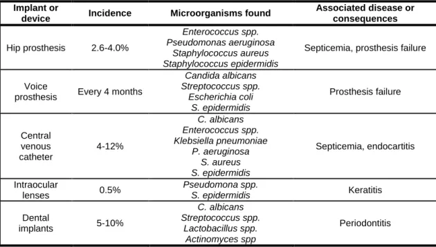
Table 1.1| Incidence and microorganisms most commonly isolated from infections on different biomedical
implants and devices. Adapted from Roosjen et al. 2006, Maathuis et al. 2007.
Implant or
device Incidence Microorganisms found
Associated disease or consequences Hip prosthesis 2.6-4.0% Enterococcus spp. Pseudomonas aeruginosa Staphylococcus aureus Staphylococcus epidermidis
Septicemia, prosthesis failure
Voice
prosthesis Every 4 months
Candida albicans Streptococcus spp. Escherichia coli S. epidermidis Prosthesis failure Central venous catheter 4-12% C. albicans Enterococcus spp. Klebsiella pneumoniae P. aeruginosa S. aureus S. epidermidis Septicemia, endocartitis Intraocular lenses 0.5% Pseudomona spp. S. epidermidis Keratitis Dental implants 5-10% C. albicans Streptococcus spp. Lactobacillus spp. Actinomyces spp Periodontitis
4 CHAPTER 1
GENERAL INTRODUCTION
Influence of a Quaternary Ammonium Compound on the Cell Structure of Bacteria using AFM
The occurrence of BAI is determined by the probability of microorganisms to reach the biomaterial surface. Biomaterials in contact with the outer part of the body, such as contact lenses and intravenous catheters, are colonized as soon as they are placed on tissue surfaces exhibiting therefore a higher incidence of BAI than fully implanted biomaterials, such as hip or knee implants (0.5% - 100% versus 0.1% - 7%) (Gottenbos
et al. 2000). Microorganisms can reach a biomaterial implant in several ways and at
different times post-implantation (Subbiahdoss et al. 2009). The most common route of infection is the direct contamination of the biomaterial implant during its insertion (intra-operative contamination) by microorganisms inevitably present in the operating theatre or microorganisms from the skin commensal microflora (Davis et al. 1999). Contamination can also occur during hospitalization (post-operative contamination) when the infected material contacts with the wound or after using invasive devices like catheters. Since microorganisms have the ability to stay at a low metabolic state on a biomaterial surface (Gottenbos et al. 2000) inside the human body for several years, they can be responsible for the occurrence of an infection (BAI) years after its insertion. A third possible route of infection, but less likely to occur, is late haematogenous contamination when bacteria from local infections elsewhere in the body are spread through blood.
Whenever microorganisms reach the biomaterials surface, initial microbial adhesion which is mediated by physicochemical properties of both bacterium and biomaterial surface can occur. Once adhered, the microorganisms are protected against phagocytosis, as the microorganism and biomaterial together are too large to ingest. After adhesion, most microorganisms start secreting slime and embed themselves in a slime layer, the glycocalix, which is an important virulence factor for BAI (Branda et al. 2005). The complex structure of a biofilm on current biomaterials makes it resistant against antibiotic treatment and host immune system (Prakash et al. 2003). Even high concentrations of antibiotics have been reported to fail in eradicating mature biofilms (Anwar et al. 1992). As a consequence, the only solution to an infected implant device is often its surgical removal at the expense of considerable costs and patient‟s suffering (Francolini and Donelli 2010). The best approach to overcome this problem is to prevent biofilm formation on the biomaterial surface. In order to accomplish this purpose, a better understanding of mechanisms underlying the biofilm development is required.
Influence of a Quaternary Ammonium Compound on the Cell Structure of Bacteria using AFM
1
1
.
.
1
1
.
.
1
1
|
|
B
B
IIOOFFIILLMMSS:
:
AA SSUURRVVIIVVAALL SSTTRRATATEEGGYY OOFF MMIICCRROOOORRGGAANNIISSMMSSIn the history of Microbiology, microorganisms have been primarily characterized as unicellular life forms, living as planktonic, free-suspended cells (Donlan 2002), and their description on the basis of their growth characteristics in nutritionally rich culture medium provided important insights about microbial pathogenesis and physiology (Davey and O‟Toole 2000). However, it is now widely recognized that microorganisms have a marked and natural tendency to interact with surfaces and interfaces, where they are found in the form of multicellular aggregates enclosed in a matrix of primarily polysaccharide material, commonly referred as biofilms (Flemming and Wingender 2010).
The discovery of microbial biofilms can be attributed to Antoni van Leeuwenhoek, who first observed microorganisms on tooth surfaces using his simple microscopes, in the 17th century. Afterwards, Hukelekian and Heller (1940) and Zobell (1943) demonstrated that bacterial growth and activity were substantially enhanced by the incorporation of a surface to which these organisms could attach, and the number of bacteria on surfaces was dramatically higher than in the surrounding medium (seawater). The study of Characklis (1973) about microbial slimes in industrial water systems showed that they were not only very tenacious, but also highly resistant to disinfectants such as chlorine. However, it was Costertan et al. (1978) that postulated a theory of biofilms explaining the mechanisms by which microorganisms adhere to surfaces and the benefits derived by this mode of growth.
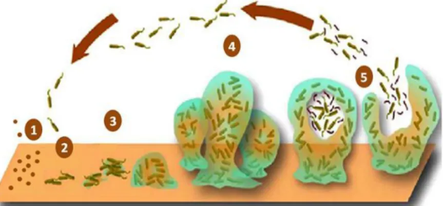
The development of a biofilm is characterized by a series of complex and well-regulated steps (Figure 1.1): adsorption of a conditioning film (1), transport of microbial cells towards the surface followed by reversible adhesion (2), irreversible adhesion (3), biofilm maturation (4) and detachment of individual bacteria or aggregates (5) (Dunne, 2002).
Figure 1.1| Schematic representation of the steps involved in biofilm formation on a surface: conditioning
film formation (1), reversible attachment (2), irreversible attachment (3), maturation (4) and detachment (5). Adapted from Stoodley and Dirckz 2003.
6 CHAPTER 1
GENERAL INTRODUCTION
Influence of a Quaternary Ammonium Compound on the Cell Structure of Bacteria using AFM
Prior to biofilm formation, the surface is first covered with a layer of proteins and glycoproteins such as fibronectin, vitronectin, fibrinogen, albumin and immunoglobulins, which are present in the surrounding aqueous environment. Biofilm formation starts with the transport of bacteria to the surface-liquid interface, which is governed by a combination of transport mechanisms, including Brownian motion, gravity, diffusion, convection or the intrinsic motility of a microorganism (Roosjen et al. 2006). The bacterium approaches the surface so closely that its motility is slowed, and it forms a reversible association with the surface and/or other microbes previously attached to the surface (Prakash et al. 2003). In this process non-specific interactions which are governed by physicochemical properties are involved, such as surface charge, hydrophobicity and chemical structure of both bacterium and surface. Reversible adhesion of bacteria changes afterwards to irreversible, since the attachment of adhering microorganisms is strengthened through extracellular polymeric substances (EPS) production, unfolding of cell structures and protein-protein interactions. EPS are biopolymers that form hydrogels with water and provide a stable structure to the biofilm. Most of these biopolymers are polysaccharides consisting of sugars such as glucose, galactose, mannose and fructose, but also traces of proteins, lipids and nucleic acids are present. Adhering bacteria grow and divide, forming microcolonies that are considered to be the basic organizational units of a biofilm. Entrapment of other planktonic bacteria in the EPS also occurs, resulting in a multi-layered and mature biofilm. The last step is the detachment of individual bacteria or aggregates caused by occasionally high fluid shear or other detachment forces operative, which enables bacteria to disseminate into other areas for further surface colonization. In the clinical setting, this last step usually leads to severe systemic infections (Katsikogianni and Missirlis 2004).
The final structure and composition of the biofilm are determined by the characteristics of the system where it was developed. Factors such as the type of microorganisms, the hydrodynamic environment, surface roughness, nutrients available, attraction and adhesion to other microorganisms from the surrounding environment regulate biofilm formation (Costerton et al. 1987, Hall-Stoodley et al. 2004). Biofilm formation is an important survival strategy for bacterial cells. In Nature, more than 99% of bacteria exist as biofilms. The ubiquity of these structures on several and different ecosystems demonstrates the strong survival and selective advantage of sessile communities over planktonic cells (Dunne 2002). In fact, it is estimated that bacterial cells growing as biofilms are up to 1000-fold more resistant to antibiotics and can cope much better with unfavourable external conditions, as the host immune system, than their planktonic counterparts (Falagas et al. 2009). This resistance can be attributed to a number of
Influence of a Quaternary Ammonium Compound on the Cell Structure of Bacteria using AFM
factors observed in biofilm populations, including restricted penetration, decreased growth rate, a distinct genetic phenotype (Handke et al. 2004, Harrison et al. 2004, Schierholtz and Beuth 2001), the expression of resistance genes (Maira-Litran et al. 2000) and the presence of biofilm persistent cells (Roberts and Stewart 2005).
1
1
.
.
1
1
.
.
2
2
|
|
C
C
OAOAGGUULLAASSEE-
-
NENEGGAATTIIVVEE SSTTAAPPHHYYLLOOCCOOCCCCII AASS NNOOSSOOCCOOMMIIAALL PPAATTHHOOGGEENNSSStaphylococci are Gram-positive bacteria belonging to the family Staphylococcaceae characterized by round cells (coccus or spheroid shaped), with about 1 m in diameter and found as single cells, in pairs or, more frequently, in clusters that resemble clusters of grapes (Huebner and Goldmann 1999, Fey and Olson 2010). They are clustering, non-motile and non-spore forming cocci, and facultative anaerobes that produce catalase. Although more than 30 species of the genus Staphylococcus have been described, S. aureus and S. epidermidis are the most significant in their interactions with humans (Gotz 2002). Staphylococci are divided into positive and coagulase-negative strains depending on its ability or inability to clot blood plasma. Coagulase-negative staphylococci (CNC) have been regarded as harmless skin commensals, but during the past few decades their importance as human (predominantly nosocomial) pathogens has been recognized (Vuong and Otto 2002). From 1990 to 1995, the National Nosocomial Infection Surveillance (NNIS) program reported that CNS were responsible for 11% of all nosocomial infections reported, making this pathogen the third most common nosocomial isolate (Huebner and Goldmann 1999).
S. epidermidis is currently the most significant member of the CNS and constitutes the
most widespread and persistent species found on the human skin and mucous membranes, representing an important part of its normal microflora (comprises 65% of the 90% of all staphylococci isolated from these environments) (Vuong and Otto 2002, Otto 2009). In recent years, S. epidermidis emerged as one of the most important and frequently causes of nosocomial infection, mainly associated with implanted medical devices (Rupp and Archer 1994). In fact, in what concerns BAI, nearly 80% of the cells involved are S. epidermidis (Gotz 2002). The indwelling medical devices mostly colonized by S. epidermidis include central venous catheters, cerebrospinal fluid shunts, prosthetic heart valves, ocular lenses implants, prosthetic joints, dialysis devices, hip prostheses and many other invasive biomaterials (Donlan, 2001).
Numerous studies have clearly reported the ability of S. epidermidis to form a thick adherent, multi-layered biofilm, strongly resistant to antibiotic treatment which is considered the most important virulence factor involved in its pathogenesis. Biofilm
8 CHAPTER 1
GENERAL INTRODUCTION
Influence of a Quaternary Ammonium Compound on the Cell Structure of Bacteria using AFM
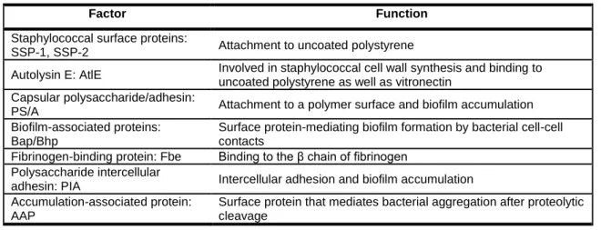
formation by staphylococci proceeds in two fundamental steps, namely attachment of bacterial cells to the biomaterial surface and cells accumulation to form multilayered cell clusters enclosed in an exopolimeric matrix (Figure 1.2).
Figure 1.2| Schematic representation of the phases involved in Staphylococcus epidermidis biofilm
formation and bacterial factors involved. Taken from von Eiff et al. 2002.
The first step is influenced by numerous factors, such as hydrophobicity and surface charge (Ziebuhr et al. 2006), as well as by means of cell wall teichoid acids and proteins that interfere with matrix proteins, like collagen and fibronectin (McCann et al. 2008). S.
epidermidis adhesion to a foreign body surface is followed by biofilm accumulation which
requires the production of factors that mediate intercellular adhesion, such as polysaccharide intercellular adhesion (PIA). PIA is a homoglycan composed of β-1,6-linked 2-amino-2-deoxy-D-glucopyranosyl residues, containing positive charged amino groups as well as negative charges (Rohde et al. 2010). Table 1.2 summarizes some of the factors involved in the biofilm formation by staphylococci.
Table 1.2| The major factors contributing to Staphylococcus epidermidis biofilm formation and their function.
Adapted from von Eiff et al. 2002 and Ziebuhr et al. 2006.
Factor Function
Staphylococcal surface proteins:
SSP-1, SSP-2 Attachment to uncoated polystyrene
Autolysin E: AtlE Involved in staphylococcal cell wall synthesis and binding to uncoated polystyrene as well as vitronectin
Capsular polysaccharide/adhesin:
PS/A Attachment to a polymer surface and biofilm accumulation Biofilm-associated proteins:
Bap/Bhp
Surface protein-mediating biofilm formation by bacterial cell-cell contacts
Fibrinogen-binding protein: Fbe Binding to the β chain of fibrinogen Polysaccharide intercellular
adhesin: PIA Intercellular adhesion and biofilm accumulation Accumulation-associated protein:
AAP
Surface protein that mediates bacterial aggregation after proteolytic cleavage
Influence of a Quaternary Ammonium Compound on the Cell Structure of Bacteria using AFM
1
1
.
.
2
2
|
|
T
T
R
R
E
E
A
A
T
T
M
M
E
E
N
N
T
T
A
A
N
N
D
D
P
P
R
R
E
E
V
V
E
E
N
N
T
T
I
I
O
O
N
N
O
O
F
F
B
B
I
I
O
O
M
M
A
A
T
T
E
E
R
R
I
I
A
A
L
L
A
A
S
S
S
S
O
O
C
C
I
I
A
A
T
T
E
E
D
D
I
I
N
N
F
F
E
E
C
C
T
T
I
I
O
O
N
N
S
S
Whenever a BAI is detected there are two possible modes of action, namely the removal of the device and/or the initialisation of an antimicrobial treatment. The therapy of choice, especially due to the easiness to change devices such as short term peripheral catheters, is the removal of the infected device and its replacement if still needed (von Eiff et al. 2005). However, removing the infected device is not always possible, easy to perform and/or without risk. Therefore, the recovery of the device is sometimes the preferred option. Once a BAI is established it is very difficult to treat it as the minimal inhibitory concentration (MIC) of antimicrobial agents, necessary to kill microorganisms, is significantly higher for microorganisms in a biofilm as compared to their planktonic counterparts (Costerton et al. 1999). Although much research has been done to turn biofilms more susceptible to antimicrobial agents, the best approach to overcome this issue is to prevent biofilm formation (Simões et al. 2010).
Over the past years several strategies have been used to prevent bacterial adhesion and biofilm formation on biomaterials. In general, these strategies are based in the reduction of the attractive force between microorganisms and a biomaterial surface by optimizing the physicochemical surface properties of the biomaterial (Kazmierska and Ciach 2009). For instance, extremely hydrophobic surfaces (Everaert et al. 1999), more negatively charged biomaterials (Hogt et al. 1986), biomaterials coated with albumin or heparin (El-Asrar et al. 1997), have shown to attract fewer bacteria. Nevertheless, as biomaterial surfaces are often covered with a conditioning film consisting of proteins that can be anchors for microorganisms to adhere to, another approach to prevent biofilm formation can include the application of antimicrobial agents near the biomaterial surface in order to prevent the growth of adhering microorganisms. Gentamicin-loaded bone cements and silver-loaded catheters (Carlsson et al. 1978) are two examples in which this approach was implemented. Some drawbacks of such applications are their lifetime (few days to weeks), the limited amount of antibiotic released and the development of antibiotic resistant microbial strains (Gottenbos et al. 2002). A better approach to prevent BAI is to render the biomaterial surface antimicrobial properties by functionalizing it with antimicrobial agents covalently attached (Vasilev et al. 2009). This strategy can only be employed with antimicrobial agents working at the level of the cell wall or membrane, since they can only reach the outside of the microbial cells. Quaternary ammonium compounds (QACs) work at the membrane level, and are one of the few known antibacterial molecules that retain their bactericidal properties when covalently bound to a surface (Flemming et al. 2000, Kenawy et al. 1998). In this approach, no antimicrobial
10 CHAPTER 1
GENERAL INTRODUCTION
Influence of a Quaternary Ammonium Compound on the Cell Structure of Bacteria using AFM
agents are leaching from the surface, providing long term protection against bacterial colonization, and reducing the risk of developing antimicrobial resistant microbial strains, since the concentration of antimicrobial groups is constantly above the MIC.
1
1
.
.
2
2
.
.
1
1
|
|
T
T
HEHE RROOLLEE OOFFQ
Q
UAUATTEERRNNAARRYYA
A
MMMMOONNIIUUMMC
C
OOMMPPOOUUNNDDSS OONN PPRREEVVEENNTTIINNGGB
B
A
A
I
I
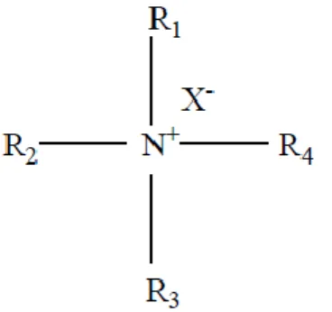
QACs constitute a family of surfactants that have been widely used in domestic, agricultural, healthcare, and industrial applications (Garcia et al. 1999, Patrauchan and Oriel 2003). Surfactant is an abbreviation for surface active agent that literally means active at a surface, and is characterized by its ability of lowering surface and interfacial tensions of liquids, which comprise the ability to wet surfaces, penetrate soil and solubilise fatty materials (Christofi and Ivshina 2002, Pereira et al. 2007). These agents are composed by two distinct structural elements: a hydrophobic (water repellent) group and a hydrophilic (water attractive) group (Figure 1.3).
Figure 1.3| General chemical structure of a surfactant (R represents a functional group and X- represents a counter ion such as Cl-, Br- or NO3
-).
Depending on the basis of the charge or absence of ionization of the hydrophilic group, surfactants can be classified into cationic, anionic, non-ionic, and ampholytic or amphoteric compounds. Of these, the cationic agents, as exemplified by QACs, are the most useful antiseptics and disinfectants. In addition to their detergent properties, QACs also present remarkable antimicrobial properties being used for a variety of clinical purposes, such as pre-operative disinfection of undamaged skin, application to mucous membranes and disinfection of noncritical surfaces (Simões et al. 2005b).
Influence of a Quaternary Ammonium Compound on the Cell Structure of Bacteria using AFM
1
1
.
.
2
2
.
.
1
1
.
.
1
1
|
|
Q
Q
A
A
C
C
’
’
SS MMEECCHHAANNIISSMMSS OOFFA
A
CTCTIIOONNAlthough a wide variety of studies on the interaction between microbial cell surface and QACs have been reported (Ioannou et al. 2007, Marcotte et al. 2005), the details of their mechanism of action are not completely understood. Two hypotheses have been reported to explain the susceptibility of a broad range of species to these compounds. The most quoted theory hypothesizes that sufficiently long cationic polymers penetrate cells and thereby disrupt the membrane like a needle bursting a balloon (Milovic et al. 2005). Several observations indicate that QACs, given their cationic nature, act at the membrane level which is negatively charged at physiological pH and thus, for many decades, such compounds have been designated as membrane active agents. When microorganisms are exposed to cationic agents the following sequence of events occur (Ikeda et al. 1984): (i) adsorption and penetration of the agent into the cell wall; (ii) reaction with the cytoplasmic membrane followed by membrane disruption; (iii) leakage of intracellular low-molecular weight material; (iv) degradation of proteins and nucleic acids and (v) cell wall lysis caused by autolytic enzymes. At a molecular level, QAC‟s action involves an association of the positively charged quaternary nitrogen with the head groups of acidic-phospholipids within the membrane (Figure 1.4 a, b), and the hydrophobic tail is then inserted into the hydrophobic membrane core (Figure 1.4 b, c). At QAC‟s low concentrations, such interaction is responsible for the increase of surface pressure with a subsequent decrease of membrane fluidity and increase of phase transition temperature. As a consequence, the membrane undergoes a transition from solid to liquid crystalline state losing many of its physiological functions (Figure 1.4 d). For higher concentrations, QACs form micellar aggregates that solubilise hydrophobic membrane compounds, such as lipid A and phospholipids (Figure 1.4 e, f) (Gilbert and Moore 2005).
12 CHAPTER 1
GENERAL INTRODUCTION
Influence of a Quaternary Ammonium Compound on the Cell Structure of Bacteria using AFM
Figure 1.4| Schematic representation showing the mechanism of action for QACs. Progressive adsorption of
the quaternary head group to acidic phospholipids in the membrane with increasing QAC exposure/concentration leads to decreased fluidity of the bilayers and the creation of hydrophilic voids in the membrane. Taken from Gilbert and Moore 2005.
The second hypothesis, proposed by Kugler et al. (2005), states that a highly charged surface can induce what is essentially an ion exchange between the positive charges on the surface and structurally critical mobile cations within the membrane, such as calcium and magnesium. When approaching to a cationic surface, divalent cations are free to diffuse out of the membrane without performing their role in charge neutralization of the membrane components, which results in a loss of membrane integrity. An attempt to study the contribution of both hypotheses was performed by Murata and co-workers (2007). QACs were used to prepare antimicrobial polymer brushes on inorganic surfaces. By variation of the chain length containing the quaternary ammonium group and the surface density in a gradient manner, the authors could conclude that the density of surface quaternary ammonium groups, and thus the density of cationic surface charges, is a key parameter. However, the authors did not exclude that membrane insertion of alkyl chains may also be a possible mechanism of action.
Influence of a Quaternary Ammonium Compound on the Cell Structure of Bacteria using AFM
1
1
.
.
2
2
.
.
1
1
.
.
2
2
|
|
A
A
NTNTIIMMIICCRROOBBIIAALL AANNDD AANNTTII-
-
ADADHHEESSIIVVEE AACCTTIIVVIITTIIEESS OOFFQ
Q
A
A
C
C
S SQACs are organic compounds that contain four functional groups attached covalently to a central nitrogen atom (Figure 1.3). These functional groups (R) include at least one long chain alkyl group, and the rest are either methyl or benzyl groups. Their antimicrobial activity depends on their structure and size, but especially on the length of the long-chain alkyl group. In fact, it was reported that there is a parabolic relationship between their antibacterial properties and their hydrophobic character, which means that there is a linear relationship between activity and alkyl chain length with increased carbon number up to a maximum of between 12 an 14, at which region could be observed a decrease in activity (Tomlinson et al. 1977).
The antimicrobial activity of several QACs against Gram-positive and Gram-negative bacteria in planktonic cultures, as well as in biofilms, has been reported. For instance, the cationic surfactant cetyltrimethylammonium bromide (CTAB) was investigated by Simões et al. (2005a) for its ability to control mature Pseudomonas fluorescens biofilms formed under laminar and turbulent flow in flow cell reactors. The authors found that CTAB by itself did not cause the detachment of biofilms, but it reduced the respiratory activity of the biofilm cells. Total respiratory inactivation was not achieved and, in almost all the cases studied, it was observed that the biofilm respiratory activity was recovered over time. However, the same authors, in another study (2005c) reported that the synergistic action of CTAB with the application of high shear stress to mature biofilms (formed in a rotating device) increased its detachment.
In the last decade, continuous effort has been made to develop polymers with antimicrobial properties because polymeric antimicrobials comprise the following advantages: are non volatile, chemically stable, have long-term antimicrobial activity and are hard to permeate through the skin. There are several studies reporting the efficacy of polymers containing quaternary ammonium salt groups against both Gram-positive and Gram-negative bacteria. Kenawy et al. (1998) concluded that quaternary ammonium and phosphonium copolymers based on the modified poly(glycildyl methacrylate-co-hydroxyethyl methacrylate) materials could be promising candidates for preventing BAI, after evaluating their antimicrobial activity in vitro against Gram-positive (Bacillus subtilis and Bacillus cereus) and Gram-negative (E. coli, P. aeruginosa, Shigella sp. and
Salmonella typhae) bacteria.
QACs have also been found to inhibit the adhesion of pathogenic organisms to solid surfaces, thus prior adhesion of these compounds to the surfaces might constitute a new and effective means of avoiding colonization by pathogenic microorganisms. Such
14 CHAPTER 1
GENERAL INTRODUCTION
Influence of a Quaternary Ammonium Compound on the Cell Structure of Bacteria using AFM
approach was implemented by Tiller et al. (2001) that covalently attached by different methods long chains of N-alkylated poly (4-vinylpyridine) (PVP) to a glass slide. The resultant glass slides were able to kill by contact several airborne Gram-negative and Gram-positive bacteria. Such surface modifications can be readily performed with a number of other materials, thus making this approach useful for the coating of various medical devices. The same research group (Lin et al. 2002) reported the efficiency of these functionalized surfaces against wild-type and mutant, including antibiotic-resistance, strains of the ubiquitous pathogenic bacterium S. aureus. These observations support the assertion of a possible role in preventing microbial adhesion and their potential in developing anti-adhesive biological coatings for implant materials.
Silicone rubber with covalently coupled 3-(trimethoxylsilyl)-propyldimethyloctadecylammonium chloride (a quaternary ammonium silane, QAS) showed antimicrobial properties against adhering bacteria, both in vitro and in vivo (Gottenbos et al. 2002). In another study, reported by Oosterhof et al. (2006), two QAS were used to coat silicone rubber tracheoesophageal shunt prostheses in order to evaluate their inhibitory effects against the development of a mixed fungal and bacterial biofilm. This study reported for the first time that both yeasts and bacteria in mixed biofilms are affected by QAS coatings on silicone rubber. Because QAS coatings are non toxic, its clinical use could increase the lifetime of tracheoesophageal shunt prostheses. However, it is important to notice that the relevance of the current findings also extends to all biomedical and environmental applications where mixed biofilms can occur and be detrimental.
1
1
.
.
3
3
|
|
M
M
I
I
C
C
R
R
O
O
S
S
C
C
O
O
P
P
Y
Y
T
T
E
E
C
C
H
H
N
N
I
I
Q
Q
U
U
E
E
S
S
T
T
O
O
S
S
T
T
U
U
D
D
Y
Y
T
T
H
H
E
E
A
A
N
N
T
T
I
I
M
M
I
I
C
C
R
R
O
O
B
B
I
I
A
A
L
L
E
E
F
F
F
F
E
E
C
C
T
T
S
S
Understanding the interactions between bacterial cells and antimicrobial compounds is crucial to develop solutions that envisage the prevention of infections associated to biomaterials. Over the past, several techniques have been taken into consideration to study the antimicrobial effects of different compounds (Katsikogianni, 2004). Among them, microscopy-based methodologies (Table 1.3) have proved to be particularly useful since visualization of the changes undergone by microorganisms when subjected to antimicrobial treatment can provide new and important insights about the agents‟ mechanism of action (Hannig et al. 2010, Torrent et al. 2010).
Influence of a Quaternary Ammonium Compound on the Cell Structure of Bacteria using AFM
Table 1.3| Comparison between the most commonly used techniques used to visualize the effect of antimicrobials on bacteria.
Technique Description Advantages Disadvantages Applications References
Optical Microscopy
Visible light is used as a source of illumination. A system of optical lenses is used to magnify the sample.
Measurements performed in liquid, air or vacuum.
Resolution limited by the wavelength of source light.
Evaluation of early stages of oral Streptococci biofilm growth and the effect of antimicrobial agents. (Cortizo and Lorenzo 2007) Electron Microscopy (SEM and TEM) A beam of high-energy electrons is used as a source of illumination. A system of electromagnetic lenses is used to magnify the sample.
High-resolution.
Higher magnification and larger depth of focus.
Very time consuming and complex technique.
Sample preparation procedure requires freeze drying and gold sputtering.
Measurements performed under vacuum.
Time consuming sample preparation.
Evaluation of different bacterial species not possible.
TEM study of antibiotic action on Klebsiella pneumonia biofilm. SEM study on the effect of doxycylcine and vancomycin on enterococcal induced biofilm.
(Zahller and Stewart 2002, Somayaji et al. 2010) Confocal Laser Scanning Microscopy (CLSM)
A laser beam passes through a light source aperture and then is focused by an objective lens into a small focal volume within or on the surface of the sample.
Visualization of cells in situ. Measurements performed in liquid, air or vacuum.
Ability to control depth of field.
Limited number of excitation wavelengths with common lasers. Harmful nature of high-intensity laser irradiation on living cells and tissues.
Assess the bactericidal effect of chlorhexidine on dental biofilm. (Zaura-Arite et al. 2001) Atomic Force Microscopy (AFM)
A sharp tip scans over the surface of a sample while senses the
interaction between them.
Measurements performed in liquid, air or vacuum.
High resolution (up-to sub-nanometer).
Minimal sample preparation. Visualization of cells in real time.
Long image acquisition time.
Action of the antimicrobial peptide CM15 on individual Escherichia coli cells.
(Fantner et al. 2010)
16 CHAPTER 1
GENERAL INTRODUCTION
Influence of a Quaternary Ammonium Compound on the Cell Structure of Bacteria using AFM
The atomic force microscopy (AFM) belongs to the broad family of scanning probe microscopes (SPM), in which a probe is used for investigating surface topography and properties with sub-nanometer resolution. This technique, initially developed in 1986 by Binning, Quate and Gerber to overcome the limitations of its ancestor (scanning tunnelling microscope, STM) in imaging non-conducting samples, immediately attracted the attention of the biophysical community (Santos and Castanho 2004). In biological applications, the most appealing advantage of the AFM when compared with other techniques such as SEM and TEM is that it allows the characterization of biological samples regarding their structure and mechanical properties under physiological conditions in real-time with sub-nanometer resolution, avoiding complex sample preparation procedures and the associated artefacts. Since its invention, the significant improvements obtained both at the instrumental and sample preparation levels, as well as recording conditions, led to a revolution of the way in which biologists explore microbial surfaces (Dufrêne 2002).
1
1
.
.
3
3
.
.
1
1
|
|
B
B
AASSIICC PPRRIINNCCIIPPLLEESS OOFFA
A
F
F
M
M
AANNDDA
A
F
F
M
M
IIMMAAGGIINNGG MMOODDEESSThe basis of AFM is the measurement of the interaction between the sample surface and a tip located very close to it (Figure 1.5). The sample is mounted on a piezoelectric scanner which can move precisely in three dimensions and the tip is assembled under an extremely flexible cantilever (Liu and Wang 2010). The cantilever can be simple or triangular, usually between 23-300 µm length, 10-30 µm width and 0.5-3 µm thick (Santos and Castanho 2004). The most commonly used are made of silicon nitride (Si3N4), with spring constants raging from 0.01 to 100 N/m. Any interaction between the
tip and the sample leads to a bending of the cantilever that will be proportional to the interaction force. To detect this bending, AFM uses a small laser focused on the back of the cantilever. The reflection of the laser beam is focused on a photo-detector that measures any minimal bending of the cantilever, and thus the interaction of the tip with the sample (Rajasekaran 2008, Butt et al. 2005, Reich et al. 2001).