CHARACTERIZATION OF ENDOPHYTIC Fusarium
SPECIES FROM COWPEA SEEDS
ANTÔNIA ALICE COSTA RODRIGUES1 MARIA MENEZES2
1Universidade Federal Rural de Pernambuco, Recife, Pernambuco. 2Academia Pernambucana de Ciência Agronômica, Recife, Pernambuco.
_______________ ABSTRACT
IDENTIFICATION AND PATHOGENIC
CHARACTERIZATION OF ENDOPHYTIC Fusarium SPECIES FROM COWPEA SEEDS
Isolates of Fusarium were obtained and identified from seeds of cowpea, Vigna unguiculata (L.) Walp., by means of blotter tests and slide cultures. Species were differentiated according to the morphology of the macroconidia, microconidia and their arrangement in chains or false heads, the size and type of conidiophore, and the presence or absence of chlamydospores. The species were identified as F. semitectum, F. equiseti, F. oxysporum, F. solani, F. anthophilum, F. sporotrichioides, F. moniliforme, and Fusarium sp. Among the species, F. semitectum was the most frequently detected. None of these species were pathogenic when inoculated in susceptible cowpea cultivar (BR 17–Gurgueia). But, an isolate of F. oxysporum f. sp. tracheiphilum used as a standard of comparison for pathogenicity (control) induced symptoms of yellowing, vascular wilting, and death of a susceptible cowpea cultivar under the same environmental conditions.
Index terms: cowpea cultivars, Fusarium, seed, Vigna unguiculata.
RESUMO
IDENTIFICAÇÃO E CARACTERIZAÇÃO PATOGÊNICA DE ESPÉCIES ENDOFÍTICAS DE Fusarium DE SEMENTES DE
CAUPI
Isolados de Fusarium foram obtidos e identificados de sementes de caupi, Vigna unguiculata (L.) Walp., pelos métodos do papel de filtro e culturas em lâminas. As espécies foram diferenciadas de acordo com a morfologia dos
macroconídios, microconídios e disposição destes em cadeias ou em “falsas cabeças”, tamanho e tipo de conidióforo, bem como da presença ou ausência de clamidósporos. As espécies identificadas foram F. semitectum, F. equiseti, F. oxysporum, F. solani, F. anthophilum, F. sporotrichioides, F. moniliforme e Fusarium sp. Dentre as espécies, F. semitectum foi a mais freqüentemente detectada. Nenhuma dessas espécies mostrou-se patogênica quando inoculadas em plantas susceptíveis de caupi (cultivar BR17-Gurgueia). Entretanto, um isolado de F. oxysporum f. sp. tracheiphilum usado como padrão para comparação de patogenicidade (controle) induziu sintomas de amarelecimento, murcha vascular e morte de plantas da cultivar de caupi susceptível, submetidas as mesmas condições ambientais.
Termos para indexação: cultivares de caupi, Fusarium, sementes, Vigna
unguiculata.
1. I
NTRODUCTIONThe genus Fusarium Link comprises inhabitants of the soil and of organic substrata and is widely distributed throughout the world (Burgess, 1981). Like many soil fungi, this genus is endowed with several means of survival, amongst which is its quick capacity for change, both morphological and physiological, when faced with environmental changes (Booth, 1971). There are pathogenic and non–pathogenic forms. The latter can colonize the cortex of roots of plants without causing symptoms of disease (Appel & Gordon, 1994), and survive in live tissue, as also exercising antagonism between the pathogenic forms in the soil (Edel et al., 1997). The phytopathogenic species affect a wide range of hosts and cause root rot, vascular wilting, yellowing and foliar necrosis (Ramachandran et al., 1982; Nelson & Hansen, 1997).
Many pathogenic isolates are included in the F. oxysporum Schlechtend.: Fr. These can be subdivided into formae speciales, characterized by their ability to cause diseases in specific hosts, and in race, according to their reaction with a group of differentiating cultivars (Gordon & Martyn, 1997).
In the culture of cowpea, one of the main diseases is Fusarium Wilt, caused by F.
oxysporum Schl. f. sp. tracheiphilum (E. F. Smith). The symptoms in the plants begin
with a slight change in the colouring of the older leaves, from green to yellow, on one of the sides of the leaves or of the plant. When the infection progresses, defoliation occurs as does the darkening of the vessels and later death of the plants (Kendrick, 1931; Kendrick & Snyder, 1942). The disease is responsible for losses of
production in the areas where this legume is grown in Brazil. In Nigeria and the United States, plant mortality can reach levels above 50% (Pio–Ribeiro & Assis Filho, 1997). The pathogen can also be transmited by cowpea seeds, important vehicle for dissemination.
Currently, three races of F. oxysporum f. sp. tracheiphilum are known: Races 1 and 2 were described in South Carolina. Race 1, in addition to cowpea also attacks soybean [Glycine max (L.) Merr.] and chrysanthemum (Chrysanthemum morifolium Ramat), according to Armstrong & Armstrong (1950; 1965), and Race 3 was described in Mississippi (Hare, 1953).
Just as in the soil, the presence of Fusarium in cowpea seeds does not necessarily result in transmission of disease. The fungus can remain endophytic in the seeds as dormant mycelium or chlamydospores without causing disease (Menezes, 1988). According to Champion (1997), many fungi considered to be saprophytes, pathogens or others that lost their ability to cause disease, can survive in latency inside of the seeds, becoming active when these seeds germinate. The pathogenic forms may result in pre– or postemergence damping–off. This, in turns, results in a poor plant stand in the field (Agarwal & Sinclair, 1997).
The detection of seedborne fungi can be easily done using the blotter test which permits mycelium growth and formation of fruiting bodies on the seed surface (Neergaard, 1979; Maude, 1996), becoming possible the identification of fungal species by morphology. The inclusion of a pathogenicity test is important to seed health diagnostic.
The economic importance of cowpea to Northeastern Brazil, and the frequent presence of Fusarium spp., has stimulated our interest to know which species occur in cowpea seeds and their role as possible agents of plant disease, under conditions of Pernambuco State. Then, the purpose of this study was to detect and identify the species of Fusarium endophytically associated with cowpea seeds, as well their pathogenicity when inoculated in plants of a susceptible cowpea cultivar. This study provides basic information on pathogenic and nonpathogenic Fusarium species as a contribution to programmes of seed quality control.
2. M
ATERIALSANDM
ETHODS2.1. Obtaining and identifying the isolates
The Fusarium isolates were obtained from the internal part of seeds of cowpea cultivars from two counties (Serra Talhada and Caruaru) of Pernambuco State, Brazil. A total of 4,000 seeds, distributed in samples of 400 seeds per cultivar, were analysed using the blotter test (Neergaard, 1979), with prior surface sterilization in a solution of sodium hypochlorite with 1.5% of active chlorine. After the fungi grow on the seeds, fragments of mycelium were transferred to potato–dextrose–agar medium (Windews, 1993) to facilitate the morphological analysis of the colonies.
To identify the species of Fusarium, the technique of slide culture was used (Tuite, 1969) which allows the direct microscopic observation of morphological structures of taxonomic value. The technique consisted of inoculating a bit of fungus at the sides of a small cube of agar (1 cm2) maintained in the center or extremity of
a slide and covered with a glass cover. The slide cultures were kept on a support to avoid direct contact with the humid base of the Petri dish. After incubating for 48 hours, the microcultures were examined in preparations with Amann blue.
2.2. Morphological differentiation of the species
The criteria used for identification of species were based on the form of macroconidia and microconidia produced in the aerial mycelium, on monophialides or polyphialides, as well as of macroconidia formed on sporodochium or microconidia produced in chains or in false heads, on monophialides and/or polyphialides. To differentiate the species, the following were also considered: the presence or absence of chlamydospores, and, in some cases, the size or the branching of the conidiophore. When necessary, the specialized literature used to make identifications included Snyder & Hansen (1940), Booth (1971; 1977), Gerlach & Nirenberg (1982), and Nelson et
The relative frequency (Rf) of the Fusarium species was evaluated using the formula: Rf = (n/N) × 100, where n = the number of colonies presented by each species; N = the total number of colonies of all species.
2.3. Pathogenicity of Fusarium species on cowpea – Hosts used
The pathogenicity tests were carried out in greenhouse conditions. The lineage L–288004 and the cultivar BR–17 Gurgueia from IPA (Empresa Pernambucana de Pesquisa Agropecuária) were used as standard cultivars for resistance and susceptibility to F. oxysporum f. sp. tracheiphilum, respectively. The experimental unit was represented by plastic pots, with a capacity for 2 kg, containing sterilized soil, with four plants per pot.
– Preparation of the inoculum and inoculation:
Isolates of eight species of Fusarium obtained from cowpea seeds, plus one standard pathogenic isolate of F. oxysporum f. sp. tracheiphilum from cowpea plant were used. Initially, the isolates were grown in Petri dishes, containing PDA, and later transferred to Erlenmeyer flasks containing 80 mL of Armstrong liquid medium (Armstrong & Armstrong, 1948), and incubated for five days under continuous light from two fluorescent lamps, with a luminous intensity of 2,400 lux, and at temperature of 27 oC, being the cultures stirred manually twice a day. After the
incubation period, the inoculum was homogenized in a mixer for two minutes, and the concentration of the suspension was adjusted in a Neubauer chamber to 1×10⁶
conidia per mL of sterile water. The inoculation of plants was made by 5–day old wounded roots, when 20 mL of the suspension were added per plant, in according to Menezes (1972). Seven days later, the second inoculation was carried out, using the same procedure.
The experimental design was completely randomized block with four replications for each treatment, represented by the Fusarium species and two cultivars (resistent
and susceptible). The pathogenicity of the isolates was evaluated 30 days after inoculation of the plants by presence or absence of symptoms conform the species involved.
3. R
ESULTSANDD
ISCUSSION3.1. Identification of Fusarium species
Out of a total of 211 isolates of Fusarium obtained from cowpea seeds, eight species were identified in the following relative frequency: F. semitectum (47.39%), F.
equiseti (22.27%), F. oxysporum (16.59%), F. solani (3.79%), F. anthophilum (2.37%), F. sporotrichioides (1.42%), F. moniliforme (0.95%), and Fusarium sp. (5.21%). (Table 1).
From the results, F. semitectum stands out from the others by having the greatest frequency and is followed by F. equiseti and F. oxysporum. The seeds of IPA–202 from
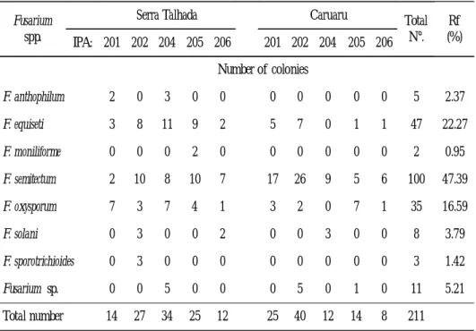
Table 1.
-
Endophytic Fusarium species from seeds of different cowpea cultivars grown in two counties of Pernambuco state, BrazilFusarium spp.
Serra Talhada Caruaru Total N°. Rf (%) IPA: 201 202 204 205 206 201 202 204 205 206 Number of colonies F. anthophilum 2 0 3 0 0 0 0 0 0 0 5 2.37 F. equiseti 3 8 11 9 2 5 7 0 1 1 47 22.27 F. moniliforme 0 0 0 2 0 0 0 0 0 0 2 0.95 F. semitectum 2 10 8 10 7 17 26 9 5 6 100 47.39 F. oxysporum 7 3 7 4 1 3 2 0 7 1 35 16.59 F. solani 0 3 0 0 2 0 0 3 0 0 8 3.79 F. sporotrichioides 0 3 0 0 0 0 0 0 0 0 3 1.42 Fusarium sp. 0 0 5 0 0 0 5 0 1 0 11 5.21 Total number 14 27 34 25 12 25 40 12 14 8 211
Caruaru county showed a greater number of colonies of F. semitectum in relation to the same cultivar from Serra Talhada. In this study, the species were identified under standardized conditions in order to avoid influence of the environment on the phenotypical expression. The species were distributed in the following Sections:
– Section Liseola
Fusarium anthophilum (A. Braun)Wollenweber (Figure 1)
Identification was based on microconidia of various types, such as fusoid, allantoid, oval, pear–shaped or globular, formed on monophialides and on polyphialides. Macroconidia, when present, are slightly curved or fusoid, with three to five septate, and a pedicellate basal cell. Chlamydospores absent. Colony on PDA is white or yellowish, violet–like colouring with purple on the back.
Fusarium moniliforme Sheldon (Figure 2)
Identification was based on the abundant presence of microconidia, unicellular, oval to club–shaped, formed in chains on monophialides. Macroconidia, when present, are equilaterally fusoid, fine wall and basal cell is foot–shaped. Chlamydospores absent. Colony on PDA, initially presents white aerial mycelium, becoming pale pink, or tinged with purple, salmon pink on undersurface.
– Section Gibbosum
Fusarium equiseti (Corda) Saccardo (Figure 3)
Identification was based on the morphology of macroconidia septate, falcate, with a distinctive curvature, and the foot–shaped basal cell, with the apical cell very elongated. Conidiophores are either branched or unbranched monophialides. Chlamydospores are produced in abundance, smooth or roughened walls, formed in clumps or chains. Colony on PDA develop rapidly with white aerial mycelium at first, becoming tan to brown as the culture ages. However, one of the isolates displayed totally white color, and thereby differed notably from the others.
– Section Elegans
Fusarium oxysporum Schlecht (Figure 4)
Identification was based on the microconidia produced on short monophialides as a false head, mostly unicellular, varying from oval–ellipsoid to cylindrical and from straight to curved. Macroconidia are also formed in abundance, with an attenuated apical cell and a pedicellate basal cell, generally with 3–5 septate, produced
in short branched or unbranched monophialides or sporodochia. Another striking characteristic observed was the constant presence of chlamydospores, with a smooth wall, the most formed singly, with intercalated or terminal location. Colony on PDA initially with white aerial mycelium, becoming salmon, with a tendency towards violet, and a purple back.
– Section Arthrosporiella
Fusarium semitectum Berk. & Rav. (Figure 5)
Identification was based on the presence of two types of macroconidia, the primary ones formed in aerial mycelium on conidiophores in polyphialides, from zero to five septate, slightly curved and without a pedicellate basal cell. The secondary ones, which are formed in sporodochia, displaying from 3–7 septate, slightly curved, with the basal cell slightly pedicellate or apiculate. Chlamydospores often globose, intercalary, single or in chains, with smooth walls. Colony on PDA display rapid growth, with dense aerialmycelium that is peach to brown colored. The back varies from tan to dark brown over time.
Fusarium sporotrichioides Sherb. (Figure 6)
Identification was based on the presence of microconidia, formed at intervals in branchings of the aerial mycelium on conidiogenic polyphialides cells, and displaying an ellipsoidal or ovoid shape, unicellular or bicellular. Macroconidia also on conidiophores formed in aerial mycelium or in sporodochia, which appear as the cultures ages, displaying a sickle–like or fusoid shape, without distinct pedicellate basal cells. Chlamydospores are formed singly, in chains or in clumps. Colony on PDA shows abundant aerial mycelium, fluffy and white, becoming pink or carmine red, and undersurface showing the same color.
– Sections Martiella and Ventricosum
Fusarium solani (Mart.) Saccardo (Figure 7)
Identification was based on microconidia formed from lateral long monophialides, narrowing at the apex, unicellular, oval or kidney–shaped. Macroconidia are generally cylindrical almost the entire length, with 3–5 septate. Chlamydospores are formed singly or in pairs, globular or oval, with a smooth or wrinkledwall. Colony on PDA has abundant aerial mycelium, cream surface or purple–coloured with undersurface showing a dark violet color or colorless.
Figure 1–7. — Fusarium species from cowpea seeds. 1. F.
anthophilum: a. Microconidia oval to globose; b. Microscopic aspects. 2. F. moniliforme: a. Microconidia formed in chain, oval to club–shaped, macroconidium slightly sickle–shaped with the basal cell in foot–shaped, b. Microscopic aspects. 3. F. equiseti: a. Macroconidium with elongated apical cell, and sporodochium, b. Microscopic aspects. 4. F. oxysporum: a. Macroconidium slighly sickle–shaped with a foot–shaped basal cell, short phialides with microconidia in false head, chlamydospores globose, singly or in pairs, b. Microscopic aspects. 5. F. semitectum: a. Macroconidia, chlamydospores in chain, and sporodochium, b. Microscopic aspects. 6. F. sporotrichioides: a. Macroconidium formed in polyphialides, b. Microscopic aspects. 7. F. solani: a. Microconidium formed in long monophialides, chlamydospores singly or in pairs, macroconidia variable in size, b. Microscopic aspects.
3.2. Pathogenicity of Fusarium species
The isolates of the species of Fusarium, F. equiseti (ISO–1, ISO–2, ISO–3, ISO– 4), F. solani (ISO–5, ISO–6), F. semitectum (ISO–7, ISO–8), F. oxysporum (ISO–9, ISO–10), F. anthophilum (ISO–11, ISO–12), F. sporotrichioides (ISO–13, ISO–14), and
F. moniliforme (ISO–15), when inoculated in the cowpea cultivar BR–Gurgueia–17
(susceptible) and in line L 288004 (resistant) did not display pathogenicity to susceptible cultivar. But F. oxysporum f. sp. tracheiphilum (ISO–16) used as term of comparison (control) induced symptoms of the disease, characterized by wilting, defoliation, and later death of plants, when inoculated on BR–Gurgueia–17.
Among the species of Fusarium detected in cowpea seeds, F. equiseti, F. oxysporum,
F. solani, and F. semitectum could be considered as potential pathogens for cowpea.
Ramachandran et al. (1982) reported a necrosis of the top in cowpea caused by F.
equiseti, and the progress of disease to the stem that can result in plant death. Shama et al. (1988) analysed cowpea seeds commonly used by producers and observed the
presence of F. oxysporum, F. moniliforme, F. semitectum, F. solani and F. equiseti as components of the microflora of these seeds, located mainly in the cotyledons, causing rot or giving rise to abnormal plantlets with damage to the plumule. Jindal & Thind (1990) detected in cowpea seeds F. equiseti and F. semitectum, associated with wrinkled and withered seeds. Barros (1981) observed that cowpea plants inoculated with F. semitectum displayed internal and external symptoms of disease.
Although F. oxysporum is a causal agent of vascular wilting in several hosts, including cowpea, it commonly occurs colonizing the roots of plants yet without demonstrating symptoms of the disease (Edel et al., 1997). This may be due to the incapacity of some nonpathogenic isolates to penetrate the vascular system (Gordon & Martyn, 1997). On the other hand, the plant can tolerate a limited growth of the fungus in its interior, without it setting off a chain defensive response. This constitutes an endophytic association. In this genus, other species possess the ability to establish themselves systemically in the host’s xylem vessels (Machardy & Beckman, 1981), as was observed with F. solani f. sp. phaseoli from agricultural soil. This fungus survives in the absence of the primary host, with a temporary supply of nutrients in nonsusceptible plants and also in plant residues (Schroth & Hendrix, 1962). At the same time, isolates of this pathogen obtained from soybean seeds, when inoculated in the soybean plants (Glycine max) showed themselves to be pathogenic, and induced root rot and foliar symptoms, causing the death of the plants (Nelson & Hansen,
From the results obtained in this study, in relation to pathogenicity of Fusarium spp., we conclude that the presence of these species inside cowpea seeds does not always show their ability to cause disease under field conditions. This suggests that intrinsic or extrinsic factors may be involved in the process, resulting in loss of pathogenicity of the endophytic isolates. This observation seems accurate when comparing the results obtained with Fusarium isolated from cowpea seeds and that of F. oxysporum f. sp. tracheiphylum (control) from cowpea plants, when all species were inoculated in a susceptible cowpea cultivar, where only the last species showed a pathogenic behavior, under the same environmental conditions studied. Based on Maude (1996), some fungal species may be seedborne but not seedborne pathogens; such fungi are mostly saprophytes or have lost their pathogenic capacity. This lack of pathogenicity constitutes a symptomless endophytic infection, is characteristic of the infection described here and referred as an endophytic association.
Then, seed health testing should be accurate, principally when the purpose is to establish the pathogen levels in a lot of seeds. According to Agarwal & Sinclair (1997), before establishing the inoculum thresholds for seeds it is essential to distinguish pathogens from other nonpathogenic fungi to avoid a false positive assessment of seed health quality. So, it is important to include of a pathogenicity test with the fungal species taken from seeds, and to compare the results with a known infected control standard to permit an accurate evaluation of the seed health quality.
4. B
IBLIOGRAPHICR
EFERENCESAGARWAL, V.K. & SINCLAIR, J.B. Principles of seed pathology. Bocca Raton. CRC Press. 1997.
APPEL, D.J. & GORDON, T.R. Local e regional variation in populations of Fusarium oxysporum from agricultural field soils. Phytopathology 84:786–791. 1994.
ARMSTRONG, G.M. & ARMSTRONG, J.K. Biological races of Fusarium causing wilt of cowpeas and soybeans. Phytopathology 40:181–193. 1950.
ARMSTRONG, G.M. & ARMSTRONG, J.K. Nonsusceptible host as carries of wilt Fusaria. Phytopathology 38:808–826. 1948.
ARMSTRONG, G.M. & ARMSTRONG, J.K. Wilt of chrysanthemum caused by Race 1 of the cowpea Fusarium. Plant Disease 49:673–676. 1965.
BARROS, S.T. Fungos de sementes de feijão macassar, Vigna unguiculata (L.) Walp. Recife. UFRPE/Ed. Universitária. 1981.
BOOTH, C. Fusarium: Laboratory guide identification of the major species. Kew. Commonwealth Mycological Institute. 1977.
BOOTH, C. The genus Fusarium. Kew. Commonwealth Mycological Institute. 1971. BURGESS, L.W. General ecology of the Fusaria. In: Nelson, P.E., Toussoun, T.A. & Cook, R.J. (eds.). Fusarium disease, biology, and taxonomy. London. Pensylvania University Press. 1981.
CHAMPION, R. Identifier les champignons transmis par les semences. Paris. INRA. 1997. EDEL, V., STEINBERG, C., GAUTHERON, N. & ALABOUVETTE, C. Populations of non pathogenic Fusarium oxysporum associated with roots of four plant species compared to soilborne populations. Phytopathology 87:693–697. 1997.
GERLACH, W. & NIRENBERG, H. The genus Fusarium: A pictorial atlas. Forstw. Mitt Biol Bundesanst. 1982. 209p.
GORDON, T.R. & MARTYN, R.D. The evolutionary biology of Fusarium oxysporum. Annu Rev Phytopathol 35:111–128. 1997.
HARE, W.W. A new race of Fusarium causing wilt of cowpea. Phytopathology 43: 291. 1953.
JINDAL, K.K. & THIND, B.S. Microflora of cowpea seeds and its significance in the biological control of seedborne infection of Xanthomonas campestris pv. Vignicola. Seed Sci & Technol 18:393–403. 1990.
KENDRICK, J.B. & SNYDER, W.C. Fusarium yellows of beans. Phytopathology 32:1010– 1014. 1942.
KENDRICK, J.B. Seed transmission of cowpea wilt. Phytopathology 21:979–983. 1931. MACHARDY, W.E. & BECKMAN, C.H. Vascular wilt Fusaria: Infection and pathogenesis. In: Nelson, P.E., Toussoun, T.A. & Cook, R.J. (eds.) Fusarium disease, biology, and taxonomy. London. Pennsylvania University Press. 1981.
MAUDE, R.B. Seedborne diseases and their control: Principles and practice. Cambridge. CAB International. 1996.
MENEZES, M. Aspectos diagnósticos na detecção de Fusarium em sementes. 3o Simpósio
MENEZES, M. Relações entre Fusarium oxysporum f. sp. vasinfectum (ATK) Snyd. & Hans. e diferentes hospedeiros não susceptíveis. (Dissertação de Mestrado). Piracicaba. Escola Superior de Agricultura Luiz de Queiroz/Universidade de São Paulo. 1972.
MESSIAEN, C.M. & CASSINI, R. Taxonomy of Fusarium. In: Nelson, P.E., Toussoun, T.A. & Cook, R.J. (eds.) Fusarium disease, biology, and taxonomy. London. Pennsylvania University Press. 1981.
NEERGAARD, P. Seed pathology. London. MacMillan Press. 1979.
NELSON, B.D. & HANSEN, J.M. Reaction of soybean cultivares to isolates of Fusarium solani from the Red River Valley. Plant Disease 81:664–668. 1997.
NELSON, P.E., TOUSSOUN, T.A. & MARASAS, W.F.O. Fusarium species: An illustrated manual for identification. London. Pensylvania State University Press. 1983.
PIO–RIBEIRO, G. & ASSIS–FILHO, F.M. Doenças do caupi. In: Kimati, H., Amorim, L., Bergamin Filho, A., Carmargo, L.E.A. & Resende, J.A.M. (eds.) Manual de Fitopatologia. São Paulo. Agronômica Ceres. 1997.
RAMACHANDRAN, P., SUMMANWAR, A.S. & LAL, S.P. Cowpea top. Necrosis–caused by Fusarium equiseti (Corda) Sacc. Current Science 51:475–477. 1982.
SCHROTH, M.N. & HENDRIX, F.F.J.R. Influence of non susceptible plants on the survival of Fusarium solani f. sp. phaseoli in soil. Phytopathology 52:906–909. 1962.
SHAMA, S., RAGHUNATHAN, N.A. & SHETTY, H.S. Seed mycoflora of cowpea (Vigna unguiculata (L.) Walp.) and their pathogenic importance. Seed Sci & Technol 16:541–548. 1988.
SNYDER, W.C. & HANSEN, H.N. The species concept in Fusarium. Amer. J. Botany 27:67–80. 1940.
TUITE, J. Plant pathological methods: Fungi and bacteria. Minnesota. Burgess Publishing. 1969.
WINDELS, C.E. Fusarium. In: Singleton, L.L., Mihail, J.D. & Rush, C.M. (eds.). Methods for research on soil borne phytopathogenic fungi. Minnesota. CAPS Press. 1993.