HUMORAL IMMUNE RESPONSE IN CATTLE EXPERIMENTALLY
INFESTED WITH LARVAE OF
DERMATOBIA HOMINIS
RESPOSTA IMUNE HUMORAL EM BOVINOS INFESTADOS EXPERIMENTALMENTE COM LARVAS DE DERMATOBIA HOMINIS
Celso Guimarães Barbosa1 Argemiro Sanavria2 Ronald Bastos Freire3
1Professor Adjunto, PhD., em Parasitologia Veterinária, Universidade Federal Rural do Rio de Janeiro, BR 465, km 7, CP 74506,
23851-970, Seropédica, RJ, Brasil. E-mail: celsogb@ufrrj.br. Autor para correspondência.
2Professor Adjunto, PhD. em Parasitologia Veterinária, Universidade Rural do Rio de Janeiro, RJ. SUMMARY
Six bovines were infested with 60 first instar larvae of Dermatobia hominis. The animals were bleed weekly, and their antibodies levels to D. hominis L1, L2 and L3 instars meas-ured during the time, following the infestation course. The anti-sera were submitted to a titration against optimal dilutions of antigen coated wells of microplates, previously sensitized with L1, L2 and L3 preparations, respectively. The ELISA assay was used to test single dilutions of antisera, which results were compara-tively analyzed with a control of not infested animals. Antibodies against L1 were detected between the first and 21st day post-infestation (DPI) and, from the 42nd DPI on. Anti-L2 antibodies, could be detected on the 21st DPI and from the 35th DPI until approximately the 49th DPI, when it was observed a decreasing of antibodies titration equivalent to the control group. No antibodies were detected against the L3 instar-antigens. Antibodies levels against L1 showed absorbance higher than 1.500 O.D. at 492nm in the ELISA assay, when compared to the 0.096 O.D. observed to the negative animals. High anti-L2 antibodies were also detected on the 21st DPI, where two animals showed O.D. of 0.450 and 0.900 at 492nm, with a cut-off estimated on 0.110 O.D. It was also demonstrated a rising of anti-L2 antibodies in the same four animals, which presented antibodies response against L1 instar. The obtained results, with an estimated prevalence of 50%, were comparatively evaluated, taking the double diffusion immunoas-say precipitation test as a standard, and showed a concordance of 98%. The association between infestation and presence of specific antibodies was also discussed.
Key words: Dermatobia hominis, myiasis, humoral immune
response, ELISA.
RESUMO
Foram estudadas as alterações imunológicas em um grupo de seis bovinos infestados, experimentalmente, com 60
larvas de primeiro ínstar (L1) de Dermatobia hominis por ani-mal, enquanto que outro grupo de seis animais foi utilizado como controle. Amostras de sangue tomadas, semanalmente, durante a infestação experimental, foram analisadas para se detectar anticorpos anti-L1, anti-L2 e anti-L3 de D. hominis, utilizando-se a técnica de ELISA, na qual utilizou microplacas contendo amostras de soro dos animais e preparações antigênicas de L1, L2 e L3. Observou-se nos animais infestados, o aparecimento de anticorpos anti-L1 desde o primeiro até o 21o dia pós-infestação (DPI) e após o 42o DPI, enquanto que os anticorpos anti-L2 foram detectados no 21o DPI, e no período do 35o ao 49o DPI, seguido de declínio até atingirem valores semelhantes aos ani-mais controles. Não se detectaram níveis expressivos de anticor-pos para antígenos de L3. Os níveis de anticoranticor-pos anti-L1 dos animais infestados revelaram absorbância (D.O.) alta, ou seja, acima de 1,500 a 492nm quando comparado ao valor médio de 0,096 obtido nos animais controles, o que também ocorreu com os níveis de anticorpos anti-L2 no 21o DPI, nos quais dois animais apresentaram valores de D.O. de 0,450 e 0,900 a 492nm, em relação ao valor discriminante (cut-off) estimado em 0,110 dos animais controles. Verificou-se um aumento dos níveis de anti-corpos anti-L2 em quatro animais que também apresentaram resposta contra antígenos de L1. Os resultados obtidos com a técnica de ELISA, pressupondo-se uma prevalência de 50%, quando comparados com a técnica padrão de imunodifusão dupla, revelaram uma concordância de 98%. Discutiu-se ainda a associação entre infestação e presença de anticorpos específicos.
Palavras-chave:Dermatobia hominis, miíase, resposta imune
humoral, ELISA.
INTRODUCTION
in Latin America’s tropical and subtropical regions and is one of the most important ectoparasites in-festing domestic animals. Its importance in cattle breeding is related to the economic losses caused by its larval forms (ANDERSEN, 1960; LELLO et al., 1982; SANCHO, 1988). Generally the life cycle of D. hominis in cattle last 35 days, but it varies ac-cording to environmental conditions and to the host, and might extend to over 100 days (LELLO et al., 1982).
The larval development of this fly in-cludes first (L1), second (L2), and third (L3) instars,
growing from 1 to 25mm in length and 0.3 to 10mm in width, producing nodules that are easily detected in the host. The literature is limited regarding the host’s immune response to this parasite. MOTA et al. (1980) has demonstrated that the larval antigens ofD. hominisare immunogenic in rabbits, whereas anti-D. hominis antibodies were detected up to 13 weeks post infestation (WPI). CORONADO-FONSECA (1989) has observed an increase in D. hominis antibody titres in cattle up to day 12 post infestation (DPI), suggesting a secondary immune response. LELLO et al. (1980) observed inflamma-tory reactions caused by D. hominis larvae in rabbits that had been immunized. They verified that the inflammatory reactions surrounding the parasite occured earlier and with greater intensity in immu-nized rabbits.
This study represent the first ELISA in-vestigation of the kinetics of the bovine humoral immune response to D. hominis. The aim of this paper was to verify the evolution of the immune response on non-immunized cattle infested with larvae of this parasite.
MATERIAL AND METHODS
Raising of D. hominis L1 larvae was
car-ried out on the Institute of Veterinary at Universi-dade Federal Rural do Rio de Janeiro, Brazil. Fre-quent collections of L3 from naturally infested cattle
were made from hide of cattle slaughtered as well as by extracting the warble from living cattle. L3
weighing, over 400mg, were placed in glass flasks, containing moistened sawdust, and kept in an incu-bator (Fanem, Brazil) at 25 ± 2ºC with 70 ± 10% relative humidity. After a period of 26 to 31 days, emerged adults were placed inside wood cages and allowed to mate. Adults of Musca domestica (L.), the housefly, were also placed inside the cage and served as vectors for the oviposition of D. hominis adult females. In each cage a ratio of 10 to 30 D. hominis adults to 50 to 100 carrier insects was used. The housefly vectors serving as carriers of D.
homi-nis eggs were captured, conditioned in assay tubes and placed in an incubator under the conditions previously mentioned. After the incubation period, which varied from 4 to 6 days, the L1 already at the
phase of hatching were kept in an incubator at 20 ±
1°C, until the experimental animal infestation. Twelve crossbred (Bos taurus x Bos indi-cus) male calves free from infestation by D. hominis were used which aged 12 to 18 months and weighted 120 to 140kg. The calves were kept in a stable for an adjustment period of 25 days fed on commercial concentrated feed (Purina brand), chopped “elephant” grass (Pennisetum purpureum, Schum.), and fresh water ad libitum. They were
treated with levamisol chlorohydrate (Ripercol, Cyanamid) at 1 ml/20 kg l.w. and sprayed with deltametrin (Butox, Hoechst). After 25 days post drugs treatment, infestation was carried out. At this period, the calves were randomized in two groups of six calves, a control group (non-infested) and an infested group. Infested animals were held and their hairs removed on different spots of the dorsal region. The infestation methodology used was the same as carried out by SANAVRIA et al. (1987), wherein the L1 were encouraged to hatch and were
individu-ally removed with a thin point paintbrush and placed on the dorsal area of the calves. Calves were infested with 60 L1 ofD. hominis per calf.
The animals were bleed in the mornings of days -7, 0, 7, 14, 21, 28, 35, 42, 49, and 63 after infestation. Blood collection was made by jugular venipuncture, and after clotting, sera were obtained by centrifugation at 3000 rpm for 10 minutes. Sera samples were frozen at -20°C until use.
The ELISA (Enzyme-Linked Immuno-sorbent Assay) was carried out according classic methodology previously described (SÁNCHEZ-VISCAÍNO & CAMBRA-ALVAREZ, 1987; CROWTHER, 1995, 1998). Antigens were obtained by processing D. hominis larvae of three different ages: L1 newly-emerged from eggs, L2 (14 days) and
L3 (28 days) obtained from cattle artificially
in-fested. L1 antigens were prepared by sonication of L1
at 50 Hz in saline solution (0.85%) for 30 minutes (Fisher, USA). L2 and L3 antigens were prepared by
Polystyrene microtiter plates, with 96 flat-bottom wells, were sensitized by the addition of 100µl (2.0µg/ml) of each of the three antigens di-luted in carbonate buffer, pH 9.6, into each well and incubated at 5 ± 1°C overnight. The sensitized plates were washed twice with PBS 0.01M, pH 7.2 with tween 20 (polyoxyethylene sorbitan monolaurate, Vetec, Brazil) 0.05% (PBS-T) and dried for poste-rior use. Sensitized plates could be used for ap-proximately 30 days when maintained at 4°C. Test sera were diluted 1:500 in PBS-T containing 1% powdered milk, instead of casein, for blocking ir-relevant spaces, according methodology previously described (CROWTHER, 1998). Each serum was added to 100µl into each well in triplicate. Plates were incubated at 37 ± 1°C for 45 minutes in a hu-mid incubator, then washed three times with PBS-T. The anti-bovine conjugate (FIOCRUZ-RJ) diluted at 1:500 was added to 100µl into each well. The reac-tion was incubated at 37 ± 1°C for 45 minutes in a humid incubator. The plates were then washed for three times with PBS-T and, 100 µl of substrate (phosphate-citrate buffer, pH 3.5, orthophenylenedi-amine and hydrogen peroxide, Merck) added to each well. The material was kept in the dark for 15 min-utes. Color development (absorbance) was read at 492nm in a ELISA-reader (Labsystems, USA). The negative samples, obtained from the control group, constituted of not infested animals, served as a tool for constructing a cut-off, which was based on the average added of two standard deviations for each assessed antigen. A value higher than the cut-off was considered as a really positive. The obtained results were comparatively studied taking precipitation by double immune diffusion as a standard method (CROWTHER, 1995). The correlation of co-positivity and co-negativity was evaluated. The independent variants were also estimated (TOMAN, 1981).
Assays with animals were planned con-sidering a two-way design (infestation status - either present or absent - and periods of evaluations) and six replications per treatment. For assessment of the results regarding the levels of antibodies anti-L1,
anti-L2 and ant-L3 of D. hominis, the following
statistic evaluations were done: ANOVA, Tukey’s test and polynomial regression analysis (SNEDECOR & COCHRAN, 1976).
RESULTS AND DISCUSSION
The main aim of this paper was to estab-lish an immunoassay to measure bovine antibodies, which could react against antigenic preparations obtained from different instars of D. hominis. For
this reason, it was established two groups of ani-mals: the first one, constituted of six bovines which were experimentally infested with 60 L1; and the
second, with six control animals, which were not infested at this time. The animals were bleed weekly, and their antibodies levels against soluble antigens of D. hominisL1, L2 and L3 instars
meas-ured during the time, following the infestation course of the experimentally inoculated animals. The antisera were submitted to a titration against optimal dilutions of antigen coated wells of microplates, previously sensitized with L1, L2 and L3
prepara-tions, respectively. The reacting antibodies detection was carried out by addition of a constant amount of an antispecie conjugate. Such assays were evaluated fully, from the diagnostic point of view where num-bers of animals and experimental antisera, from each experimental group were available. Therefore, the ELISA assay was used to test single dilutions of antisera, and the tests could be adequately controlled using standard positive and negative antisera (CROWTHER, 1995, 1998). The averages of levels of antibodies anti-L1, anti-L2 and anti-L3 of D.
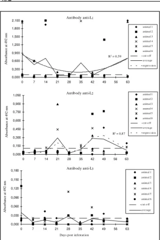
hominis observed in the infested animals are found in figure 1. The averages of L1 and L2 preparations,
expressed in absorbance values at 492nm, as ob-served in the infested animals, were significantly higher (P<0.01) than those observed in the control animals. Although it was observed a great variation on the antibodies levels from the experimentally infested group, the anti-L1 and anti-L2 qualities
could be detected in different periods post-infestation. Antibodies against L1 were detected
during the period between the first and 21st day
post-infestation (DPI) and, from the 42nd DPI on. Anti-L 2
antibodies, by their turn, could be detected on the 21st DPI and from the 35th DPI until approximately
the 49th DPI, when it was detected a decreasing of
antibodies titration which were equivalent to the control group. No antibodies were detected against the L3 instar-antigens on the evaluated period. Some
infested animals presented a tendency of rising anti-bodies levels against L1 and L2 antigenic
prepara-tions. Such phenomena was noticed, manly to the L1
instar, where four animals showed very high anti-bodies levels, whose absorbance was higher than 1.500 O.D. at 492nm in the ELISA assay, when compared to the cut-off, estimated in 0.096 O.D. Except for two animals, which did not present cir-culating antibodies, all the others infested animals were always positive against the L1 instar.
Never-theless, between the 21st and 28th DPI period and, on
the 42nd DPI, they presented results very close
21st DPI, where two animals showed O.D. of 0.450 and 0.900 at 492nm, respectively, with a cut-off estimated on 0.110 O.D. It was also demonstrated a rising of anti-L2 antibodies in the same four animals,
which presented antibodies response against L1
instar, on the 42nd and 49th DPI, when titration were
higher than 0.300 O.D. The obtained results, with an estimated prevalence of 50%, were comparatively evaluated, taking the double diffusion immunoassay precipitation test as a standard, and showed a con-cordance of 98%. On the other hand, the predictive values of sensitivity, specificity and accuracy, sug-gested that ELISA assay against different instars of D. hominis (L1 and L2) should be of relevance to
epidemiological studies, carried out at large inci-dence regions. The high specificity (95%) of the antigenic preparations showed a positive predictive value of 86%. Such value risen according to the estimated occurrence of positive animals: 93% to a 70% prevalence and 98% to a prevalence of 90%. Otherwise, its accuracy could be assured manly in low incidence regions, whose prevalence should be estimated below 50%, when the accuracy was 60%.
Taking on consideration that a good accuracy to this method should be higher than 70%, it should be of great utility in a 37% prevalence region. In spite of this, more detailed studies have to be made in order to identify better antigenic preparations, once the estimated sensitivity was only 28%. There is a need of a strong research base to obtain scientific insights and valuable reagents to allow more routine applica-tions. The method outlined shows the flexibility of the experimental systems. Recent advances in sci-ence have given ways to improving the sensitivity and specificity of the ELISA assay to D. hominis instars. It should be pointed out that the ability to develop ELISA against the different instars depends on a closer understanding on the immunological-serological-biochemical knowledge of the highest antibodies affinity of antigenic markers present in the antigenic preparations used (ROGAN, 1997). Such experiments have been made, actually, at our laboratories. It was noticed that three infested ani-mals presented antibodies against L1 and L2 instars
before infestation. Although such results suggest similar determinants could be presented in both, L1
and L2preparations, once the tested sera were
sub-mitted to 1:500 dilutions before assaying in order to avoid cross reactions, there is a possibility of previ-ous contact. The total absence of similar results on the control group corroborates with this last hy-pothesis, once these animals were restrained and free of infestations for a long period of time. The pres-ence of one animal which presented antibodies against the L3 instar at 28 DPI should be related to a
cross reaction, or any contaminant of different instar present during the antigen preparation. Other possi-bility should be related to the presence of reactive antibodies to the L3 instar, which development
oc-curred independently of the other instars, once such transformations were not synchronized on each infested animal. Previous work on D. hominis adaptive immune response on cattle showed the same tendency, with 90% of the animals presenting antibodies against D. hominis prior to experimental infestation (CORONADO-FONSECA, 1989). SANDEMANN et al. (1995), as well as BARON & NELSON (1985), seeking for antibodies titres to Lucilla cuprina and Melophagus ovinus, respectively, in experimentally infested sheep ob-served gradual augments of serum antibodies, which decreased on few weeks after exposition. These authors concluded that adaptive immune response against L. cuprina and M. ovinus should occurs only when larval infestation was active. Similar results were obtained by PERAÇOLI et al. (1980), LELLO & BOULARD (1990) and LELLO & PERAÇOLI (1993) working with rabbits
experi-Antibody anti-L2
0,000 0,150 0,300 0,450 0,600 0,750 0,900 1,050
0 7 14 21 28 35 42 49 56 63
Absorbance at 492 nm
animal 1 animal 2 animal 3 animal 4 animal 5 animal 6 cut-o ff ave ra ge regres s io n
R2 = 0,87
Antibody anti-L3
0,000 0,030 0,060 0,090 0,120 0,150 0,180
0 7 14 21 28 35 42 49 56 63
Days post infest at ion
Absorbance at 492 nm
animal 1 animal 2
animal 3
animal 4
animal 5
animal 6 cut-o ff
ave rage Antibody anti-L1
R2 = 0,59
0,000 0,300 0,600 0,900 1,200 1,500 1,800 2,100
0 7 14 21 28 35 42 49 56 63
Absorbance at 492 nm
animal 1 animal 2 animal 3 animal 4 animal 5 animal 6 cut-o ff ave rage regre s s io n
Figure 1 - Averages of levels of antibodies anti-L1, anti-L2 and
anti-L3 of Dermatobia hominis observed in the
Ciência Rural, v. 30, n. 3, 2000.
mentally infested with D. hominis. The present study has a significant importance concerned to the capability of making specific antibodies following a D. hominis infestation, independent of class or iso-type, which lasted for two months. Such immune response, although needing more detailed work on the antigenic components of each larval instar, sug-gests the L1 antigen as a representative marker to
infestation on bovines, once there was a straight correlation between infestation and presence of specific antibodies. These levels of antibodies sug-gest that the antigens produced by L1 were more
immunogenic than those produced by the other lar-val instars. The observed average of antibodies titra-tion against L1 was 0.373 (O.D.). Such average was
significantly higher (P<0.05) than those observed for antibodies anti-L2 (O.D. = 0.140) and anti-L3 (O.D.
= 0.013). Finally, it can be concluded that ELISA against D. hominis larval antigens is an important tool for studies of occurrence of infestations in dif-ferent regions and periods of time.
REFERENCES
ANDERSEN, E.H. Biology, distribution and control of
Dermatobia hominis. Veterinary Medicine, Chicago, v.55,
n.1, p.72-78, 1960.
BARON, R.W., NELSON, W.A. Aspects of the humoral and cell-mediated immune response of sheep to the ked
Melophagus ovinus (Diptera: Hippoboscidae). Journal of
Medical Entomology, Lanham, v.22, n.5, p.544-549, 1985.
CORONADO-FONSECA, A.J. Aspectos imunológicos, atividade antibacteriana e efeito de várias doses de ivermectina sobre larvas de Dermatobia hominis. Itaguaí – RJ, 1989. 23p. Dissertação (Mestrado em Parasitologia Veterinária) – Curso de Pós-graduação em Medicina Veterinária, Universidade Federal Rural do Rio de Janeiro, 1989.
CROWTHER, J.R. ELISA – theory and practice. In: WALKER, J.M. Methods in molecular biology. New Jersey : Humana, 1995. V.42. p.1-114.
CROWTHER, J.R. Enzyme-linked immunosorbent assay (ELISA). In: RAPLEY, R., WALKER, J. M. Molecular
biomethods handbook. New Jersey : Humana, 1998.
p.595-618.
HUDSON, L., HAY, F.C. Practical immunology. 3 ed. London: Blackwell, 1989. 507p.
LELLO, E., BOULARD, C. Rabbit antibody responses to experimental infestation with Dermatobia hominis. Medical
and Veterinary Entomology, Oxford, v.4, n.4, p.303-309,
1990.
LELLO, E., MOTA, N.G.S., PERAÇOLI, M.T.S. Reação inflamatória causada pelo berne, em coelhos imunizados ou não com extrato antigênico de Dermatobia hominis (Diptera: Cuterebridae). Ciência e Cultura, São Paulo, v.32, n.4, p.458-461, 1980.
LELLO, E., PERAÇOLI, M.T.S. Cell-mediated and humoral immune responses in immunized and/or Dermatobia hominis
infested rabbits. Veterinary Parasitology,Amsterdam, v.47,
n.1-2, p.129-138, 1993.
LELLO, E., PINHEIRO, F.A., NOCE, O.F. Epidemiologia de miíases no Município de Botucatu, S. P., Brasil. Arquivo da Escola de Veterinária da Universidade Federal de Minas Gerais,Belo Horizonte, v.34, n.1, p.93-108, 1982.
MOTA, N.G.S., PERAÇOLI, M.T.S., LELLO, E. Anticorpos circulantes em coelhos imunizados com antígenos obtidos de larvas de Dermatobia hominis, Linnaeus (Diptera: Cuterebridae). Ciência e Cultura, São Paulo, v.32, n.4, p.453-457, 1980.
PERAÇOLI, M.T.S., LELLO, E., MOTA, N.G.S. Comportamento da resposta imune-humoral em coelhos imunizados com antígenos de Dermatobia hominis Linnaeus, frente às larvas desse parasita (Diptera: Cuterebridae).
Ciência e Cultura,São Paulo, v.32, n.11, p.1537-1541, 1980.
ROGAN, M.T. Immunological analysis of parasite molecules. In: ROGAN, M. T. Analytical parasitology. Berlin :
Springer-Verlag, 1997. Cap.10. p.320-359.
SANAVRIA, A., LOPES, C.W.G., MOYA BORJA, G.E. Histopatologia da pele de bovino na infecção experimental por
Dermatobia hominis. Arquivos da Universidade Federal Rural do Rio de Janeiro,Itaguaí, v.10, n.1, p.9-23, 1987. SÁNCHEZ-VISCAÍNO, J.M., CAMBRA-ALVAREZ, M.
Enzyme immunoassay techniques, ELISA, in animal and plant diseases. 2 ed. Paris : O I E, 1987. 55p.
SANCHO, E. Dermatobia, the neotropical warble fly.
Parasitology Today, Amsterdam, v.4, n.2, p.242-246, 1988. SANDEMANN, R. W., CHANDLER, R. A., TURNER, N. et al.
Antibody degradation in wound exudates from blowfly infections on sheep. International Journal of Parasitology,
Oxford, v.25, n.5, p.621-628, 1995.
SNEDECOR, G. W., COCHRAN, W. G. Statistical methods. 6 ed. Ames : Iowa, 1976. 593p.