mercury contamination in the Brazilian Amazon
MARÚCIA I. M. AMORIM1, DONNA MERGLER2, MARCELO O. BAHIA3 HÉLÈNE DUBEAU4, DANIELA MIRANDA1, JEAN LEBEL2
ROMMEL R. BURBANO1and MARC LUCOTTE5 1Depto. de Biologia do CCB, Universidade Federal do Pará (UFPA)
2Centre pour l’Étude des Interactions Biologiques entre la Santé et l’Environnement (CINBIOSE) Université du Québec à Montréal (UQAM)
3Depto. de Patologia do CCB, Universidade Federal do Pará (UFPA) 4Depto. de Ciências Biológicas, Université du Québec à Montréal (UQAM)
5Chaire de Recherche en Environnement H-Q / CRSNG / Université du Québec à Montréal (UQAM)
Manuscript received on May 2, 2000; accepted for publication on July 24, 2000; presented byLewis J. Greene
ABSTRACT
The mercury rejected in the water system, from mining operations and lixiviation of soils after deforesta-tion, is considered to be the main contributors to the contamination of the ecosystem in the Amazon Basin. The objectives of the present study were to examine cytogenetic functions in peripheral lymphocytes within a population living on the banks of the Tapajós River with respect to methylmercury (MeHg) contamina-tion, using hair mercury as a biological indicator of exposure. Our investigation shows a clear relation between methylmercury contamination and cytogenetic damage in lymphocytes at levels well below 50 mi-crograms/gram, the level at which initial clinical signs and symptoms of mercury poisoning occur. The first apparent biological effect with increasing MeHg hair level was the impairment of lymphocyte proliferation measured as mitotic index (MI). The relation between mercury concentration in hair and MI suggests that this parameter, an indicator of changes in lymphocytes and their ability to respond to culture conditions, may be an early marker of cytotoxicity and genotoxicity in humans and should be taken into account in the preliminary evaluation of the risks to populations exposedin vivo. This is the first report showing clear cytotoxic effects of long-term exposure to MeHg. Although the results strongly suggest that, under the conditions examined here, MeHg is both a spindle poison and a clastogen, the biological significance of these observations are as yet unknown. A long-term follow-up of these subjects should be undertaken.
Key words:Brazilian Amazon, Mercury, Mitotic Index, Cytogenetics.
INTRODUCTION
Since the late seventies extensive gold mining op-erations using techniques based on amalgation with mercury have developed in the Amazon Basin. In close association with these mining activities, there
Correspondence to: Marcelo de Oliveira Bahia E-mail: mbahia@ufpa.br
has been important deforestation for population set-tlements and development of agriculture. The mer-cury rejected in the water system from mining oper-ations and lixiviation of soils after deforestation is considered to be the main contributor to the contam-ination of the ecosystem (Roulet et al. 1998a).
Tapajós River, a major affluent of the Amazon River, have shown that mercury is present in all environ-mental compartments (water, soil, plant) (Roulet et al. 1988b, c; Castilhos et al. 1998, Malm 1998). The biotransformation of inorganic mercury into methylmercury in aquatic environments is a well known process that makes human exposure possible through consumption of contaminated fish (WHO 1990, 1991). In the region of the Tapajós River, where fish is the dietary mainstay, methylmercury exposure levels in humans, measured in hair ranges from a fewµg/g up to 150µg/g; the median values
reported are in the order of 10 to 20µg/g (Nakanishi
1992, Pfeiffer et al. 1993, Grandjean et al. 1993, Lebel et al. 1998).
In vitrostudies have shown that methylmercury is a cytotoxic agent; reduced mitotic index and chro-mosomal aberrations are induced in human lympho-cytes treatedin vitrowith methylmercury (Bahia et al. 1999, Nakatsuru et al. 1985, Verschaeve et al. 1985, Betti et al. 1992, 1993a, b). Few studies have been performed on possible cytotoxic effects in human populations with methylmercury contam-ination and authors, reviewing these studies, have pointed out that the findings are borderline or in-conclusive (Nickle 1999, De Flora et al. 1994). Although there are methodological problems with each of the studies (small sample size, no control group, confounding factors not taken into account), the overall picture suggests that there may be an association between methylmercury exposure and cytogenetic deficiencies.
In the early seventies, two studies from Swe-den, which included 9 and 23 exposed persons re-spectively, suggested correlations between blood mercury levels and lymphocyte structural chromo-somal aberrations (Skerving et al. 1970, 1974). In 17 patients with Minamata Disease, Kato and Naka-mura (1976) reported a 18.7% frequency of some breakage and a 2.6% frequency of chromo-some reunions and rearrangements, however, there was no control group. A study of 16 fish-eating subjects from a polluted area in Colombia found no differences in the frequency of sister-chromatid
exchanges compared to controls; the frequency of chromosomal aberrations was higher in the exposed group only when achromatic lesions were included (Monsalve & Chiape 1987). In a larger scale study, Wulf et al. (1986) analyzed sister-chromatid ex-changes in lymphocytes taken from 147 persons whose diet included seal meat; they showed mean frequencies 1.7 times higher in the heavy seal-meat eaters as compared to those who ate less; more-over, the frequency of sister-chromatid exchanges increased with increasing blood mercury levels. They did not report hair mercury levels, which may be a better indicator of longterm exposure.
The objectives of the present study were to examine cytogenetic functions in peripheral lym-phocytes (mitotic index, polyploidal aberrations and chromatid breaks) among a population living on the banks of the Tapajós River with respect to methylmercury contamination, using hair mercury as a biological indicator of exposure.
MATERIAL AND METHODS
Population
Cytogenetic study of the analysed population was aproved by the Ethics Committees from the Con-selho Nacional de Desenvolvimento Científico e Tec-nológico(CNPq) and theUniversidade Federal do Pará, Belém – Pará, Brasil. The persons who par-ticipated in this study live in a small village, Brasília Legal, situated on the bank of the Tapajos River, an effluent of the Amazon (3◦59′00′′S, 55◦30′00′′W).
The village is approximately 250 km downstream from gold-mining operations and the villagers are not exposed to mercury vapours. Brasília Le-gal is accessible only by water, a 12 hour boat trip from Santarém, a city of several hundred thousand. There are minimal health facilities in Brasília Le-gal and electricity for only 3 hours every evening. The adult population (≥ 15 years) is made up of approximately 250 persons.
objec-tives were explained and villagers were invited to come to the Health Center where blood and hair samples were taken, and a questionnaire in-cluding socio-demographic information, smoking and drinking habits, medical and work history was administered by interview.
A total of 98 adults, ranging in age from 15-81 years, participated in the study. Their age distribu-tion, which is similar to the overall populadistribu-tion, is presented in Table I with other relevant characteris-tics: sex, smoking and drinking habits, a history of malaria and previous work in the gold mining region. For the current smokers, the number of cigarettes per day ranged from 1 to 4; 4% (3 persons) of the current drinkers reported drinking more than once or twice a week and only 2 persons reported previously us-ing marijuana. One third (33.3%) of the men and two of the women had worked in the gold mining region, where they would have been exposed to mer-cury vapours. It is in this area that most of the cases of malaria were contracted; of the 24 persons with a history of malaria, 15 (79%) had worked in the gold mining region.
Cytogenetic Techniques
Blood was obtained in heparinized vacutainers by venupuncture. In order to avoid potential loss of vi-ability associated with transport and delay, cultures were prepared on the boat, using electricity provided by a generator. The blood was stored immediately in a refrigerator and within a maximum of 4 hours, two independent cultures were set up by adding 12 drops of whole blood in 5ml of RPMI 1640 (GIBCO), containing 20% fetal calf serun (Cultilab) with an-tibiotics (100 IU penicilin/ml and 100µg
strepto-mycin/ml, GIBCO) and 4% phytohemoagglutinin (Cultilab). Cultures were incubated in a controlled water-bath at 37◦C for 48h. Colchicine (0.8mM,
Sigma) was added to the cultures 2h before harvest to obtain a maximum of cells at metaphase.
Cells were harvested by centrifugation (1000 rpm/min) and treated during 10 minutes with KCl (0,075 M, MERCK). They were fixed with Carnoy fixative 1:3 (glacial acetic acid:absolute methanol).
TABLE I
Characteristics of the study participants.
n %
age category
≥15<25 35 35.7
≥25<35 24 24.5
≥35<45 17 17.3
≥45<55 12 12.3
≥55 10 10.2
sex
women 46 46.9
men 52 53.1
smoking habits
non-smoker 51 52.0
ex-smoker 29 20.4
smoker 27 27.6
alcoholic beverages
non-drinker 37 37.8
drinker 61 62.2
malaria
no 72 74.2
yes 25 25.8
gold mining region
never 78 80.4
yes 19 19.6
Slides were prepared, air-dried and stained for 10 minutes with 3% Giemsa stain (MERCK) diluted in buffer solution (pH = 6.8).
Hair Sampling and Analyses
Hair strands from the root were taken from the oc-cipital region and then placed in plastic bags, with the root end stapled. Analyses for mercury deter-mination were conducted in the laboratories of the Environmental Research Chair of the University of Québec in Montréal, using Cold Vapor Atomic Flu-orescence spectrophotometry (CVAF).
Hair strands were cut in one cm segments and each segment was analyzed for total mercury, ac-cording to the procedure described by Bloom and Fitzgerald (1988) and adapted for hair. Inorganic mercury determination was done on the first segment using the methods described by Farant et al. (1981) and adapted for CVAF. Analytical quality was en-sured by including a Health Canada sample of pow-dered hair in the series. In the present analysis, the mean value for total mercury of the 2 first centime-ters was used.
RESULTS
Hair Mercury Levels
Total hair mercury levels for this population was log normally distributed and ranged from 0.57µg/g
to 153.8µg/g. The latter however was an outlier
and is treated separately in the analyses, the next highest value was 71.85µg/g. The median level
was 13.50µg/g, with 7.95µg/g and 22.19µg/g, the
25th and 75th quartile values, respectively. Women have significantly lower levels (median: 10.8µg/g)
as compared to men (median: 17.08µg/g;
Mann-Whitney U =823.5; p < 0.05). There was no
relation between hair mercury levels and smoking status, alcohol consumption, age, having worked in the garimpos or having suffered from malaria.
Mitotic Index
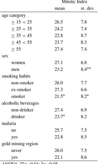
For the participants in this study, the mitotic index ranged from 8 to 36 per 1000 cells, with a mean of 25.20±7.8. Table II presents the mean mitotic
indices with respect to the relevant characteristics of the group. Women had a significantly higher in-dex as compared to men. Although smokers
pre-TABLE II
Mean mitotic index per 1000 cells with respect to the characteristics of the study participants.
Mitotic Index mean st. dev. age category
≥15<25 26.5 7.8
≥25<35 24.2 7.4
≥35<45 22.8 8.7
≥45<55 23.7 8.3
≥55 27.4 7.4
sex
women 27.1 6.8
men 23.2 8.4∗∗
smoking habits
non-smoker 26.0 7.7
ex-smoker 27.3 6.6
smoker 21.5∗ 8.2∗
alcoholic beverages
non-drinker 27.4 6.9
drinker 23.7∗ 8.2
malaria
no 25.7 7.5
yes 22.8 8.5
gold mining region
never 26.0 7.5
yes 22.1 8.6
ANOVA.∗∗p<0.01;∗p<0.05.
sented a significantly lower mitotic index as com-pared to ex-smokers and non-smokers, when sex was entered into the model, this difference was no longer significant (2 factor ANOVA: sex: F = 5.05; p = 0.03; smoking: F = 0.70; ns). The same pat-tern was observed for alcohol consumption, with drinkers having a lower mitotic index as compared to non-drinkers, however, when sex was included in the model, neither variable was significant (2 factor ANOVA: sex: F = 1.85; ns; alcohol consumption: F = 1.85; ns). No significant differences were ob-served with age, malaria and having lived in the gold mining area.
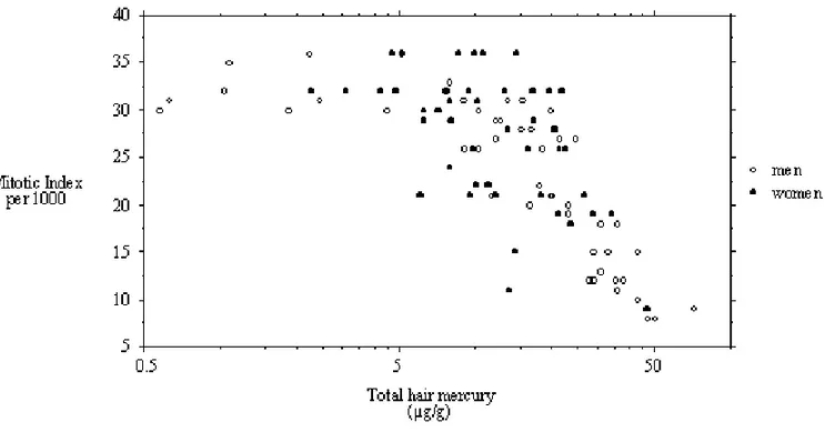
and men with respect to total hair mercury is pre-sented in Figure 1. There appears to be an initial plateau where mitotic indices vary between 30 and 36; the relation is best described by a second degree polynomial (r2= 0.64; F=84.5; p<0.001) The outlier (not in the graph), whose HHg is 153.8µg/g
has a mitotic index of 12.
Polyploides
The frequency of polyploides per 1000 lymphocyte cells ranged from 0 to 16. The majority of partici-pants (63.9%) did not present this aberration. The distribution of persons with polyploidal aberrations (PA) with respect to the characteristics of the group are presented in Table III. Although proportionally more men than women presented PA, the difference did not attain statistical significance. Polyploidal aberrations were observed significantly more fre-quently among alcohol drinkers as compared to non-drinkers and among those who had lived in the gold mining region as compared to those who had not. When stratified according to sex, these differences are no longer present.
Figure 2 shows the relation for women and men for PA frequency and hair mercury level. The low-est level at which PAs are observed is 7.25 µg/g
HHg. Above this level there is an increase in both the number of aberrations observed as well as the relative frequency of persons presenting them. Us-ing 7.25 as a cut-off point, the simple regression line between the frequency of polyploidal aberra-tions and total hair mercury level is highly signifi-cant (r2=0.41;F =51.7;p <0.001). At HHg≥ 20µg/g, the prevalence of persons with polyploidal
aberrations is 86.7% as compared to 18.8% for those with HHg≥10µg/g <20µg/g and 8.8% for those
with levels below 10. The differences are highly significant (Chi square: 48.9, df=2;p <0.001). A
total of 16 polyploides per 1000 cells were observed for the outlier.
Chromatid Breaks
Between 1-3 breaks were observed in lymphocytes for 14 persons (14.6%); for 11 there was only one
TABLE III
Distribution of persons with polypoidal aber-rations with respect to the characteristics of the study participants.
Polyploidal aberrations
n %
age category
≥15<25 9 25.7
≥25<35 13 54.2
≥35<45 7 43.8
≥45<55 4 33.3
≥55 2 20.0
sex
women 12 26.1
men 23 45.1
smoking habits
non-smoker 16 45.7
ex-smoker 6 17.1
smoker 13 37.1
alcoholic beverages
non-drinker 8 21.6
drinker 27 45.0∗
malaria
no 22 31.0
yes 13 52.0
gold mining region
never 23 29.9
yes 11 57.9∗
Chi square:∗p <0.05.
break, for 2 there were 2 breaks and 1 person pre-sented 3 breaks. The only characteristic that distin-guishes those with breaks from those without is the level of HHg; those with chromatid breaks have significantly higher levels of HHg as compared to those without (30.46µg/g ± 10.7 vs 14.5µg/g ± 11.6; ANOVA F=23.3; p<0.001). Among those
with HHg≥20µg/g, the prevalence of persons with
breaks is 37.9%, as compared to 9.4% for those with HHg≥10µg/g<20µg/g; none of the persons with
Fig. 1 – Relation between mitotic index for men and woman with respect to total hair mercury.
Fig. 2 – Relation for men and woman for polyploidal aberrations frequency and hair mercury level.
DISCUSSION
The findings of this study show a clear rela-tion between methylmercury contaminarela-tion and cy-togenetic damage in lymphocytes at levels well be-low 50µg/g, the level at which initial clinical signs
of cell-cycle progression and/or loss of proliferative capacity. Duringin vitroexperiments on chromo-somal aberrations, variation in MI is used to mon-itor induced cellular toxicity. This information on the degree of cytotoxicity is essential to adequately select harvest time and test concentration, and is es-pecially important when the results are used in risk assessment of compounds to which humans may be exposed. Mitotic index suppression has been sug-gested for use in dose selection for cytogenetic test-ing for regulatory purposes (UKEMS 1990). How-ever, this parameter is rarely considered forin vivo studies.
The relation between mercury concentration in hair and MI suggest that this parameter, an indi-cator of changes in lymphocytes and their ability to respond to culture conditions, may be an early marker of cytotoxicity and genotoxicity in humans and should be taken into account in the preliminary evaluation of the risks to populations exposed in vivo. In a study of chronic exposure to another metal, arsenic (As), impairment of lymphocyte prolifera-tion was observed in exposed individuals as com-pared to controls (Ostrosky-Wegman et al. 1991, Gonsebatt et al. 1994).
In this Amazonian population, no specific ef-fect of age on proliferative ability of lymphocytes was observed. These results are similar to other human studies which reported no variation in lym-phocyte proliferative rate with age in a control group from the United Kingdom (Anderson et al. 1988). Similarly, the sex-related differences in mitogen-induced lymphocyte proliferation observed here are in conformity with others. In non-exposed individ-uals, Anderson et al. (1991), observed higher blas-togenic transformation in PHA stimulated lympho-cytes from women as compared to men. Although Gonsebatt et al. 1994 report a greater impairment of lymphocytes in women exposed to arsenic in drink-ing water as compared to men, they have no measure of dose, which may prove to be higher in women. Sager et al. (1984) reported that the number of cells in the developing cerebellar external layer in 2-day-old mice was significantly reduced after
methylmer-cury treatment in males but remained unaltered in females.
The fact that exposure was associated with changes in the proliferative potential of lympho-cytes raises the question of possible immunologi-cal implications. There is a large experimental data base on the immunosuppressive properties of met-als, their inorganic salts and organometallic compo-nents (Koller 1980). Methylmercury, together with calcium, lead and As, have shown to cause immuno-logical changes in laboratory animals (NRC 1992). The observed reduced lymphocyte proliferation as-sociated with low levels of mercury may translate into reduced resistance to disease in this Amazonian population.
The positive results found for polyploidy and chromatid breaks with mercury exposure and their appearance following MI perturbations are in agree-ment with the molecular mechanism of action in-ferred fromin vitro observations. The genotoxic-ity of mercury is generally attributed to its binding with tubulin-SH, causing the impairment of spindle function and chromosome segregational error dur-ing cell division (Onfelt 1983, Sager et al. 1983). This has been regarded as the cause of the increased frequency of polyploidy and aneuploidy in Allium and Drosophila (Ramel & Magnusson 1969, 1979, Ramel 1969, Fiskesjo 1988). Moreover, in vitro polymerization and depolymerization of micro-tubules is affected by MeHg (Vogel et al. 1985). This could explain some of the multiple effects of MeHg on the cell cycle, including a lengthened G1 and decreased transition probability after short term exposure of cycling cells, and a G2 accumulation after a longer term exposure (Vogel et al. 1986). Studies on the potentiating effects of organic and inorganic mercuries on clastogen-induced chromo-some aberrations induced in CHO cells suggest that they inhibit some of the protein activities involved in the DNA repair process (Yamada et al. 1993).
and manganese may be genotoxic through genera-tion of free oxygen radicals (Ochi et al. 1983, Can-toni et al. 1984a, b, Snyder 1988). Methylmercury is known to generate free radicals through induction of lipid peroxidation in animal tissue (Ganther 1978, Shinada et al. 1990). Therefore the observed breaks could result from free radical damage.
In contrast to inorganic mercury, methylmer-cury easily passes the placenta barrier at least in some species. The occurrence of prenatal intoxica-tion in children shows that this is the case also in humans. It remains to be investigated whether tissues from exposed fetuses have an increased fre-quency of cells with polyploids or chromatid breaks. It is not yet known whether it is associated with an increased frequency of spontaneous abor-tions. Theoretically, methylmercury-induced chro-mosome damage in germline cells could give rise to abnormal offspring.
This is the first report showing clear cytotoxic effects of long-term exposure to methylmer-cury. The group was sufficiently large with a wide range of mercury exposure, based on a well-known biological marker, hair mercury. In the situation of this riverbank population on the Tapajós River, it is estimated that exposure to mercury has been present over the past twenty years. Although the re-sults strongly suggest that, under the conditions ex-amined here, MeHg is both a spindle poison and a clastogen, the biological significance of these obser-vations is as yet unknown. A long-term follow-up of these subjects should be undertaken. It is notewor-thy that lower MIs preceded appearance of chromo-somal endpoints such as polyploidy and chromatid breaks suggesting that MI may prove to be a valuable early indicator for this health hazard.
ACKNOWLEDGEMENTS
This work was supported by the International De-velopment and Research Center (IDRC) of Canada as part of the CARUSO project (grant #96-1052-01/001300-01) and Universidade Federal do Pará. The authors are grateful to Ms. Glorita Santos,
"Seu" Humberto da Silva, Francisco Borges (in memoriam), and Ana Maria Rodrigues for technical help.
REFERENCES
Anderson D, Jenkinson PC, Dewdeney RS, Francis AJ, Godbert P & Butterworth KR.1988. Chro-mosome aberrations, mitogen-induced blastogenesis and proliferative rate index in peripheral lymphocytes from 106 control individuals of the U.K. population. Mutat Res204:407-420.
Anderson D, Francis AJ, Godbert P, Jenkinson PC & Butterworth KR.1991. Chromosome aberra-tions (CA), sister-chromatid exchanges (SCE) and mitogen-induced blastogenesis in cultured peripheral lymphocytes from 48 control individuals sample 8 times over 2 years.Mutat Res250:467-476.
Bahia MO, Amorim MIM, Burbano RR, Vincent S & Dubeau H.1999. Genotoxic effects of mercury on in vitro cultures of human cells.An Acad Bras Ci
71(3-I): 437-443.
Betti C, Davini T & Barale R.1992. Genotoxic activ-ity of methylmercury chloride and dimethyl mer-cury in human lymphocytes. Mutat Res281: 255-260.
Betti C, Barale R & Pool-Zobel BL.1993a. Com-parative studies on cytotoxic and genotoxic effects of two organic mercury compounds in lymphocytes and gastric mucosa cells of Sprague-Dawley rats. Env Mol Mutag22:172-180.
Betti C, Davini T, HE J & Barale R.1993b. Liq-uid holding effects on methylmercury genotoxicity in human lymphocytes.Mutat Res301:267-273.
Bloom N & Fitzgerald WF.1988. Determination of volatile mercury species at the picogram level by low temperature gas chromatography with cold va-por atomic fluorescence detection. Anal Chim Acta
208:151-161.
Cantoni O, Christie NT, Swann A, Drath DB & Costa M.1984a. Mechanism of HgCl2 citotoxic-ity in cultured mammalian cells.Mol Pharmacology
26:360-368.
1984b. Characterization of DNA lesions produced by HgCl2in cell culture systems.Chem Biol Interact
49:209-224.
Castilhos ZC, Bidone ED & Lacerda LD.1998. In-crease of the background human exposure to mercury through fish consumption due to gold mining at the Tapajós River region, Pará State, Amazon. Bull En-viron Contam Toxicol61(2): 202-9.
De Flora S, Benniceli C & Bagnasco M.1994. Geno-toxicity of mercury compounds. A review.Mutat Res
317:57-79.
Farant JP, Brisset D, Moncion L, Brigasand L & Chartrand A.1981. Improved cold vapor atomic absorption technique for the microdetermination of total and inorganic mercury in biological samples.J Anal Toxicol5:47-51.
Fiskesjo G.1988. The Allium test. An alternative in environmental studies: the relative toxicity of metal ions.Mutat Res197(2): 243-60.
Ganther HE.1978. Modification of methylmercury toxicity and metabolism by selenium and vitamin E: possible mechanisms. Environ Health Perspect25: 71-76.
Gonsebatt ME, Vega L, Montero R, Garcia-Vargas G, Del Razo LM, Albores A, Cebrian ME & Ostrosky-Wegman P.1994. Lymphocyte replicat-ing ability in individuals exposed to arsenic via drink-ing water.Mutat Res313(2-3): 293-9.
Grandjean P, Cardoso B & Guimarães G.1993. Mer-cury poisoning.Lancet342:991.
IPCS (International Programme on Chemical Safety). 1990. Methylmercury Environmental Health Criteria, 101. World Health Organization, Geneva, p. 144.
Kato R & Nakamura A.1976. Chromosome breakage associated with organic mercury in human leukocytes “in vitro” and “in vivo”. Jpn J Human Genet20: 256-257.
Koller LD.1980. Immunotoxicology of heavy metals. Int J Immunopharmacol2:269-279.
Lebel J, Mergler D, Branches F, Lucotte M, Amorim M, Larribe F & Dolbec J.1998.
Neu-rotoxic effects of low-level methylmercury contami-nation in the Amazonian Basin.Environ Res Section A.79:20-32.
Malm O.1998. Gold mining as a source of mercury ex-posure in the Brazilian Amazon.Environ Res77(2): 73-8.
Monsalve MV & Chiappe C.1987. Genetic effects of methylmercury in human chromosomes: I. A cyto-genetic study of people exposed through eating con-tamined fish. Environ Mol Mutagen10:367-376.
Nakanishi J. 1992. Mercury pollution: Minamata, Canada and Amazon.Water Rep2:4-5.
Nakatsuru S, Oohashi J, Nozaki H, Nakada S & Imura N.1985. Effect of mercurials on lymphocyte functions “in vitro”.Toxicology36(4): 297-05.
NRC (National Research Council). 1992. Com-mission on Life Sciences, Board on Environmental Studies and Toxicology, Subcommittee on Immuno-toxicology Committee on Biological Markers. Bio-logical Markers in Immunotoxicology, National Aca-demic Press, Washington, D.C., pp. 68-71.
Nickle RA.1999. Mercury. Top of the hit parade for eight years.Drug Chem Toxicol22(1): 129-142.
Ochi T, Takahashi K & Ohsawa M.1983. A mech-anism for the stimulation by inorganic mercury of [3H] thymidine incorporation into DNA in cultured Molt-4F cells.Jpn J Exp Med53:187-94.
Onfelt A.1983. Spindle disturbances in mammalian cells. I. Changes in the quantity of free sulfhydryl groups in relation to survival and C-mitosis in V79 Chinese hamster cells after treatment with colcemid, diamide, carbaryl and ethyl mercury.Chem Biol In-teract46:201-17.
Ostrosky-Wegman P, Gonsebatt ME, Montero R, Vega L, Barba H, Espinosa J, Palao A, Corti-nas C, Garcia-Vargas G, Del Razo LM & Ce-brian M.1991. Limphocyte proliferation kinetics and genotoxic findings in a pilot study on individual chronically exposed to arsenic in Mexico.Mutat Res
250:477-482.
gold-mining in the Brazilian Amazon. Environ Rev1: 26-37.
Ramel C.1969. Genetic effects of organic mercury com-pounds. I. Cytological investigations on Allium roots. Hereditas 61(1): 208-230.
Ramel C & Magnusson J.1969. Genetic effects of or-ganic mercury compounds. II. Chromossome segre-gation inDrosophila melanogaster. Hereditas61(1): 231-54.
Ramel C & Magnusson J.1979. Chemical induction of nondisjunction inDrosophila. Environ. Health Perspec31:59-66.
Rojas E, Herrera LA, Sordo M, Gonsebatt ME, Montero R, Rodriguez R & Ostrosky-Wegman P.1993. Mitotic index and cell proliferation kinetics for the identification of antineoplastic activity. Anti-Cancer Drugs4:637-640.
Roulet M, Lucotte M, Farella N, Serique G, Coelho H, Sousa Passos CJ, de Jesus da Silva E, Scavone de Andrade P, Mergler D, Guimarães J-RD & Amorim M.1998a. Effects of recent human colonization on the presence of mercury in Amazo-nian ecosystems. Water, Air and Soil Pollution00: 1-17.
Roulet M, Lucotte M, Saint-Aubin A, Tran S, Rheault I, Farella N, de Jesus da Silva E, Dezencourt J, Sousa Passos C-J, Santos Soares G, Guimarães J-RD, Mergler D & Amorim M.1998b. The geochemistry of mercury in central Amazonian soils developed on theAlter-do-Chão for-mation of the lower Tapajós River Valley, Pará state, Brazil.Sci T Env223:1-24.
Roulet M, Lucotte M, Canuel R, Rheault I, Tran S, de Freitas GOG YG, Farella N, Souza do Vale R, Sousa Passos C-J, de Jesus da Silva E, Guimarães J-RD, Mergler D & Amorim M.1998c. Distribution and partition of total mercury in waters of the Tapajós River Basin, Brazilian Amazon.Sci T Env213:203-211.
Sager PR & Syversen TL.1984. Differential responses to methylmercury exposure and recovery in neurob-lastoma and glioma cells and fibroblasts.Exp Neurol
85:371-82.
Sager PR, Doherty RA & Olmsted JB.1983. Interac-tion of methylmercury with microtubules in cultured cells and “in vitro”.Exp Cell Res146:127-37.
Shinada M, Takizawa Y & Muto H.1990. Effect of mercuric chloride on phospholipid peroxidation in rat.Nippon Koshu Eisei Zasshi37:1010-4.
Skerfving S, Hansson K & Lindsten J.1970. Chro-mossome breakage in human exposed to methylmer-cury through fish consumption.Arch Environ Health
21:133-139.
Skerfving S, Hansson K, Mangs C, Lindston J & Ryman N.1974. Methylmercury-induced chromos-some damage in man.Environ Res7:83-98.
Snyder RD.1988. Role of active oxygen species in metal induced DNA strand breakage in human diploid fi-broblasts.Mutat Res193:237-246.
UKEMS.1990. The third United Kingdom Environmen-tal Mutagen Society collaborative trial.Mutagenesis
(Sup. 5):1-88.
Verschaeve L, Kirsch-Volders M, Hens L & Susanne C.1985. Comparative “in vitro” cytogenetic studies in mercury-exposed human lymphocytes.Mutat Res
157:221-226.
Vogel DG, Margolis RL & Mottet NK.1985. The effects of methylmercury binding to microtubules. Toxicol Appl Pharmacol80:473-86.
Vogel DG, Rabinovitch PS & Mottet NK. 1986. Methylmercury effects on cell cycle kinetics. Cell Tissue Kinet19(2): 227-42.
WHO, International Program on Chemical Safety. 1990. Environmental Health Criteria 101: Methyl-mercury. Geneva: World Health Organization, 144p.
WHO, International Program on Chemical Safety. 1991. Environmental Health Criteria 118: Mercury. Geneva: World Health Organization, 168p.