http://www.uem.br/acta ISSN printed: 1679-9275 ISSN on-line: 1807-8621
Doi: 10.4025/actasciagron.v34i4.15062
Leaf anatomy of orchids micropropagated with different silicon
concentrations
Joyce Dória Rodrigues Soares*, Moacir Pasqual, Aparecida Gomes de Araujo, Evaristo Mauro de Castro, Fabricio José Pereira and Francyane Tavares Braga
Departamento de Agricultura, Universidade Federal de Lavras, Cx. Postal 3037, 37200-000, Lavras, Minas Gerais, Brazil. *Author for correspondence. E-mail: [email protected]
ABSTRACT. Research on anatomical modifications under in vitro culture is essential to the definition and
understanding of the development of micropropagated plants. Likewise, such research is essential to improve the steps of the acclimatization process. Accordingly, the objective of the present study is to verify the differences in the leaves anatomical traits of micropropagated orchids under calcium silicate concentrations. Seedlings of in vitro-germinated seeds, measuring 0.5 cm in length, were inoculated in 250 cm3 pots with 60 mL MS culture
medium and different silicon concentrations (0, 0.5, 1.0 and 2.0 mg L-1) in different culture environments
(natural environment, in a greenhouse and an artificial environment in a growth chamber) and in all statistical combinations. The pH of the culture medium was fixed at 5.8 ± 0.1 and gelified with 5.5 g L-1 of agar before
autoclaving at 121ºC and 1 atm for 20 min. After 150 days, an anatomical analysis was performed on cross-sections of the plant leaves. A complete randomized design was used. Modifications occurred on the plants of those treatments containing silicon compared to those without silicon, and between environment fo artificial and natural light. Plants showed larger growth at the artificial light treatment with 0.5 and 2.0 mg L-1 of calcium
silicate respectively for the native and hybrid plants Treatments without silicate application induced chlorenchyma and epidermis deformation compared to silicon containing treatments, this effect can affect directly or indirectly plant growth at no silicon conditions.
Keywords: microscopy, Orchidaceae, in vitro culture, calcium silicate.
Anatomia foliar de orquídea micropropagada com diferentes concentrações de silício
RESUMO. A realização de pesquisas a respeito das modificações anatômicas decorrentes do cultivo in vitro são
fundamentais para o melhor entendimento e elucidação do desenvolvimento das plantas micropropagadas. Dessa forma, tal pesquisa é essencial para melhorar as etapas do processo de aclimatização. Sendo assim, o objetivo deste trabalho foi identificar diferenças nas características anatômicas em folhas de orquídeas micropropagadas sob concentrações de silicato de cálcio. Plântulas oriundas de sementes germinadas in vitro com aproximadamente 0,5
cm de comprimento foram inoculadas em frascos com capacidade para 250 cm³ contendo 60 mL de meio de cultura MS, acrescido de silicato de cálcio (0; 0,5; 1,0 e 2,0 mg L-1) em ambientes de cultivo (natural, em casa de
vegetação e artificial e, em sala de crescimento), em todas as combinações possíveis. O meio de cultura teve seu pH ajustado para 5,8 ± 0,1 e geleificado com 5,5 g L-1 de ágar, previamente ao processo de autoclavagem (121ºC)
por 20 minutos. Após 150 dias de cultivo, as folhas das plantas foram submetidas à avaliação anatômica por meio de secções transversais. O delineamento experimental foi o inteiramente casualizado. Ocorreram modificações nos tratamentos contendo silício em comparação com aqueles isentos de silício, e entre os ambientes de luz artificial e natural. As plantas apresentaram maior crescimento em luz artificial e nas doses de 0,5 e 2,0 mg L-1 de
silicato de cálcio para as plantas nativa e híbrida, respectivamente. Os tratamentos sem a aplicação de silicato induziram deformações no clorênquima e epiderme, ao comparar com o tratamento contendo silício, efeito que pode ter afetado direta ou indiretamente o crescimento das plantas nas condições sem silício.
Palavras-chave: microscopia, Orchidaceae, cultivo in vitro, silicato de cálcio.
Introduction
Despite the advantages of micropropagation, plants cultivated in vitro present certain anatomical and
physiological characteristics intrinsic to the culture environment. These characteristics include reduced epicuticular wax deposits, a relatively low
thickness of the epidermis and cuticle, lower phloem development, abundant intercellular spaces, rudimentary vascular bundles and deficient stomatal mechanisms (ROMANO; MARTINS-LOUÇÃO, 2003).
may develop structures that are better adapted and more efficient for the plant in the final of acclimatization stage. After plants are transferred to field conditions, the characteristics induced during the in vitro culture usually change (EVERT; ESAÚ,
2006). An examination of leaf anatomy would contribute to the understanding of the physiological and structural modifications for micropropagated plants and furnish information applicable to the plants adaptation to the new ex vitro environment
(MARIN, 2003).
Silicon added to the culture medium can benefit the plants by increasing the hemicellulose and lignin content, thereby increasing the hardness of the cell wall. These changes increase the plants survival rate during acclimatization (VALENTE et al., 2004). The direct effects of silicon are accompanied by several indirect effects, including an increase in photosynthetic capacity, reduction of transpiratory rates, greater plant growth and increased mechanical resistance of the cells (VALENTE et al., 2004).
The aim was to assess the modifications in leaf anatomy of micropropagated plants of a native (Brassavola perrinii) and hybrid orchid ((Laeliacattleya
Culminant “Tuilerie” x Laeliacattleya Sons Atout
Rotunda) x Brassolaeliacattleya Startifire Moon Beach)
in different culture environment and CaSiO3
concentrations.
Material and methods
General characterization
The study was conducted from March through August 2008 in the Tissue Culture Laboratory and annexes of the Department of Agriculture at the Federal University of Lavras in the municipality of Lavras, Minas Gerais State, Brazil.
Plant material
Seedlings of a native (Brassavola perrinii) and
hybrid orchid (Laeliacattleya Culminant “Tuilerie” x
Laeliacattleya Sons Atout Rotunda) x
Brassolaeliacattleya Startifire Moon Beach), obtained
from in vitro germination of seeds from
self-pollination (native) or cross-self-pollination (hybrid), were used in this study. Seeds were germinated in Knudson C (KNUDSON, 1946) culture medium modified with ZnSO4.7H2O (from 0.331 to 6.62 mg
L-1), H
3BO3 (from 0.056 to 1.4 mg L-1) and
MnSO4.H2O (from 7.5 to 15 mg L-1) where they
remained for three months. After this period, each seedling, measuring approximately 0.5 cm of length, was inoculated in a 250 cm² flask containing 60 mL
of MS culture medium (MURASHIGE; SKOOG, 1962). CaSiO3 (0, 0.5, 1.0 and 2.0 mg L-1) was
added, pH was adjusted to 5.8 ± 0.1, and the medium was geleified with 5.5 g L-1 of agar before
autoclaving at 121ºC during 20 minutes. The cultures were maintained in natural light (greenhouse) and artificial light (growth chamber) using all the possible combinations of experimental conditions. Two identical experiments, but independents, were conducted, one for the native plants and the other for the hybrid.
Characterization of the culture environment
The artificial environment consisted of a growth chamber with illumination supplied by special daylight fluorescent lights (OSRAM 20 W) with 42 W m-2 of mean irradiance, a 16 hour photoperiod
and at 25 ± 2oC. The natural light environment
consisted of a greenhouse with natural illumination and 70% shading (Sombrence®) with the following
environmental parameters: maximum, minimum and mean temperatures of 26ºC/32ºC, 16ºC/16ºC and 20ºC/23ºC and maximum, minimum and mean irradiance levels of 93.95 W m-2/199.69 W m-2, 11.13
W m-2/10.66 W m-2 and 49.38 W m-2/99.43 W m-2
(typical of cloudy and clear days, respectively), during the experimental period. Data on the daily incident solar radiation at the height of the flasks in the growth chamber and greenhouse were collected with radiation probes (LI-200SA, Li-cor, Lincoln, Nebraska, USA) attached to a Li-cor LI-1400 recording system. These values were measured every half hour for 11 hours (from 7 a.m. to 6 p.m.). Only one radiation measurement was made in the growth chamber over a six-hour period because the environment was controlled. A thermo-hygrograph was used to measure and record the minimum, mean and maximum weekly temperatures.
Biometric and electronic microscopy assessments
The shoot length was measured after 150 days. Assessments were performed with an electron and light microscope and the best available biometric methods. The anatomical assessments were carried out at the Plant Anatomy Laboratory at the Federal University of Lavras (UFLA) Department of Biology. For the light microscopy, five leaves were used (the second completely expanded leaf in the tip-to-base direction). These leaves were collected from five plants in each treatment. Leaves were fixed in ethanol 70% (v v-1). The assessments were made
cross-sections obtained with a desk microtome (LPC model) using a stainless steel razor blades. Cross sections were clarified in a solution of 50% sodium hypochlorite for 10 minutes and washed in distilled water for 10 minutes and stained with safrabau (safranin 1% m v-1 and astra blau 0.1%
m v-1 at the rate of 7.5:2.5) following the
methodology decribed by Kraus and Arduin (1997). The sections were then mounted in 50% glycerin and microscopy slides. The thicknesses of the adaxial and abaxial surfaces epidermis and the phloem were measured using a Ken-a-vision 2100 light microscope equipped with a micrometric ocular and a 40x lens. Five field measurements were taken on each slide.
Scanning electron microscopy analysis was performed in one leaf for replicate, totalizing five leaves for treatment. Samples of 0.5 cm length of leaves blades were fixated in Karnovisk’s modified solution (pH of 7.2) for a 24 hours, and then transferred into a glycerol 30% for 30 minutes. Samples were than sectioned with razor blades in transversal and longitudinal in liquid nitrogen. Sections obtained were transferred to a 1% osmium tetroxide (OsO4) solution for 1 hour and
subsequently dehydrated in an ascending series of acetone baths (30, 50, 70, 90 and 100%) and taken to the drier until critical point was achieved. The specimen sections were mounted on aluminum stubs and taken to Sputtering for surface coating with gold (200 Å) to increase the specimens’ resistance and conductivity. The samples were observed using a scanning electron microscope, model LEO EVO 40XVP.
Experimental design and statistical analyses
A randomized complete design was used for the experiment with the native plants (Brassavolaperrinii)
and for the experiment with the hybrid ((Laeliacattleya Culminant “Tuilerie” x Laeliacattleya
Sons Atout Rotunda) x Brassolaeliacattleya Startifire
Moon Beach). The design consisted of a 2 x 4 factorial arrangement with two environments (natural and artificial), four CaSiO3 concentrations
(0, 0.5, 1.0 and 2.0 mg L-1) and 12 seedlings per
treatment. Data was submitted to one-way anova and means were compared by Tukey test at 5% level of significance or by regression analysis according to model adjustment. Sisvar 4.3 statistical software (FERREIRA, 2011) was used for the data analysis.
Results and discussion
Biometric analysis
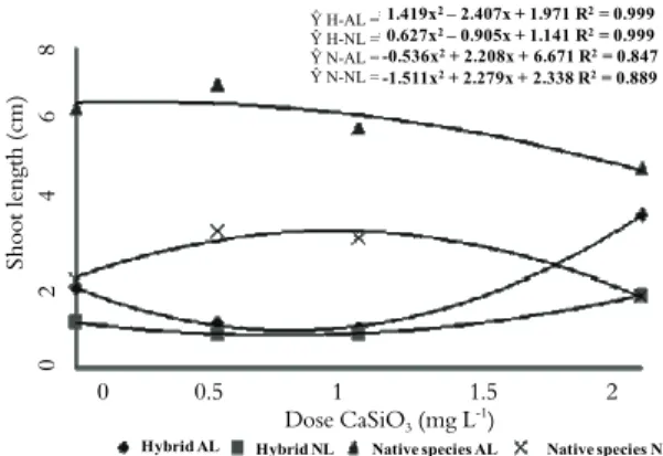
The shoot length values showed a significant interaction between the CaSiO3 concentrations and
the culture environments. The shoot length was larger in the artificial light environment for both the native and hybrid plants. Hybrid plants showed average shoot length for the best CaSiO3 53.8%
greater in artificial light than in the natural environment. The corresponding value for the native plants was 99.9% (Figure 1).
8
6
4
2
Shoo
t lengt
h (c
m)
0
Y H-AL= 1.419x2– 2.407x + 1.971 R2= 0.999 Y H-NL= 0.627x2– 0.905x + 1.141 R2= 0.999 Y N-AL= -0.536x2+ 2.208x + 6.671 R2= 0.847 Y N-NL= -1.511x2+ 2.279x + 2.338 R2= 0.889
0 0.5 1 1.5 2 Dose CaSiO3 (mg L-1)
Hybrid AL Hybrid NL Native species AL Native species NL
Figure 1. Shoot length (cm) in relation to the experimental
treatments of artificial light (AL), natural light (NL) and CaSiO3
doses for the hybrid and native species.
Plants in a shady or low-irradiation environment can etiolate as a strategy to reach higher light levels (TAIZ; ZEIGER, 2004). The effect observed in the present study may have been related to the low level of light in the red range of the spectrum furnished by the lamps used in the growth chamber. These levels may not have been sufficient to activate the phytochrome response and entirely inhibit etiolation. The increase in length observed may also be associated with the understory habitat. This habitat is typical for Brassavola, Cattleya and Laelia
orchids (ZANEGA-GODOY; COSTA, 2003), including the hybrid and native plants investigated in the present study. Certain characteristics of these understory plants may have favored the development of etiolation, but the understory environment also provides good conditions for the development of the orchid under study. One of these conditions may be related to photoinhibition of photosynthesis by excess of radiation, present in the natural light environment. Plants micropropagated can exhibit structural modifications causing prejudices to photosynthesis by reducing chlorenchyma development and aclimatization stage is necessary to correct development of this tissue in micropropagated
Ŷ H-AL =
Ŷ H-NL =
Ŷ N-AL =
plants (BRAGA et al., 2009; ROMANO; MARTINS-LOUÇÃO, 2003). So, shady environment can be favorable to development of the studied orchids, avoiding photoinhibition until complete development of chlorenchyma after acclimatization.
Adding silicon to culture medium promoted higher growth of the hybrid and the native plants in both environments. In the hybrid, the greatest shoot length were observed at the 2.0 mg L-1 of calcium
silicate. Under artificial light, the shoot length in the hybrid was 42.3% higher than the corresponding length in the treatment without silicon. Under natural light, the shoot length in the hybrid was 61.4% higher compared to the treatment without silicon. In the native plants, the greatest shoot length occurred at the 0.5 mg.L-1 concentration. Under
artificial light, the canopy length was 9.2% greater compared to the treatment without silicon. Under natural light, the canopy length was 69.9% higher than those found in the treatment without silicon (Figure 1). In this plants, higher concentrations of silicon may have inhibited growth. This effect may have been related to an alteration in the coefficient of extensibility of the cell wall (JONES, 1992) resulting from the incorporation of silicon. An alteration of this type could have restringed cell expansion (SOARES et al., 2008).
Silicon effects on growth of these orchid were evident. The native nd hybrid plants responded similarly to calcium silicate in culture medium. Silicon effects was more intense under artificial light. In the hybrid, the effect under artificial light was substantially stronger than that under natural light. The positive results of this study for orchid seedling growth are consistent with the statement by Zhou (1995) that concentrations of CaSiO3 between
0.1 and 1.0 mg L-1that were added to the VS culture
medium (VACIN; WENT, 1949) produced greater shoot growth in Phalaenopsis seedlings. In potato
plants, the application of calcium silicate promotes higher growth and tuber yield (PULZ et al., 2008). These effects may be related to silicon incorporation that helped to sustain the plant by increasing the mechanical resistance of the tissues. This reinforcement may have facilitated plant growth because it permitted plant elongation and resisted the bending of plants.
The benefits of using silicon have been associated with various indirect effects, including an increase in capacity and efficiency photosynthetic, a reduction in transpiration and consequently greater plant growth (ZHOU, 1995). This result is desirable
because the growth of these orchid plants is extremely slow, even compared with other ornamental species. Therefore, it is difficult to obtain seedlings suitable for commercial use.
Anatomical analysis
Leaves analyzed in cross section had a homogeneous mesophyll with an average thickness of 342.97 μm, approximately nine cell layers, single layer epidermis with a greater thicknesses in the treatments with CaSiO3 added to the culture
medium (Table 1).
Table 1. Mean values of mesophyll thickness (μm), adaxial
epidermis thickness (μm) and abaxial epidermis thicknesses (μm) in relation to CaSiO3 application (mg L-1) in a growth chamber
environment for the native orchid species.
CaSiO3
(mg L-1) Mesophyll thickness Adaxial epidermis Abaxial epidermis
0 296.222 b 15.849 b 20.815 b 0.5 342.972 a 22.310 a 26.313 a Means followed by the same letter in columns do not differ significantly (Tukey test, 5% probability level).
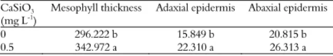
The epidermis consisted of approximately isodiametric and squared cells with few intercellular spaces and no cuticle. The hypodermis consisted of a single layer cells that were larger than epidermal cells and were also isodiametric. The leaves had collateral vascular bundles with the phloem turned to the abaxial surface. The metaxylem was poorly differentiated. The bundle sheath and mesophyll layers formed a Kranz structure. Few fibers were observed in the chlorenchyma. The bulliform cells in the leaves mid-region were had reduced diameters and showed evidence of cell collapse. The vascular bundles were reduced in number and size in the plants without silicon. In these plants, the phloem and xylem tissues were less differentiated and deformed in appearance (Figure 2).
Tetracytic stomata were observed on both adaxial and abaxial surfaces in hybrid and native plants but were more frequent on the adaxial epidermis. This characteristic classified the leaf as amphi-epistomatic (Table 2). According to Zanenga-Godoy and Costa (2003), tetracytic stomata were observed in four species of the Cattleya genus on both leaf surfaces.
Figure 2. Cross-sections of native orchid leaves in the presence (A and C) and absence (B and D) of silicon. mn= normal mesophyll, md= deformed mesophyll, fv= vascular bundle, arrow= calcium oxalate raphides. Bars= 50 μm.
Table 2. Stomatal densities (SD), polar diameters (PD), equatorial
diameters (ED), polar/equatorial diameter ratios and numbers of cells (NC) in relation to CaSiO3 concentrations (mg L-1) in a growth
chamber environment for the native orchid species.
Abaxial surface epidermis Dose CaSiO3
(mg L-1) SD
(stomata mm-2) (PD μm) (ED μm) PD/ED (epidermal cells (mmNC -2)
0 29.85a 31.09a 26.45a 1.26a 196.6a 0.5 31.04a 33.26a 27.46a 1.13a 169.8b CV (%) 26.7 9.9 5.6 8.9 7.0
Adaxial surface epidermis
0 46.57a 32.47a 24.95b 1.30a 244.2a 0.5 34.63b 30.14a 28.15a 1.07b 206.8b CV (%) 12.1 5.7 7.2 6.1 10.8
Means followed by the same letter in the column do not differ significantly (Tukey test, 5% probability level).
The ratio of polar and equatorial diameters on the adaxial epidermis indicated greater functionality in the stomatal control of aperture mechanisms. No significant difference was found on the abaxial epidermis. However, the analysis of epidermal cell number demonstrated the plant´s investiment in fewer and larger cells in the abaxial and adaxial epidermis (Table 2). This finding result is consistent with the findings of Romero-Aranda et al. (2006). Peschel et al. (2003) reported that differences in stomatal functionality are related to the ratio of polar and equatorial diameters and a reduction on this characteristic reduces stomatal functionality.
These results showed that to native plants, the silicon treatments restricted the development of the stomatal characteristics. The reduction in the stomatal density associated with a reduction in functionality may have indicated that in the absence of silicon, the stomata of this species might be more capable of capturing carbon dioxide and preventing water loss.
Close to the midrib in the mid-region of the leaf and facing the adaxial surface, bulliform cells formed groups of approximately five cells with silicon deposits close to the cell walls (Figure 3). These silicon bodies were also occasionally observed on the periclinal walls of the chlorenchyma cells. Idioblasts containing druses closer to the adaxial epidermis also occurred (Figure 3).
Figure 3. Silicon deposits (arrows in A) and druses of calcium oxalate (arrows in B) in the bulliform cells (A) and hypodermis (B) of
orchid plants in treatment containing CaSiO3. cb= bulliform cell, hd= hypodermis. Bars= 20 μm.
A
B
C D
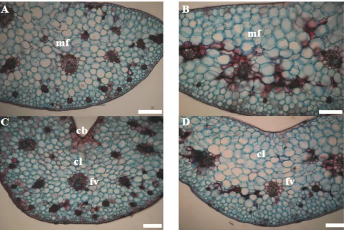
Figure 4. Scanning electron micrographies of epidermis of the native orchid plants with (A) and without silicon (B) and of the hybrid
orchid plants with (C) and without silicon (D).
In the leaves cross sections of plants from treatments without calcium silicate silicon deposits were not observed in the bulliform cells, in the chlorenchyma or in the anticlinal walls of the epidermis and hypodermis. Plants showed deformations in those chlorenchymal cells near to the
abaxial surface and thus indicated a cell collapse (Figure 2D and 4D). Mesophyll thickness was statistically smaller in plants of treatments without silicon than those found in the silicon containing treatments (Table 3). An increase in deformations was also observed in the chlorenchyma cells facing the abaxial surfaces
100 μm 100 μm
compared with the leaves from plants of treatments with CaSiO3. Leaves mesophyll from silicon treatment
was 15.8% thicker than that of the control without silicon. Adaxial epidermis was 40.7% thicker than those of the control, and the abaxial epidermis was 26.4% thicker than that of the control (Table 3).
Compared with the leaves from silicon containing treatment, control plants showed clear tissue deformation in the front view of the epidermis (Figure 4D). This characteristic may have been a result of the deformation of the internal chlorenchyma layers in the leaves of the control treatment (Figure 2). This finding indicated that the silicon may have contributed to the structural stability of the leaves. The silicon may have become incorporated in the cell walls and may have helped to maintain the leaf structure. The leaf structure is essential for the correct development of these plants because the chlorenchyma is responsible for photosynthesis (EVERT; ESAÚ, 2006). If the leaf is deformed, damage to photosynthesis and plant growth can occur. According to Marin (2003) silicon benefits to plants includes both direct effects such as structural development and indirect effects like those observed in a increased photosynthetic rate. Both of these effects can be related to a better tissue development on plant leaves under silicon treatments (VALENTE et al., 2004). A greater photosynthetic capacity is a favorable
characteristic for plant micropropagation (ARIGITA et al., 2002). This effect was shown by the restrictions in the growth of the plants without silicon (Figure 1). Thus, the presence of silicon in the culture medium may contribute substantially to the correct development of micropropagated orchid seedlings.
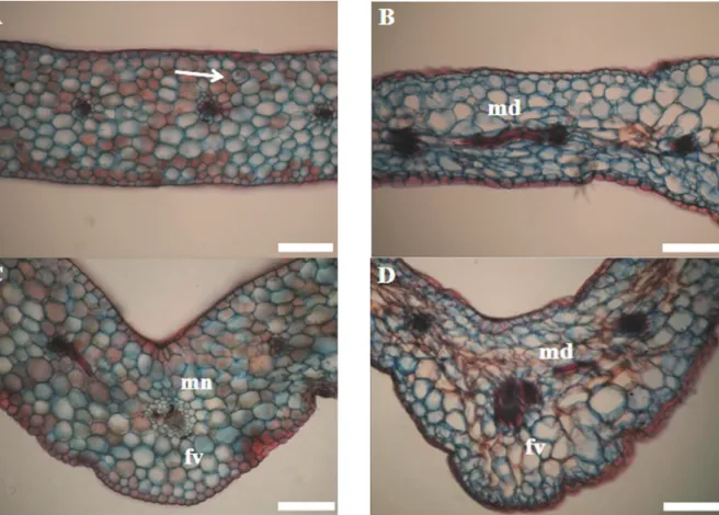
The cross sections of the hybrid orchids showed a homogeneous mesophyll with a mean thickness of 724.08 μm and 17 cell layers (Table 3 and Figure 5). The hybrid’s single layer epidermis, collateral vascular bundles, tetracytic stomata and tissue morphology were similar to those of the native species. However, the leaves of the hybrid were thicker in both the leaf-blade and the midrib region. These differences reflected a greater number of cell layers and larger cells. The hybrid’s stomata showed a more rounded shape and apparently less functional structure than those observed for the native species.
Table 3. Mean values of mesophyll thickness (μm), abaxial and
adaxial epidermis thickness (μm) in relation to CaSiO3
concentrations (mg L-1) in a growth chamber environment for the
hybrid orchid.
CaSiO3
(mg L-1) Mesophyll Adaxial epidermis Abaxial epidermis
0 558.307 b 14.868 b 16.386 b 2.0 724.084 a 19.275 a 24.078 a Means followed by the same letter in columns do not differ significantly (Tukey test, 5% probability level).
Figure 5. Cross-sections of the hybrid orchid species grown in culture medium containing silicon (A and C) and without silicon (B and
As was the case in the native species, the treatment with silicon promoted better seedling development in the hybrid. Deformation of the chlorenchyma was observed in the plants grown in the culture medium without silicon (Figures 5). Silicon deposits were observed only in the tissues of the plants cultivated in silicon containing culture media. As also observed in the native species, greater development was observed in the tissues of plants grown in the culture medium containing silicon. Mesophyll thickness was 29.7% greater in the plants cultivated with silicon compared with those without silicon. The mean thickness of the adaxial epidermis was 29.5% greater in the plants with silicon than in the plants without silicon, and the mean thickness of the abaxial epidermis was 46.9% greater in the plants with silicon than in those without silicon. Therefore, the plants cultivated in the medium containing silicon may have had a greater photosynthetic capacity control of transpiration. These differences could have produced the greater growth observed in these plants cultivated in silicon containing media.
The stomata of the hybrid were tetracytic in shape and were present on both the adaxial and abaxial epidermal surfaces. They were more frequent on the abaxial surface. These characteristics classified this orchid as an amphi-hypostomatic species. According to Evert and Esaú (2006), tetracytic stomata are frequent in many families of monocotelydons. These stomata are enclosed by four subsidiary cells. Two of these cells are parallel to the guard cells. The other pair of cells is polar and is frequently smaller. Pasqual et al. (2011) observed this same stomata type in hybrids orchids, as well as Zanenga-Godoy and Costa (2003) reported this kind of stomata in other orchids species.
The greatest stomatal density was observed in both adaxial and abaxial epidermis in plants treated with CaSiO3 (Tabela 4). The control exhibited the
most desirable value of the polar/equatorial diameter ratio. It is probable that functionality was greater in the control treatment (Table 4). This result is similar to that obtained for the native species. The number and size of the vascular bundles were lower in the plants in the culture media without silicon than in the plants in the culture media containing silicon The vascular tissues play an essential role in the distribution of water and photoassimilate (TAIZ; ZEIGER, 2004). Damage to these tissues may interfere with the development of the plants. In combination with the deficiencies observed in the chlorenchyma of these plants, damage to the vascular tissues may have contributed to the stunted development of the plants in the culture medium
without silicon compared with the plants in the culture media with silicon. Leaf vascular bundles size and density can be modified by environmental stresses such as water stress (SOUZA et al., 2010) and toxic elements on soil (PEREIRA et al., 2011) in different plant species, a a restriction of these characteristics can restrict plant tolerance to environmental stresses.
Table 4. Mean values of stomatal density (SD), polar diameter (PD), equatorial diameter (ED), polar/equatorial ratio and number of cells (NC) in relation to CaSiO3 doses (mg L-1)
concentration in a growth chamber environment for the hybrid orchid species.
Abaxial epidermis CaSiO3
(mg L-1) SD
(stomata mm -2)
PD
(μm) (ED μm) PD/ED NC (epidermal cells mm-2)
0 34.64 b 33.20 b 32.76 b 1.01 a 131 b 2.0 54.92 a 37.18 a 35.56 a 1.04 a 145.4 a CV (%) 17 6.7 3.4 6.4 6.5
Adaxial epidermis
0 19.10 b 31.22 a 27.39 b 1.14 a 101.4 b 2.0 39.40 a 31.04 a 36.47 a 0.85 b 112.4 a CV (%) 24 5.3 5.2 8.9 5.7 Means followed by the same letter do not differ significantly (Tukey test, 5% probability level).
The culture media is an important factor in orchids acclimatization stage and more researches in acclimatization culture media and substrates are necessary to increase these plant production (VENTURIERI; ARBIETO, 2011). Associated with the capacity of silicon to control some plant diseases (SANTOS et al., 2011) and the more adequate structural development and growth of native and hybrids orchids of the present study the presence of calcium silicate are important to culture media of orchids during the acclimatization stage.
Conclusion
Greater seedling growth resulted from the cultivation of the hybrid under artificial light with 2.0 mg L-1 CaSiO
3 and the cultivation of the native
species under artificial light with 0.5 mg L-1 CaSiO
3.
The addition of silicon to the culture medium produced favorable characteristics in the leaf anatomy of the orchid seedlings.
References
ARIGITA, L.; GONZALEZ, A.; TAMÉS, R. S. Influence of CO2 and sucrose on photosynthesis and transpiration of
Actinia deliciosa explants cultured in vitro. Physiologia Plantarum, v. 115, n. 10, p. 166-173, 2002.
BRAGA, F. T.; PASQUAL, M.; CASTRO, E. M.; DIGNART, S. L.; BIAGIOTTI, G.; PORTO, J. M. P.
Qualidade de luz no cultivo in vitro de Dendranthema
grandiflorum cv. Rage: características morfofisiológicas.
EVERT, R. F.; ESAÚ. Esau's Plant anatomy:meristems,
cells, and tissues of the plant body: their structure, function, and development. 3ed. Madison: Wiley Interscience, 2006. FERREIRA, D. F. Sisvar: a computer statistical analysis
system. Ciência e Agrotecnologia, v. 35, n. 6,
p. 1039-1042, 2011.
JONES, H. G. Plants and microclimate: a quantitative
approach to environmental plant physiology. 2ed. Cambridge: Cambridge University Press, 1992.
KNUDSON, L. A new nutrient solution for the
germination of orchid seed. American Orquid Society
Bulletim, v. 14, n. 14, p. 214-217, 1946.
KRAUS, J. E.; ARDUIM, M. Basic manual of methods
in plant morphology. Rio de Janeiro: UFRJ, 1997.
MARIN, J. A. High survival rates during acclimatization of micropropagated fruit tree rootstocks by increasing
exposures to low relative humidity. Acta Horticulturae,
v. 616, n. 1, p. 139-142, 2003.
MURASHIGE, T.; SKOOG, F. A revised medium for rapid growth and bioassays with tobacco tissue cultures.
Physiologia Plantarum, v. 15, n 43, p. 473-497, 1962.
PASQUAL, M.; SOARES, J. D. R.; RODRIGUES, F. A.; ARAUJO, A. G.; SANTOS, R. R. Influência da qualidade de luz e silício no crescimento in vitro de orquídeas nativas e híbridas. Horticultura Brasileira, v. 29, n. 3,
p. 324-329, 2011.
PEREIRA, F. J.; CASTRO, E. M.; OLIVEIRA, C.; PIRES, M. F.; PASQUAL, M. Mecanismos anatômicos e fisiológicos de plantas de aguapé para a tolerância à
contaminação por arsênio. Planta Daninha, v. 29, n. 2,
p. 259-267, 2011.
PESCHEL, S.; BEYER, M.; KNOCHE, M. Surface characteristics of sweet cherry fruit: stomata-number, distribution, functionality and surface wetting. Scientia Horticulturae, v. 97, n. 3, p. 265-278, 2003.
PULZ, A. L.; CRUSCIOL, C. A. C.; LEMOS, L. B.; SORATTO, R. P. Influência do silicato e calcário na nutrição, produtividade e qualidade da batata sob
deficiência hídrica. Revista Brasileira de Ciência do
Solo, v. 32, n. 4, p. 1651-1659, 2008.
ROMANO, A.; MARTINS-LOUÇÃO, M. A. Strategies
to improve rooting and acclimatization of cork oak. Acta
Horticulturae, v. 616, n. 3, p. 275-278, 2003.
ROMERO-ARANDA, M.; JURADO, O.; CUARTETO, J. Silicon alleviates the deleterious salt effect on tomato
plant growth by improving plant water status. Jounal
of Plant Physiology, v. 163, n. 8, p. 847- 855, 2006.
SANTOS, G. R.; CASTRO NETO, M. D.; RAMOS, L. N.; SARMENTO, R. A.; KORNDÖRFER, G. H.; IGNÁCIO, M. Effect of silicon sources on rice diseases
and yield in the State of Tocantins, Brazil. Acta
Scientiarum. Agronomy, v. 33, n. 3, p. 451-456, 2011.
SOARES, J. D. R.; PASQUAL, M.; RODRIGUES, F. A.; VILLA, F.; CARVALHO, J. G. Foliar fertilization with silicon in the acclimatization of a hybrid orchid. Ciência e Agrotecnologia, v. 32, n. 2, p. 626-629, 2008.
SOUZA, T. C.; MAGALHÃES, P. C.; PEREIRA, F. J.; CASTRO, E. M.; SILVA JÚNIOR, J. M.; PARENTONI, S. N. Leaf plasticity in successive selection cycles of 'Saracura'
maize in response to periodic soil flooding. Pesquisa
Agropecuária Brasileira, v. 45, n. 1, p. 16-24, 2010.
TAIZ, L.; ZEIGER, E. Plant Physiology. 3ed. Porto
Alegre: Artmed, 2004.
VACIN, E. T.; WENT, F. W. Some pH changes in nutrient solutions. Botany Gazette, v. 110, n. 1, p. 605-613,
1949.
VALENTE, A.; MORAIS, R.; COUTO, C.; CORREIA, J. H. Modeling, simulation and testing of a silicon soil moisture sensor based on the dual-probe heat-pulse
method. Sens and Actuators A, v. 115, n. 2, p. 434-439,
2004.
VENTURIERI, G. A.; ARBIETO, E. A. M. Ex-vitro
establishment of Phalaenopsisamabilis seedlings in different
substrates. Acta Scientiarum. Agronomy,
v. 33, n. 3, p. 495-501, 2011.
ZANEGA-GODOY, R.; COSTA, C. G. Leaf anatomy of
four species of genus Cattleya Lind (Orchidaceae) of the
Brasilian Central Plateau. Acta Botanica Brasilica,
v. 17, n. 1, p. 101-119, 2003.
ZHOU, T. S. The detection of the accumulation of silicon in Phalaenopsis (Orchidaceae). Annals of Botany,
v. 75, n. 1, p. 605-607, 1995.
Received on October 19, 2011. Accepted on December 31, 2011.