Printed version ISSN 0001-3765 / Online version ISSN 1678-2690 http://dx.doi.org/10.1590/0001-3765201620160030
www.scielo.br/aabc
Inhibition of proteases and phospholipases A
2from
Bothrops atrox
and
Crotalus durissus
terrificus
snake venoms by ascorbic acid, vitamin E, and B-complex vitamins
CArlos H.M. olIvEIrA, AndErson A. sIMão, MArCus v.C. TrEnTo, PEdro H.s. CésAr and sIlvAnA MArCussI
Departamento de Química, Laboratório de Bioquímica, Universidade Federal de Lavras/ UFLA, Campus UFLA, 37200-000 Lavras, MG, Brasil
Manuscript received on January 21, 2016; accepted for publication on May 26, 2016
ABsTrACT
The enzyme inhibition by natural and/ or low-cost compounds may represent a valuable adjunct to traditional serotherapy performed in cases of snakebite, mainly with a view to mitigate the local effects of envenoming. The objective of this study was to evaluate possible interactions between vitamins and enzymes that comprise Bothrops atrox and Crotalus durissus terrificus venoms, in vitro. Proteolysis
inhibition assays (substrates: azocasein, collagen, gelatin and fibrinogen), hemolysis, coagulation,
hemagglutination were carried out using different proportions of vitamins in face of to inhibit minimum effective dose of each venom. The vitamins were responsible for reducing 100% of breaking azocasein by C.d.t. venom, thrombolysis induced by B. atrox and fibrinogenolysis induced by both venoms. It is suggested the presence of interactions between vitamin and the active site of enzymes, for example the interactions between hydrophobic regions present in the enzymes and vitamin E, as well as the inhibitions exercised by antioxidant mechanism.
Key words: antiophidian properties, antioxidants, blood cells, hemostatic processes, natural inhibitors.
Correspondence to: Anderson Assaid Simão E-mail: [email protected]
InTroduCTIon
The Bothrops atrox species can be found frequently
in the Amazon region and is responsible for
about 80% of snake bites in the region. Studies
indicate that the envenoming by B. atrox venom
is characterized by a strong inflammatory response
triggering rupture of blood vessels, production
of cytokines, leukocyte migration and cell death.
Importantly, the lethality of the venom is associated
with inflammatory factors induced by enzymes that
act as thrombin-like, which are primarily proteases
(Barros et al. 1998).
Accidents with snakes of the genus Crotalus
(Viperidae family) are less frequent, but because of
the severity of the systemic response, they present
high lethal potential, and the envenoming by these
snakes is characterized by neurotoxic, coagulant
and myotoxic action (Sgrignolli et al. 2011).
Crotalus durissus is a species that inhabits
areas of the Central Brazilian Cerrado and arid
and semi-arid regions of Northeast and fields and
durissus terrificus is prevalent in the southeastern and southern regions (Melgarejo 2003).
Among the components present in the venom of snakes, those showing the greatest potential as biotechnological agents and responsible for the most serious disorders within the envenoming are the phospholipases A2 (PLA2), proteases, lectins type C, hyaluronidases, disintegrins and L-amino acid oxidases.
The PLA2s present in snake venoms are respon-sible for pharmacological effects involving blood incoagulability, myonecrosis, edema formation, in-hibition of platelet aggregation and hypertension. These enzymes are also present in the pancreatic secretions, being widely distributed in animals and plants in nature (Burke and Dennis 2009).
Among the proteases present in snake venoms are the serineproteases that act selectively on factors of the coagulation cascade, with effect on
platelet aggregation, fibrinolysis and coagulation
(Braud and Wisner 2000) and metalloproteinases, dependent on the zinc ion, responsible for bleeding,
tissue damage and local inflammation (Telesi and
Machado 2008).
In literature, little has been discussed about the venom-vitamins interaction. Mukherjee et al. (1997) have shown that vitamin E inhibited the lipid peroxidation of the cell membrane of human erythrocytes induced by the Vipera russeli
venom. Mohamed et al. (2013) obtained similar results with the synthesis of a lipophilic ascorbic acid derivative capable of inhibiting the activity of PLA2 isolated from Vipera russelli venom. Soon the investigation of the interaction between vitamins and snake venoms seems to be necessary because vitamins are molecules that are closely linked to the performance of enzymatic functions present in organisms. In addition, various enzymes, such as phospholipases and proteases present in the venoms show functional and structural homology with enzymes present in the human body, enabling similarities between the inhibition of enzymes
resulting from venoms and the likely effects on human endogenous enzymes.
Vitamins are the primary antioxidants obtained by feeding, being involved in processes such as cell migration, energy metabolism, vasomotor control, transportation of metal ions and glucose, amino acid formation, maturation of blood cells, cholesterol metabolism, decreased lipid membrane degradation - thereby reducing the formation of eicosanoids, repair of damage in DNA molecule, stimulation of the immune system and production of hormones (You et al. 2009, Du et al. 2011, Traber and Stevens 2011, Coquille et al. 2012).
The aim of this study was to evaluate the interaction between micronutrients (vitamins of B complex, vitamin E and ascorbic acid - together, and ascorbic acid alone) and enzymes present in the venom of B. atrox and C.d.t. snakes species, in vitro, focusing on the inhibition of proteases and PLA2s acting on human cells and molecules.
MATErIAls And METHods
SNAkE vENOMS
The venoms of B. atrox and C.d.t. were commercially purchased from Bio-agents serpentarium (Batatais, SP, Brazil).
HUMAN bLOOD
The coagulant, thrombolytic, hemagglutinating and hemolytic activities were performed using human blood collected in tubes containing sodium citrate or without anticoagulant (thrombolytic activity).
The experiment was conducted in accordance with the standards of the Ethics Committee on Human research (COEP) from Universidade Federal de Lavras, and has been approved by this committee (Nº CAAE/15258413.0.00000.5148).
vITAMINS
a drugstore and the isolated vitamin C was acquired from a compounding pharmacy.
azOCASEIN aCTIVITY
For purposes of this test, we made use of a methodology based on two methods previously described by Gutierrez et al. (1988) and Van der Walt and Joubert (1971) adapted by Wang et al. (2004). Thus, substrate (azocasein) concentration and dissolution conditions were maintained, obtaining proportions identical to those used by Van der Walt and Joubert (1971) to be incorporated into agarose gel (method of Gutierrez et al. (1988). The minimum dose azocaseinolytic of venoms, responsible for the formation of an activity halo of approximately 1.2 cm, has been previously determined being used in the inhibition assays. For gel preparation, azocasein, at 0.058%, was dissolved in 4 ml of Tris-HCl (pH= 9.6) and then added to the gel composition. The gels containing the samples remained in a cell culture chamber at 37°C for 12 hours. The formation of translucent halo around the opening in the gel was indicative of activity, where the halos were measured in millimeters for quantifying the proteolytic activity on azocasein. Mixtures of venoms (30µg) and vitamins previously incubated (30 minutes at 37°C) were evaluated in proportions of 1:1, 1:2.5, 1:5 and 1:10 (venom: vitamin; w/w), as well as controls containing venoms and vitamins in isolation.
PrOTEOLYTIC aCTIVITYONTHE cOLLAGENAND gELATIN
The proteolytic analysis of collagen and gelatin (at 0.058%) was carried out through the preparation of a gel identical to the one described in the previous section with the change of the substrate for one of the proteins of interest. For displaying of results, the gel was subjected to coloring in starch black solution 0.25% (w/v) dissolved in water, followed by decoloration with 10% acetic acid. The analyzed samples were incubated in the same proportions described for azocaseinolytic activity.
cLOTTING aCTIVITY
The clotting time of citrated plasma (200 µL) was evaluated in assays with addition of vitamins to the plasma to previous incubation for a 5-minute period, and subsequent addition of venom and timing, as well as preincubations of vitamins with venom, for a 5-minute period, and subsequent plasma addition and timekeeping. Controls containing solely vitamins or venoms were also performed. The citrated plasma was placed in test tubes dipped in water bath at 37°C and kept until temperature stabilization with subsequent addition of the
samples. The minimum coagulant dose was defined
as the minimum amount of venom responsible for plasma clotting in a time interval between 1 and 1.5 minutes (Seliestre et al. 1990). The proportions of venom (10µg) and vitamin within the samples were 1: 0.5, 1: 1, 1: 2.5 and 1: 5 (w/w).
tHrOMBOLYTIC aCTIVITY
Thrombolytic activity was performed by distributing the collected blood into microtiter plates (100 µl per well) to obtain the thrombus. Then, several doses of venom were tested to determine the one responsible for a complete dissolution of the
thrombus. The activity was quantified by the volume
of liquid released from the thrombus, as described by Cintra et al. (2012). Controls containing only vitamins were also performed. The inhibitory potential of vitamins was measured after previous incubation (40 minutes at 37°C) of vitamins and venom (25µg), at the proportions 1:1, 1:5 and 1:10 (venom: vitamin; w/w) and subsequent addition to the thrombus.
HEMAGGLUTINATION aSSAY
Na2HPO4, kH2PO4, NaCl), followed by three wash steps with centrifugation at 1700g for 15 minutes. Then, the erythrocytes were suspended in the same solution (1: 20 v/v blood: saline solution) and distributed into microtiter plate (volume 100 µL/ well). The venom (40µg) and vitamin samples, previously incubated (30 minutes at 37°C), were then added, followed by incubation for another hour at room temperature. The B. atrox and
C.d.t. venoms were tested in venom and vitamin proportions of 1: 0.5 and 1: 1 (w/w) for vitamins, isolated ascorbic acid and vitamin complex. The results were evaluated by a qualitative visual analysis, 2 hours after addition of samples, using
an electronic magnifier.
HEMOLYTIC aCTIVITYIN lIQUID mEDIUM
The blood collected (10 mL) in the presence of an anticlotting agent was immediately mixed with saline solution (2 mM NaH2PO4; 3 mM Na2HPO4; 154 mM NaCl; pH 7.4) and centrifuged at 700g (Fanem Baby®I Modelo 206 BL) for ten minutes. The plasma was removed, and the red cells were suspended in 5 mmol L-1 phosphate buffer, pH 7.4 and centrifuged under the same conditions. This washing procedure was repeated three times at 4°C. The 100% red blood cell concentrate was diluted to 2% hematocrit (137.33 µmol L-1 lipid, respectively), using the same buffer.
The hemolytic activity was evaluated by incubating 1 mL of erythrocyte suspension (2%) for 60 minutes at 37°C with the pre-incubated of venoms (20µg) with vitamins, at different ratios (1:1, 1:2.5, 1:5 and 1:10 w/w), followed by centrifugation at 1500g for seven minutes. The hemoglobin concentration was determined in the supernatant by measuring the absorbance at 540 nm (Shimadzu UV-160 1 PC) according to rangel et al. (1997), with the modifications of Preté et al. (2010). The controls were performed using an erythrocyte suspension in PBS (c1 = mechanical
hemolysis control) and distilled water (c2 = total hemolysis control) (Preté et al. 2010).
The hemoglobin concentration was determined using the equation:
in which Aa, Ac1 and Ac2 are, respectively, the absorbance of the sample and of the controls c1 and c2 at 412 nm.
fIBrINOGENOLYTIC aCTIVITY: POLYACrYLAMIDE gEL eLECTrOPHOrESISWITH SODIUM SULFATE dODECYLTO vISUALIzATION
The fibrinogenolytic activity was carried out with modifications (Czaikoski et al. 2010). The vitamins
were incubated with bovine fibrinogen (60 µg) for 90 minutes at 37°C in a final volume of 25 µl (PBS), as control. The samples were analyzed by polyacrylamide gel electrophoresis at 12% (acrylamide: bisacrylamide, 19:1) in the presence of SDS (SDS-PAGE) under denaturing conditions (Laemmli 1970). A sample control containing only
fibrinogen was used for the visualization of the band pattern corresponding to α, β and γ chains of fibrinogen. The minimum fibrinogenolytic dose of venom, responsible for the degradation of α and β chains from fibrinogen has been previously
determined (60µg to B. atrox and 80µg to Crotalus durissus terrificus venom) and used in inhibition assays.
The inhibitory potential of the vitamins on the proteolytic action of B. atrox and C. d. t. venoms was assessed by SDS-PAGE after preincubation of samples (1:1, 1:5 and 1:10, venom: vitamin; w/w) for 30 min. at 37ºC, with subsequent addition of
fibrinogen (60 µg) and incubation for 90 minutes at
sodium dodecyl sulfate (SDS) and 0.05% (w/v) bromophenol blue] and boiling the samples for 5 minutes in a water bath (Laemmli 1970).
Electrophoretic run was held for a period of 2 hours at 90V. After run, gel was removed and col-ored for 40 minutes in 0.1% Coomassie Blue G-250 (w/v) dissolved in water: methanol: acetic acid (40: 50: 10, v/v) and destained in 10% acetic acid.
STATISTICAL aNALYSIS
Statistical analyses were performed with GraphPad Prism software, and the Student’s t-test was chosen to compare the experimental mediums. results
with p <0.05 were considered as significant. All
assays were performed in triplicate.
rEsulTs
PrOTEOLYTIC aCTIVITY
The B. atrox venom presented proteolytic activity on substrates azocasein, collagen, and gelatin, while the C.d.t. showed activity only on azocasein.
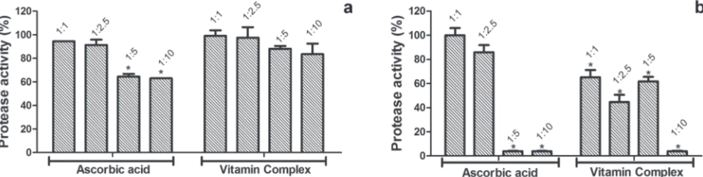
The greatest inhibitions, obtained to the pro-teolytic activity induced by B. atrox venom, were observed in a proportion of 1: 10 with reduced ac-tivity 63% and 83%, after incubation with ascorbic acid and vitamin complex, respectively (Fig. 1a).
Ascorbic acid and vitamin complex inhibited
the azocaseinolytic activity with greater efficiency
at the proportion 1: 10, where the C.d.t. venom had
its activity reduced to 5% in preincubation with ascorbic acid and vitamin complex (Fig. 1b). At the ratio of 1: 5, ascorbic acid also reduced the C.d.t. activity to 5%, while in the presence of vitamin complex activity was reduced to approximately 65%. At ratios of 1: 2.5 and 1: 1, inhibition was observed only in the presence of vitamin complex reducing the proteolytic activity to 70 and 50%, respectively (Fig. 1b).
None of the vitamin treatments proved to be effective in inhibiting proteolysis of collagen (data not shown).
The vitamin complex and ascorbic acid were effective in inhibiting gelatinolytic activity induced by B. atrox, and inhibitions were observed in all evaluated proportions (Fig. 2). The most effective ratio of 1:10 induced activity reduction to approximately 65% and 52% for vitamin complex and ascorbic acid, respectively (Fig. 2).
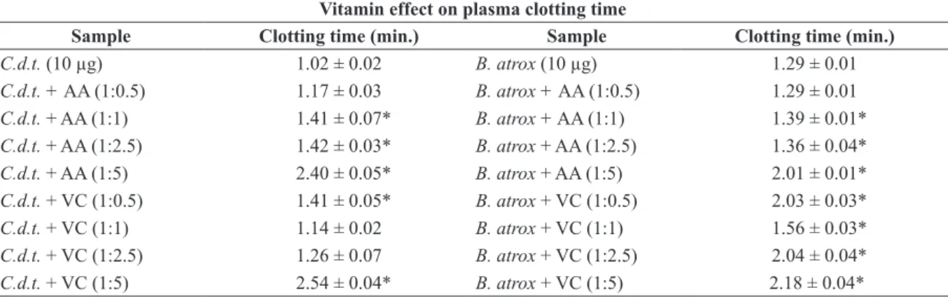
cLOTTING aCTIVITY
Either the preincubation of vitamin with plasma (Table I) and with venoms (Table II), both proposed
for coagulant testing, showed significant results
and extended clotting time for ascorbic acid and vitamin complex.
Preincubation with venom and vitamins stood out between the two treatments (Table II). This suggests, therefore, possible interactions between toxins and vitamin molecules. In addition,
the largest proportions, such as 1: 2.5 and 1: 5, promoted the highest inhibition for both venoms (Table II).
Hence, lower inhibitions were observed for preincubated vitamins with plasma, demonstrating non-significant changes in coagulation time induced by C.d.t. venom (Table I).
On the part of vitamins, higher proportions could not be assessed due to the saturation limit amounts of the reaction environment; however, variations in incubation time and concentration of ions can be further evaluated.
TABlE II
Clotting activity induced by Crotalus durissus terrificus and Bothrops atrox venom on human citrated plasma proceeding with preincubation of vitamins with the venoms in different ratios (for 5 min. at 37°C).
vitamin effect on plasma clotting time
sample Clotting time (min.) sample Clotting time (min.)
C.d.t. (10 µg) 1.02 ± 0.02 B. atrox (10 µg) 1.29 ± 0.01
C.d.t. + AA (1:0.5) 1.17 ± 0.03 B. atrox + AA (1:0.5) 1.29 ± 0.01
C.d.t. + AA (1:1) 1.41 ± 0.07* B. atrox + AA (1:1) 1.39 ± 0.01*
C.d.t. + AA (1:2.5) 1.42 ± 0.03* B. atrox + AA (1:2.5) 1.36 ± 0.04*
C.d.t. + AA (1:5) 2.40 ± 0.05* B. atrox + AA (1:5) 2.01 ± 0.01*
C.d.t. + VC (1:0.5) 1.41 ± 0.05* B. atrox + VC (1:0.5) 2.03 ± 0.03*
C.d.t. + VC (1:1) 1.14 ± 0.02 B. atrox + VC (1:1) 1.56 ± 0.03*
C.d.t. + VC (1:2.5) 1.26 ± 0.07 B. atrox + VC (1:2.5) 2.04 ± 0.04*
C.d.t. + VC (1:5) 2.54 ± 0.04* B. atrox + VC (1:5) 2.18 ± 0.04*
Results expressed in clotting time represent averages of triplicates and the standard deviation (SD). The significance asterisks result from comparative analysis of treatments with control containing only venom. AA = ascorbic acid. VC = vitamin complex.
TABlE I
Coagulant activity induced by Crotalus durissus terrificus and Bothrops atrox venoms on human citrated plasma preincubated with vitamins in different ratios (for 5 min. at 37°C).
vitamin effect on plasma clotting time
sample Clotting time (min.) sample Clotting time (min.)
C.d.t. (10 µg) 1.05 ± 0.04 B. atrox (10 µg) 1.15 ± 0.02
C.d.t. + AA (1:0.5) 1.21 ± 0.03 B. atrox + AA (1:0.5) 1.31 ± 0.05
C.d.t. + AA (1:1) 1.12 ± 0.01 B. atrox + AA (1:1) 1.34 ± 0.05
C.d.t. + AA (1:2.5) 1.31 ± 0.03* B. atrox + AA (1:2.5) 1.49 ± 0.09*
C.d.t. + AA (1:5) 1.39 ± 0.04* B. atrox + AA (1:5) 2.07 ± 0.06*
C.d.t. + VC (1:0.5) 1.18 ± 0.03 B. atrox + VC (1:0.5) 1.39 ± 0.04*
C.d.t. + VC (1:1) 1.10 ± 0.02 B. atrox + VC (1:1) 1.56 ± 0.04*
C.d.t. + VC (1:2.5) 1.16 ± 0.01 B. atrox + VC (1:2.5) 2.23 ± 0.05*
C.d.t. + VC (1:5) 1.16 ± 0.04 B. atrox + VC (1:5) 2.24 ± 0.05*
Results expressed in clotting time represent averages of triplicates and the standard deviation (SD). The significance asterisks result from comparative analysis of treatments with control containing only venom. AA = ascorbic acid. VC = vitamin complex.
Figure 2 - Vitamins effect on gelatinolytic activity induced by
tHrOMBOLYTIC aCTIVITY
Only the B. atrox venom induced lysis of the thrombus. The vitamin complex preincubated with venom at 1: 5 and 1: 10 ratios was able to reduce the thrombolytic activity to 15 and 3.3%, respectively (Fig. 3). However, ascorbic acid showed higher inhibitory potential, being responsible for reducing the venom activity to 47, 8 and 3.8% at ratios of 1: 1, 1: 5 and 1: 10, respectively (Fig. 3).
HEMAGGLUTINATION aSSAY
Only the B. atrox venom induced hemagglutination activity. This activity was completely inhibited after preincubation of the venom with ascorbic acid at the ratio of 1: 0.5, whereas in higher proportions (1: 1, 1: 2.5 and 1: 5), partial agglutination
of the erythrocytes was observed. Absence of hemagglutination was also observed after preincubating the venom with vitamin complex at a ratio of 1: 1 (data not shown).
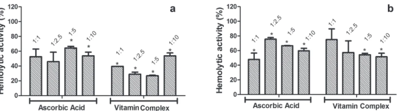
HEMOLYTIC aCTIVITYIN lIQUID mEDIUM
The two treatments evaluated, ascorbic acid and vitamin complex, reduced hemolytic activity, highlighting the vitamin complex which reduced the hemolytic activity induced by the B. atrox
venom to 27% at the proportions of 1: 2.5 and 1: 5 (Fig. 4a). Although ascorbic acid in all tested proportions has shown significant inhibition against the C.d.t. venom, the greater inhibitions of hemolysis induced by this venom were obtained after incubation with vitamin complex at ratios of 1: 5 and 1: 10, with activity reduced to about 50% (Fig. 4b).
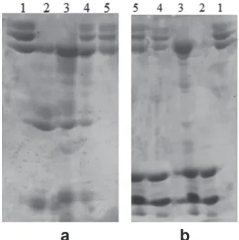
fIBrINOGENOLYTIC aCTIVITY
The two treatments evaluated were effective in
inhibiting the degradation of fibrinogen molecules,
induced by the B. atrox venom (Fig. 5a) and C.d.t.
venom (Fig. 5b). Some fibrinopeptides can be seen on line 4 of Figure 5a, which corresponds to the treatment with ascorbic acid at a ratio of 1: 5, showing partial inhibition of proteolytic activity. Figure 5b shows 100% inhibition of the activity induced by the C.d.t. venom, for both treatments.
Figure 4 - Vitamins effect on hemolytic activity. (a) Bothrops atrox venom previously incubated with ascorbic acid or vitamin complex in different ratios. (b) Crotalus durissus terrificus venom previously incubated with ascorbic acid or vitamin complex in different ratios. Data presents average of triplicate and the values obtained to the venom alone were converted to 100% activity. Figure 3 - Vitamins effect on thrombolytic activity induced by
However, smaller and larger doses were also evaluated (data not shown) and shown to be without effect, only being presented the ratio of 1: 5 and this one is considered as corresponding to the equivalent molar concentrations between
fibrinogenolytic protease and vitamins.
substantially all classes of proteases expressed in the B. atrox venom, although they have been shown to be effective in inhibiting the C.d.t. venom.
The lack of inhibition observed for both vitamin samples, evaluated on the proteolysis of collagen molecules, is probably due to a quick action of several proteases from the venom, which act on coagulation factors, platelets and bleeding induction, requiring additional tests to exploit the concentration-time relation in order to observe the inhibitory effect (Escalante et al. 2011).
The different structures of the evaluated substrates, azocasein, gelatin and collagen, suggest the action of different proteases on each of them, enabling the interaction of these with inhibitors also in a variable form, which explains the variations in inhibition percentages observed at each test.
The ascorbic acid and the vitamin complex
have drastically inhibited (inhibition of ≈ 100%)
the fibrinogenolytic activity induced by both venoms evaluated. The result is in agreement with the data obtained in coagulation assay, since the fibrinogenolysis is directly related to blood clot development (Braud and Wisner 2000). Thus, the same way as vitamins proved effective in prolonging plasma-clotting time, by partially inhibiting coagulant toxins, they were also able to
inhibit degradation of fibrinogen.
In the coagulant assay, the lower inhibition response by vitamin incubation with plasma can be related to the less interaction between vitamins and constituents of the plasma that participate in clot cascade, in comparison to the interactions between vitamins and molecules as PLA2 and proteases, present in crotalic and bothropic venoms, act on coagulation. In addition, different amounts of these molecules in the composition of venoms can interfere in the process, as well as variations in the ionic composition of the same (enzyme cofactor) that are able to interact with vitamins.
Antioxidant compounds have been suggested as being able to interact with binding sites of
Figure 5 - Vitamins effect on fibrinogenolytic activity. (a) Samples: 1- Fibrinogen (60 µg); 2- Bothrops atrox venom (60 µg); 3- Fibrinogen + B. atrox venom; 4- Fibrinogen + B. atrox
venom + ascorbic acid – AA (300 µg) and 5- Fibrinogen + B. atrox venom + vitamin complex – VC (300µg). (b) Samples: 1- Fibrinogen (60 µg); 2- Crotalus durissus terrificus venom (80 µg); 3- Fibrinogen + C. d. terrificus venom; 4- Fibrinogen + C. d. terrificus venom + ascorbic acid – AA (300 µg) and 5- Fibrinogen + C. d. terrificus venom + vitamin complex – VC (300µg).
dIsCussIon
As observed during caseinolytic activity tests, the inhibition differential can be related to the greater expression of proteases from snake venoms of the species B. atrox (69.3% serineproteases plus metalloproteases) compared to the species C.d.t.
cofactors within protease structure, and modify their structure; thus, interfering with the binding of cofactors and consequently resulting in reduced enzymatic activity (Patiño et al. 2013, Oliveira et al. 2014). For instance, thiol groups have been described with the ability of both chelating enzyme cofactors and interfering with enzymatic redox reactions in pharmacological tests induced by viperid venoms (Sunitha et al. 2011). Another
result that confirms this hypothesis is the inhibition
of proteolytic activity by rosmarinic acid, cinnamic acid and their analogues (Aung et al. 2011). Thus, the aforementioned interaction seems to be one of the inhibition mechanisms performed by vitamins with antioxidant activity on the various activities induced by different proteases, such as coagulant, proteolytic on various substrates and thrombolytic activities.
The inhibition of proteases, in addition to increasing vitamin survival time and decrease severity of toxic signs also contributes in preserving the cardiovascular system changes by inhibiting blood pressure lowering and depressed atrial contractility (Shabbir et al. 2014).
The hemagglutinating activity exerted by C-type lectins may vary in composition of the different batches of venoms used, since this protein class has increased expression in juvenile snake specimens and semi-adults and lower expression in adults (Cruz et al. 2005). Thus, the absence of activity observed for the C.d.t. venom may be related to maturity of the snakes used to obtain the samples of venom used in evaluations.
The C-type lectins are related to platelet and erythrocytes aggregation (zelensky and Gready 2005), acting by binding to carbohydrates present in cell membranes (kassab 2001). Thus, vitamins could be interacting with the glycidic ligands present on the surface of erythrocytes and preventing the lectin-binding present in the B. atrox venom, resulting in complete inhibition of hemagglutinating activity observed in this study.
Interactions of lectin present in the venom with possible inhibitory molecules are poorly explored, with no indications of possible action mechanisms of vitamins, such as lectin inhibitors (Oliveira et al. 2016).
The hemolytic activity partial inhibition, whose induction is primarily attributable to PLA2s, may represent the absence of interactions between vitamin molecules and phospholipases in
venoms, as well as the difficulties of interaction
of vitamins with membrane components, such as phospholipids, resulting in low protection against the catalytic action exerted by phospholipases.
However, there are reports in literature about a lipophilic ascorbic acid derivative, synthesized in laboratory, which due to its changes in polarity, can be effective in inhibiting PLA2 activity isolated from Vipera russelli (Mohamed et al. 2013). Moreover, venom proteases may also contribute to hemolysis induction, being, thus, necessary to inhibit several phospholipases and proteases by vitamins, hindering their action.
Vitamin E, present in the vitamin complex, can also have inhibitory effect against lipid peroxidation of the cell membrane of human erythrocytes, this being one of the possible mechanisms for hemolytic activity reduction (Mukherjee et al. 1997).
Vitamins of the B complex may contribute to repair mechanisms, acting on the damage caused to cellular membrane by free radicals generated by toxin activity (Huang et al. 2010), as well as ascorbic acid (antioxidant) which can neutralize radicals reducing damage to the membranes. This way, the vitamins E, B complex and ascorbic acid, present in the vitamin complex, probably act in synergy through several action mechanisms, reducing the hemolytic activity induced by the toxins.
The inhibitory effects provided by ascorbic acid and the B-complex vitamins, analyzed in this paper, on the biological activities induced by bothropic and crotalic venoms, have mechanisms
come from the antioxidant activity of some vitamins (e.g. ascorbic acid), as well as interactions between hydrophobic regions of others (e.g. vitamin E). Nevertheless, further studies are required to expand the mechanistic discussions about interactions between vitamins and toxins and of these with animal cells and molecules, aiming to formulate therapies complementary to serotherapy, increasing
treatment efficiency.
ConClusIons
The vitamin complex evaluated, as well as the isolated ascorbic acid, show inhibitory potential on activities, mainly the ones induced by proteases and PLA2, the major classes of toxins present in the snake venoms from the Viperidae family.
Once vitamins can be easily obtained in
pharmaceutical formulations certified by ANVISA
(Brazilian National Health Surveillance Agency), they represent a safer alternative and can bring
benefits to snakebite victims.
There is a lack of knowledge on the interaction of vitamins with the different classes of enzymes present in snake venoms, since existing studies describe only interactions between lipophilic vitamins and phospholipases.
Therefore, several additional studies should be conducted to extend the information on inhibitory mechanisms, administration forms and dosages which may be effective as adjuvants of traditional serotherapy.
ACKnoWlEdGMEnTs
The authors are grateful to the Conselho Nacional de Desenvolvimento Científico e Tecnológico (CNPq), Coordenação de Aperfeiçoamento de Pessoal de Nível Superior (CAPES) and Fundação de Amparo à Pesquisa do Estado de Minas Gerais (FAPEMIG), for financial support. They thank STTA (serviços técnicos de tradução e analises) provided the English editing of the paper.
rEsuMo
A inibição enzimática por compostos naturais e/ou de baixo custo pode representar um valioso adjuvante ao tratamento soroterápico tradicional realizado em casos
de ofidismo, com vistas a amenizar principalmente
os efeitos locais do envenenamento. Objetivou-se avaliar in vitro possíveis interações entre vitaminas e enzimas presentes nas peçonhas de Bothrops atrox e Crotalus durissus terrificus. Ensaios de inibição de proteólise (substratos: azocaseína, colágeno, gelatina e
fibrinogênio), hemólise, coagulação, hemaglutinação,
foram realizados utilizando diferentes proporções de vitaminas em face de inibir à dose mínima efetiva de cada peçonha. As vitaminas foram responsáveis por reduzir 100% a quebra da azocaseína por peçonha de C.d.t., a trombólise induzida por B. atrox e a fibrinogenólise induzida por ambas as peçonhas. Sugere-se a presença de interações de vitaminas com o sítio ativo das enzimas, como por exemplo, interações entre regiões hidrofóbicas presentes nas enzimas e vitamina E, assim como, inibição de algumas atividades por mecanismo de ação antioxidante.
Palavras-chave: propriedades antiofídicas, antioxidantes, células sanguíneas, processos hemostáticos, inibidores naturais.
rEFErEnCEs
aUNG Ht, fUrUkAWA t, nIkAI t, nIWA m AND tAkAYA y. 2011. Contribution of cinnamic acid analogues in ros-marinic acid to inhibition of snake venom induced hemor-rhage. Bioorg Med Chem 19: 2392-2396.
bArrOS Sf, frIEDLANSkAIA i, PETrICEVICH vl AND kIPNIS tl. 1998. Local inflammation, lethality and cyto-kine release in mice injected with Bothrops atrox venom. Mediat Inflamm 7: 339-346.
brAUD Sb AND wISNEr ac. 2000. Snake venom proteins acting on hemostasis. Biochimie 82: 851-859.
bUrkE Je AND dENNIS ea. 2009. Phospholipases A2 Biochemistry. Cardiovasc Drugs Ther 23: 49-59. cINTrA ac, dE tONI lg, SArTIM ma, frANCO JJ,
cAETANO rc, mUrAkAMI mt AND SAMPAIO Sv. 2012. Batroxase, a new metalloproteinase from B. atrox snake venom with strong fibrinolytic activity. Toxicon 60: 70-82. cOQUILLE Sr, fITzPATrICk c AND tHOrE tb. 2012. The last piece in the vitamin B1 biosynthesis puzzle: Structural and functional insight into yeast HMP-P synthase. JBC Papers in Press 287: 42333-42343.
crUz aH, mENDONçA rz AND PETrICEVICH vl. 2005.
Mor-phological, Functional, and Biochemical Changes in Mu-rine Macrophage. Mediat Inflamm 6: 349-359.
czAIkOSkI Pg ETAL. 2010. Anticoagulant and fibrinogeno-lytic properties of the venom of Polybia occidentalis social wasp. Blood Coagul Fibrinolysis 21: 653-659.
dU q, wANG H AND XIE J. 2011. Thiamin (Vitamin B1) Bio-synthesis and regulation: A rich Source of Antimicrobial Drug Targets?Int J Biol Sci 7: 41-52.
eSCALANTE t, orTIz n, rUCAVADO a, SANCHEz ef, rICHArDSON m, fOX Jw AND gUTIérrEz Jm. 2011. role of collagens and perlecan in microvascular stability: exploring the mechanism of capillary vessel damage by snake venom metalloproteinases. Plos One 6: e28017. fIGUEIrOS m AND lAJOLO fm. 2009. Effect of chemical
modification of Phaseolus vulgares lectins on their biological properties. J Agric Food Chem 45: 639-643. gEOrGIEVA d, öHLEr m, SEIFErT J, bErGEN mv, arNI
rk, gENOV n AND bETzEL c. 2010. Snake Venomic of
Crotalus durissus terrificus-Correlation with Pharmaco-logical Activities. J Proteome res 9: 2302-2316.
gUérCIO raP, SHEVCHENkO a, SHEVCHENkO a, lóPEz -lOzANO Jl, PABA J, SOUSA mv AND rICArT cao. 2006. Ontogenetic variations in the venom proteome of the Amazonian snake Bothrops atrox. Proteome Sci 4: 11. gUTIErrEz Jm, aVILA c, rOJAS e AND cErDAS l. 1988.
An alternative in vitro method for testing the potency of the polyvalent antivenom produced in Costa rica. Toxicon 26: 411-413.
HUANG Hm, cHEN Hl AND gIBSON ge. 2010. Thiamine and oxidants interact to modify cellular calcium stores. Neurochem res 35: 2107-2116.
JAFFé wg. 1969. Toxic constituents of plant foodstuffs. In: Liener IE. Academic Press, New York, p. 69-101. kASSAB bH. 2001. Characterization of a hemagglutinating
glycoprotein isolated from Bothrops moojeni snake venom. Protein Pept Lett 8: 13-20.
kOHLHOFF m, bOrGES mH, yArLEQUE a, cABEzAS c, rICHArDSON m AND SANCHEz ef. 2012. Exploring the proteomes of the venoms of the Peruvian pit vipers
Bothrops atrox, B. barnetti and B. pictus. J Proteomics 75: 2181-2195.
lAEMMLI uk. 1970. Cleavage of structural proteins during the assembly of the head of bacteriophage T4. Nature 227: 680-685.
mELGArEJO ar. 2003. Serpentes peçonhentas do Brasil. In: Cardoso JLC, França FOS, Wen JH, Málaque CMS and Haddad Jr V. Acidentes peçonhentos no Brasil – biologia, clínica e terapêutica dos acidentes. São Paulo, Editora Sarvier, p. 33-61.
mOHAMED r, SHIVAPrASAD Hv, JAMEEL nm, SHEkAr ma AND vISHWANATH bS. 2013. Neutralization of Lo-cal Toxicity Induced by Vipera russelli Phospholipase A2 by Lipophilic Derivative of Ascorbic Acid. Curr Top Med Chem11: 2531-2539.
mUkHErJEE ak, gHOSAL Sk AND mAITY Sr. 1997. Lyso-somal membrane stabilization by a-tocopherol against the damaging action of Vipera russelli venom phospholipase A2. Cell Mol Life Sci 53: 152-155.
oLIVEIrA cH, SIMãO aa AND mArCUSSI S. 2016. Inhibitory effects of ascorbic acid, vitamin E, and vitamin B-complex on the biological activities induced by Bothrops
venom. Pharm Biol 54: 845-852.
oLIVEIrA ec, fErNANDES cP, SANCHEz ef, rOCHA l AND fULY al. 2014. Inhibitory Effect of Plant Manilkara subsericea against Biological Activities of Lachesis muta
Snake Venom. Biomed res Int 2014: 408068.
PATIñO ac, bENJUMEA dm AND PErEAñEz Ja. 2013. Inhibition of venom serine proteinase and metalloproteinase activities by Renealmia alpinia (zingiberaceae) extracts: comparison of wild and in vitro propagated plants. J Ethnopharmacol 149: 590-956.
PrETé PS, dOMINGUES cc, mEIrELLES nc, mALHEIrOS Sv, gOñI fm, dE PAULA e AND SCHrEIEr S. 2010. Mul-tiple stages of detergent-erythrocyte membrane interaction - A spin label study. Biochim Biophys Acta 1808: 164-170. rANGEL m, mALPEzzI el, SUSINI Sm AND dE frEITAS
Jc. 1997. Hemolytic activity in extracts of the diatom Nitzschia. Toxicon 35: 305-309.
SELIESTrE HS, qUEIrOz lS, cUNHA oa, dE SOUzA ge AND gIGLIO Jr. 1990. Isolation and characterization of hemorrhagic, myonecrotic and edema-inducing toxins from Bothrops insularis (jararaca ilhoa) snake venom. Toxicon 28: 261-273.
SGrIGNOLLI lr, mENDES gef, cArLOS cP AND bUrDMAN ea. 2011. Acute kidney Injury Caused by
Bothrops Snake Venom. Nephron Clin Pract 119: 131-137. SHABBIr a, SHAHzAD m, mASCI P AND gOBE gc. 2014. Protective activity of medicinal plants and their isolated compounds against the toxic effects from the venom of Naja (cobra) species. J Ethnopharmacol 157: 222-227. SUNITHA k, HEMSHEkHAr m, SANTOSH mS, kUMAr
mS, kEMPArAJU k AND gIrISH kS. 2011 Inhibition of hemorrhagic activity of viper venoms by N-acetyl cysteine: involvement of N-acetyl and thiol groups. Curr Top Med Chem 11: 2589-2600.
tELESI m AND mACHADO fa. 2008. A influência do exercício físico e dos sistemas de antioxidantes na formação de radicais livres no organismo humano. SaBios: rev Saúde Biol 3: 40-49.
trABEr mg AND STEVENS Jf. 2011. Vitamins C and E: Beneficial effects from a mechanistic perspective. Free radic Biol Med 51: 1000-1013.
vAN dEr wALT SJ AND JOUBErT fJ. 1971. “Studies on puff adder (Bitis arietans) venom I. Purification and properties of protease A”. Toxicon9: 153-161.
activity, agkislysin, from Agkistrodon acutus venom. Biochem Biophys res Comm 324: 224-230.
yOU q, yU H, wU d, zHANG y, zHENG J AND PENG c. 2009. Vitamin B6 points PC6 injection during acupuncture
can relieve nausea and vomiting in patients with ovarian cancer. Int J Gynecol Cancer 19: 567-571.